
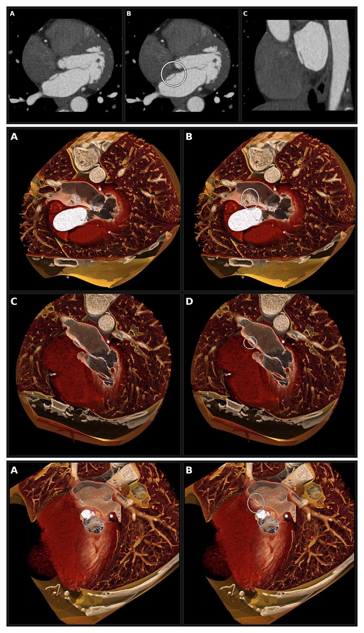
Figure 1
Conventional cross-sectional coronary computed tomography angiography. (A) Axial image at the level of the fossa ovalis. (B) Same axial image with annotated overlay (white circle) showing the septal pouch. (C) Double-orthogonal oblique sagittal reformat along the long axis of the pouch. Fenestrations are not resolved on any cross-sectional view. Figure 2 Cinematic rendering, left atrial perspective, pseudo-four-chamber-equivalent cut plane. (A) Unannotated view shows a smooth-walled left-sided atrial septal pouch superiorly, with the septum primum extending inferiorly into the fossa ovalis region. (B) Annotated overlay (white ellipse) marks the fenestrated patent foramen ovale within the inferior wall of the pouch; the multiple small dark foci correspond to discrete perforations of the septum primum. (C) Unannotated view at a more superior level through the pouch. (D) Annotated overlay (white circle) marks the pouch. Figure 3 Cinematic rendering, right atrial perspective. (A) Unannotated oblique view of the septal pouch from the right atrial side. (B) Annotated overlay (white ellipse) marks the corresponding fenestrations, confirming through-and-through interatrial communication.