
A 45-year-old woman presented with supraventricular tachycardia and frequent premature atrial and premature ventricular contractions. Electrocardiography-gated coronary computed tomography angiography (CCTA), performed to determine an ischemic etiology, showed normal coronary arteries and an incidental patent foramen ovale (PFO) with left-to-right shunting. Diastolic-phase data (65% to 85% of the R-R interval) were reconstructed at submillimeter slice thickness and post-processed with cinematic rendering, a Monte Carlo path-tracing technique, using virtual light sources positioned within the left atrium and a pseudo-four-chamber-equivalent cut plane.
Axial and double-orthogonal oblique reformats showed a left-sided atrial septal pouch (LSSP) but no discrete defect, contrast jet, or step-off in any plane (Figure 1). On cinematic rendering from the left atrium, the pouch appeared as a smooth-walled outpouching bounded inferiorly by the septum primum; in addition, the rendered septal surface displayed multiple discrete dark foci, each a perforation through which virtual illumination from the contralateral atrium passed unattenuated (Figure 2). The same fenestrations were seen independently from the right atrial perspective at the corresponding position, confirming through-and-through interatrial communication (Figure 3).
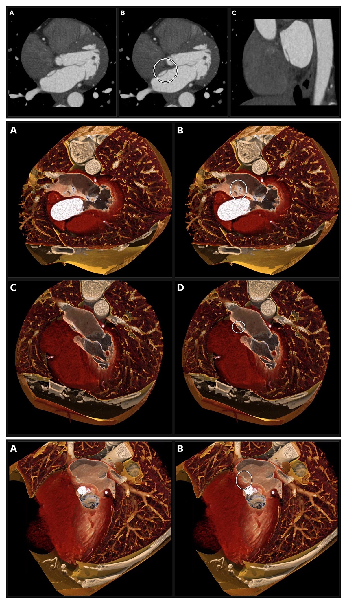
Figure 1
Conventional cross-sectional coronary computed tomography angiography. (A) Axial image at the level of the fossa ovalis. (B) Same axial image with annotated overlay (white circle) showing the septal pouch. (C) Double-orthogonal oblique sagittal reformat along the long axis of the pouch. Fenestrations are not resolved on any cross-sectional view. Figure 2 Cinematic rendering, left atrial perspective, pseudo-four-chamber-equivalent cut plane. (A) Unannotated view shows a smooth-walled left-sided atrial septal pouch superiorly, with the septum primum extending inferiorly into the fossa ovalis region. (B) Annotated overlay (white ellipse) marks the fenestrated patent foramen ovale within the inferior wall of the pouch; the multiple small dark foci correspond to discrete perforations of the septum primum. (C) Unannotated view at a more superior level through the pouch. (D) Annotated overlay (white circle) marks the pouch. Figure 3 Cinematic rendering, right atrial perspective. (A) Unannotated oblique view of the septal pouch from the right atrial side. (B) Annotated overlay (white ellipse) marks the corresponding fenestrations, confirming through-and-through interatrial communication.
Together the images show a fenestrated PFO within the fused inferior wall of an LSSP. Both entities are well described individually and are known to coexist.1,2 What these renderings add is resolution: the septum primum here is 1 to 2 mm thick, and partial volume averaging blurs perforations of this scale into adjacent septal tissue. Cinematic rendering depicts the same anatomy by transillumination of a thin structure rather than by attenuation display, resolving the defects as countable openings.3 The distinction is actionable: a multi-fenestrated septum may call for a device with a larger left atrial disc or a cribriform platform, and fenestrations remote from the dominant defect can leave residual right-to-left shunt after closure.4,5