
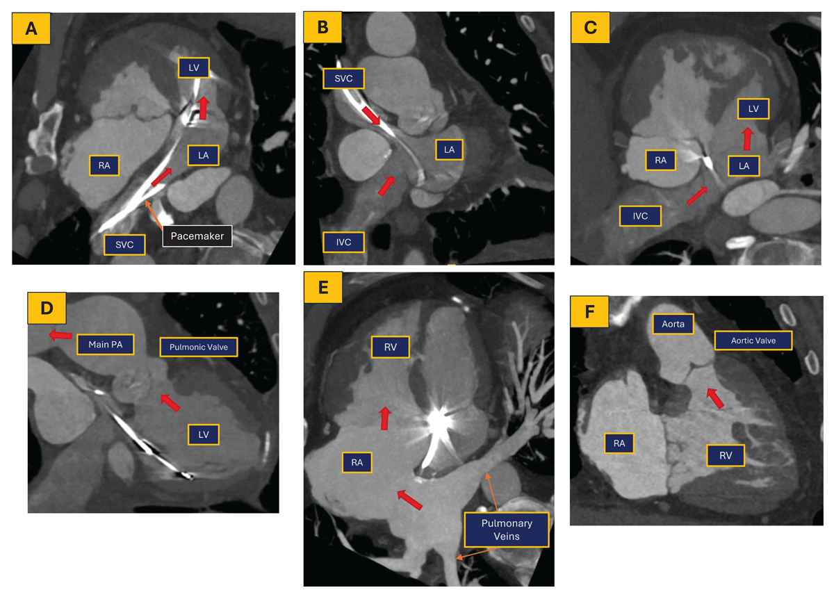
Figure 1
Patient with a history of dextro-transposition of the great arteries post atrial switch operation; coronary computed tomography angiography was performed to assess for coronary assessment and right atrial appendage thrombus assessment. The red arrow shows blood flow through the heart. LV: left ventricle, RV: right ventricle, LA: left atrium, RA: right atrium, PA: pulmonary artery, SVC: superior vena cava, IVC: inferior vena cava
Table 1
Suggested adult congenital heart disease transthoracic echocardiography protocol checklist by windows, views, and structural assessment. 2C: two-chamber; 2D: two-dimensional; 3C: three-chamber; 3D: three-dimensional; 4C: four-chamber; 5C: five-chamber; Ao: aorta; Abd Ao: abdominal aorta; AoV: aortic valve; AR: aortic regurgitation; ASD: atrial septal defect; Asc Ao: ascending aorta; CS: coronary sinus; CW: continuous-wave Doppler; DAo: descending aorta; FR: frame rate; GA: great arteries; IAS: interatrial septum; IVC: inferior vena cava; IVRT: isovolumic relaxation time; IVS: interventricular septum; LA: left atrium; LAA: left atrial appendage; LAX: long axis; LIV: left innominate vein; LPA: left pulmonary artery; LSVC: left superior vena cava; LV: left ventricle; LVOT: left ventricular outflow tract; MPA: main pulmonary artery; MR: mitral regurgitation; MV: mitral valve; PA: pulmonary artery; Pulm veins: pulmonary veins; Peric: pericardium; PLAX: parasternal long-axis view; PM: papillary muscle; PR: pulmonary regurgitation; PSAX: parasternal short-axis view; PV: pulmonary valve; PW: pulsed-wave Doppler; RA: right atrium; RAA: right atrial appendage; RCA: right coronary artery; RPA: right pulmonary artery; RV: right ventricle; RVIT: right ventricular inflow tract; RVOT: right ventricular outflow tract; SAX: short axis; SSN: suprasternal notch; SVC: superior vena cava; TAPSE: tricuspid annular plane systolic excursion; TDI: tissue Doppler imaging; TV: tricuspid valve; TR: tricuspid regurgitation; VSD: ventricular septal defect.
| ACHD VIEWS [2D, COLOR, PW, CW SEQUENCE]. ZOOM AND 3D AS NEEDED. COLOR FR > 20 | ✓ | |
|---|---|---|
| Left PLAX | ||
| 1 | PLAX: Panoramic PLAX view, zoom as needed (MV, LVOT-AoV) | |
| 2 | High PLAX: VSD patch, AoV, AR, Asc Ao, peric | |
| 3 | 2D/Color Sweep PLAX to RVIT: VSD. ASD, CS, eccentric jets | |
| 4 | PLAX RVIT: RV, TV & TR, RA, CS, IVC | |
| 5 | 2D/Color Sweep PLAX-RVOT: RVOT, PR, outlet VSD | |
| 6 | PLAX RVOT: RVOT, PV, MPA, LPA | |
| Left PSAX | ||
| 7 | 2D/Color Sweep apex to GA: LV, PM, RV IVS, MV, TV, IAS, LA, RA, AoV, RVOT, PV | |
| 8 | PSAX apex: LV, RV, IVS | |
| 9 | PSAX Mid level: LV, PM. RV, IVS | |
| 10 | PSAX MV: LV, RV, IVS, MV, TV, sweep MR | |
| 11 | PSAX GA: panoramic view | |
| 12 | PSAX GA: Zoom AoV | |
| 13 | PSAX GA: focus on LAA | |
| 14 | PSAX GA: focus on Pulm veins (ideally all) | |
| 15 | PSAX GA: focus IAS sweep | |
| 16 | PSAX GA: focus on TV, TR & IVC | |
| 17 | PSAX GA: focus on RVOT and PV, sweep PR | |
| 19 | PSAX GA: MPA. PA branches | |
| 20 | PSAX GA: coronary arteries | |
| APICAL | ||
| 21 | Apical 4C: panoramic view | |
| 22 | 2D/Color Sweep from CS to PV | |
| 23 | Apical 4C: Pulm veins, LA, MV, LV, RA, TV, RV, IAS, IVS, LV strain, TDI | |
| 24 | Apical SC: Pulm veins, LA, MV, LVOT, AoV, Asc Ao, RVOT, IVRT | |
| 25 | Apical 2C: Pulm veins, LA, LAA, MV, LV, CS, LV strain | |
| 26 27 | Apical 3C: Pulm veins, LA, MV, LV, LVOT, AoV, Asc Ao, IVS, RVOT, LV strain Apical 4C RV focused: RA, TV, RV, TDI, TAPSE, RV strain | |
| 28 | Apical RVOT @3: RVOT, PV | |
| 29 | Apical RV Oblique @1-2: SVC, RA, TV, RV, IVS, RVOT, PV, MPA, AoV | |
| 30 | Apical RV2C-@11: RA, TV, RV, IVC, CS | |
| SUBCOSTAL | ||
| 31 | Subcostal transverse @3: abominal Ao, IVC (visceroatrial situs), liver, stomach | |
| 32 | 2D/Color Sweep @3 from IVC/CS to PA | |
| 33 | Subcostal 4C @3: pulm veins, LA, LAA, MV, LV, IAS, IVS, RA, TV, RV | |
| 34 | Subcostal 5C @3: p veins, LA, LAA, MV, LV, LVOT, AoV | |
| 35 | Subcostal RVOT@3: RVOT, PV | |
| 36 | Subcostal RV oblique ~@1-2: RA, TV, RV. RVOT, PV, MPA, AoV | |
| 37 | 2D/Cotor Sweep SAX ~@1 apex to base | |
| 38 | Subcostal SAX GA ~@1: AoV, Pulm veins. LA, RA, IAS, IVS, TV, RVOT, PV, PAs | |
| 39 | Subcostal bicaval -@11: IVC, RA, RAA, SVC, LA, IAS | |
| 40 | Subcostal long-axis of IVC & hepatic veins | |
| 41 | Subcostal Abd Ao: LAX & SAX as needed | |
| SSN | ||
| 42 | 2D/Color Sweep SSN: arch sidedness and branching (SAX/LAX) | |
| 43 | SSN candy cane view ~@1 (left-sided Ao arch): LIV, Asc Ao, arch, DAo, RPA, LA | |
| 44 | SSN left tilt from @1: LPA, Dao, PDA, left pulm veins, LSVC, vertical vein | |
| 45 | SSN @3: LIV, Asc Ao, RPA, Pulm veins (crab view if possible), SVC | |
| SUPRACLAVICULAR | ||
| 46 | LT Supraclavicular ~@12: LSVC/Vertical Vein, Dao | |
| 47 | Rt Supraclavicular ~@12: RSVC, Asc Ao, AoV, RPA | |
| RIGHT PARASTERNAL–Right Lateral Decubitus position | ||
| 48 | Right Parasternal Bicaval ~@11: RAA, RA, IAS, LA, right pulm veins, SVC, IVC | |
| 49 | High Right Parasternal: AoV, asc Ao, RPA | |
| 50 | Right Parasternal @1 - 3: RAA, RA, IAS, LA, Ao. right pulm veins | |
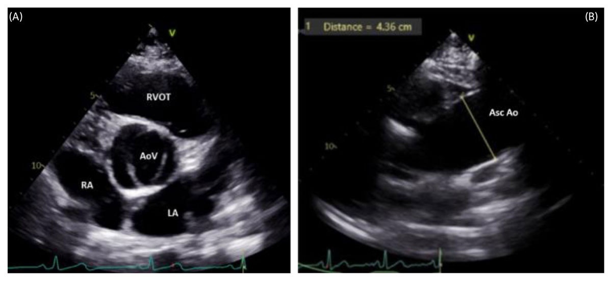
Figure 2
(A) Parasternal short-axis 2-dimensional transthoracic echocardiography (2D TTE) image in systole demonstrating a bicuspid AoV with two cusps/commissures and a characteristic “fish-mouth” appearance. (B) High parasternal long-axis 2D TTE view showing dilatation of the Asc Ao, commonly associated with bicuspid AoV. AoV: aortic valve; LA: left atrium; RA: right atrium; RVOT: right ventricular outflow tract; Asc Ao: ascending aorta
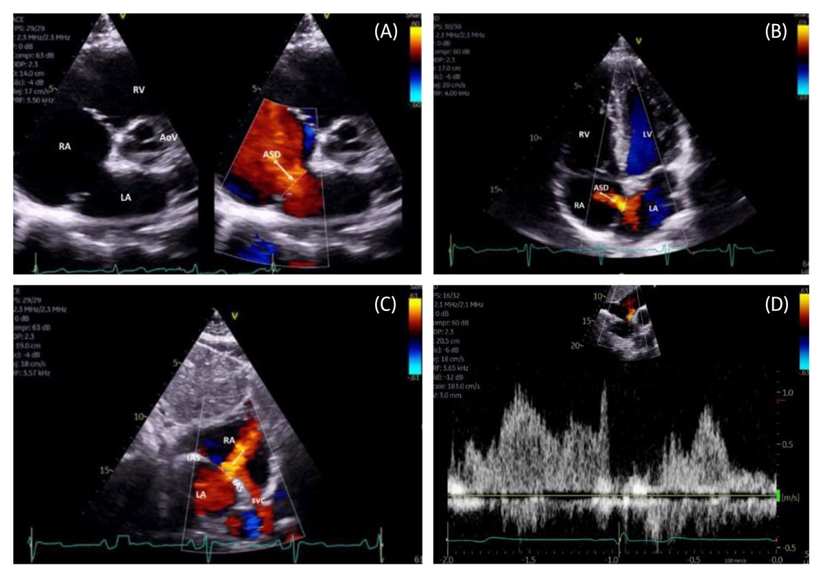
Figure 3
(A) Parasternal short-axis 2-dimensional (2D) color compare transthoracic echocardiography (TTE) image demonstrating a large ostium secundum ASD with left-to-right shunt. (B) Apical 4.chamber color Doppler TTE view showing an ostium secundum ASD with left-to-right shunt. (C) Subcostal bicaval color Doppler TTE view demonstrating an ostium secundum ASD with left-to-right shunt. (D) Subcostal short.axis pulsed.wave Doppler TTE of an ostium secundum ASD with left-to-right shunt. AoV: aortic valve; ASD: atrial septal defect (arrow/dash line); LA: left atrium; LV: left ventricle; RA: right atrium; RV: right ventricle; SVC: superior vena cava
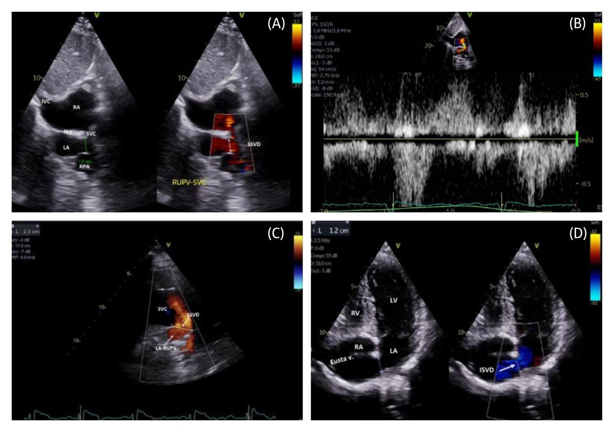
Figure 4
(A) Subcostal bicaval transthoracic echocardiography (TTE) color compare image demonstrating a large SSVD with bidirectional shunt, including a frame showing left-to-right flow from the RUPV ostium to the SVC. (B) Pulsed-wave Doppler demonstrating bidirectional shunt across the SSVD. (C) High right parasternal long-axis color Doppler TTE view of the SVC illustrating large SSVD size with left-to-right shunt from the RUPV to the SVC. (D) Apical 4-chamber view with inferior tilt, color Doppler TTE demonstrating a large inferior sinus venosus defect with left-to-right shunt below the Eustachian valve and tendon of Todaro. IAS: interatrial septum; IVC: inferior vena cava; LA: left atrium; LV: left ventricle; RA: right atrium; RV: right ventricle; RPA: right pulmonary artery; RUPV: right upper pulmonary vein ostium; SSVD: superior sinus venosus defect (green line/arrowed dash line); SVC: superior vena cava
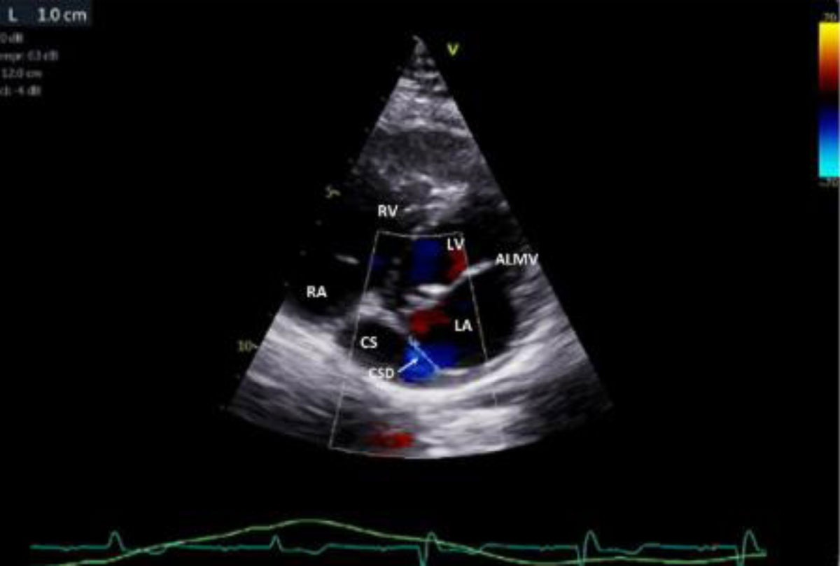
Figure 5
Intermediate parasternal short-axis view, color Doppler transthoracic echocardiography demonstrating an unrepaired coronary sinus defect with left-to-right shunt from the left atrium to the coronary sinus. ALMV: anterior leaflet mitral valve; CS: coronary sinus; CSD: coronary sinus defect; LA: left atrium; LV: left ventricle; RA: right atrium; RV: right ventricle.
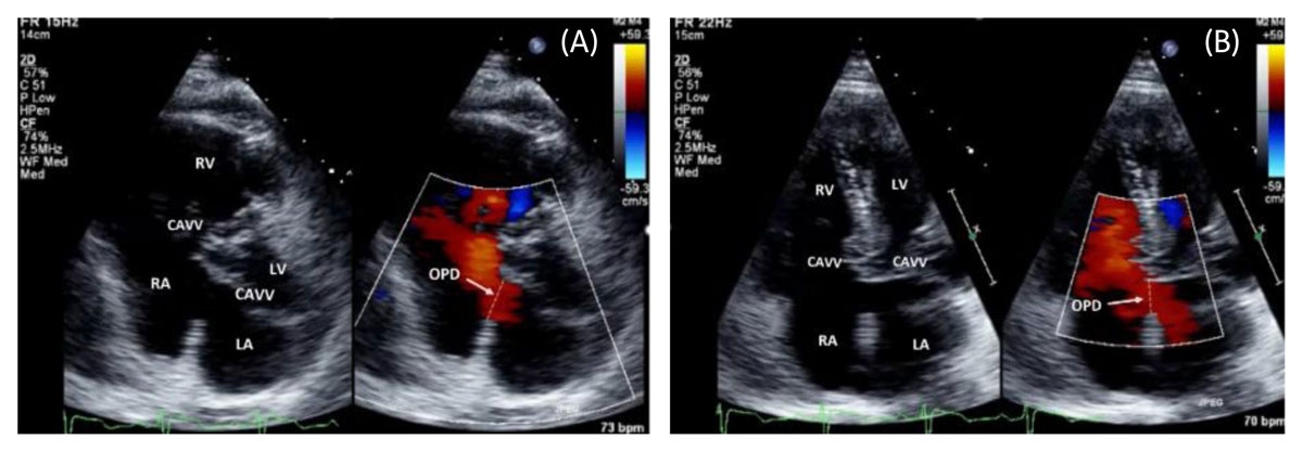
Figure 6
(A) Parasternal short-axis color Doppler transthoracic echocardiography (TTE) demonstrating an OPD with left-to-right shunt in a patient with atrioventricular septal defect. (B) Apical 4-chamber color compare TTE view demonstrating the OPD component of an atrioventricular septal defect with left-to-right shunt. CAVV: common atrioventricular valve; LA: left atrium; LV: left ventricle; RA: right atrium; RV: right ventricle; OPD: ostium primum defect (arrow, dash line)
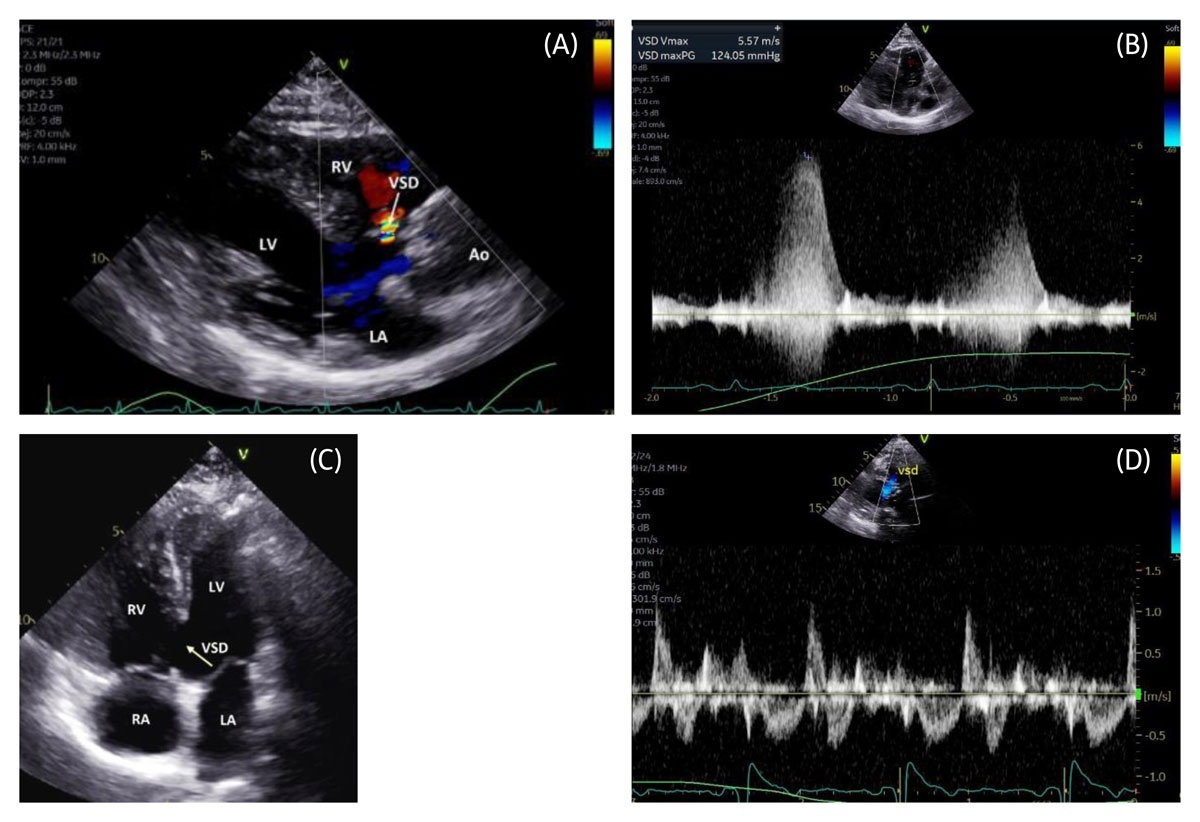
Figure 7
(A) Parasternal long-axis color Doppler transthoracic echocardiography (TTE) demonstrating a small perimembranous VSD with left-to-right shunt. (B) Color Doppler demonstrating a pressure-restrictive perimembranous VSD with left-to-right shunt. (C) Apical 4-chamber 2-dimensional TTE view demonstrating a large inlet VSD. (D) Pulsed-wave Doppler demonstrating a large bidirectional unrestrictive VSD. Ao: aorta; LA: left atrium; LV: left ventricle; RA: right atrium; RV: right ventricle; VSD: ventricular septal defect (arrow, dash line)
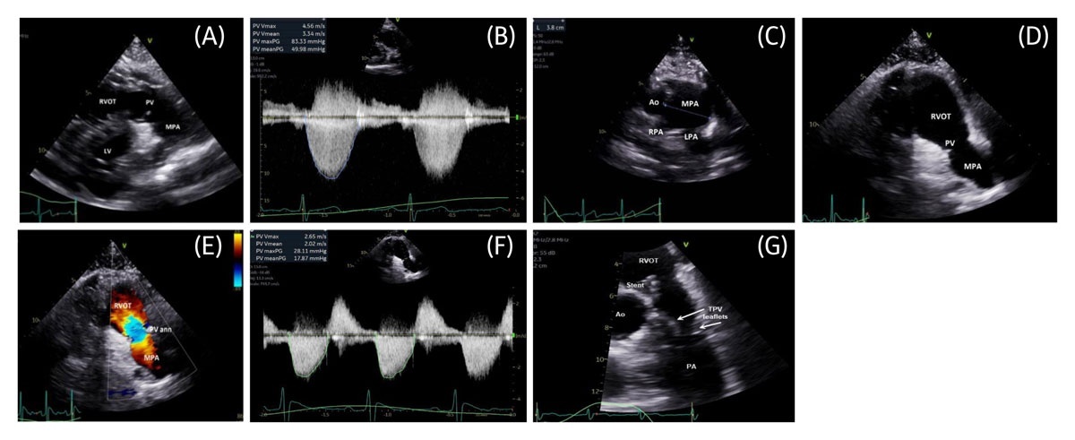
Figure 8
(A) Parasternal short-axis 2-dimensional (2D) transthoracic echocardiography (TTE) demonstrating a stenotic PV with tethered, doming leaflets. (B) Color Doppler waveform demonstrating severe congenital PV stenosis. (C) High parasternal short-axis 2D TTE demonstrating post-stenotic dilatation of the pulmonary trunk. (D) Parasternal short-axis 2D TTE demonstrating a surgically disrupted PV with near absence of leaflet tissue. (E) Parasternal short-axis color Doppler TTE demonstrating severe PV regurgitation with some degree of stenosis following valvectomy. (F) Color Doppler waveform demonstrating mild residual stenosis and severe pulmonary regurgitation. (G) 2D TTE zoomed parasternal short-axis view demonstrating a transcatheter PV replacement with stent in the RVOT and valve leaflets in systole. Ao: aorta; LV: left ventricle; LPA: left pulmonary artery; MPA: main pulmonary artery; PA: pulmonary artery; PV: pulmonary valve; PV ann: pulmonary valve annulus; RPA: right pulmonary artery; RVOT: right ventricular outflow tract; TVP: transcatheter pulmonary valve
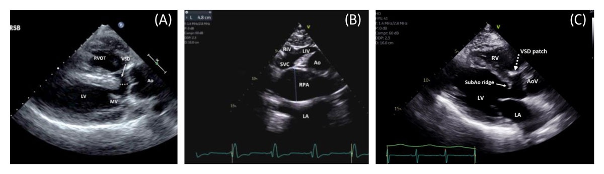
Figure 9
(A) Parasternal long-axis 2-dimensional (2D) transthoracic echocardiography (TTE) demonstrating unrepaired tetralogy of Fallot with overriding Ao and associated VSD. (B) Suprasternal notch 2D TTE demonstrating an aneurysmal RPA in tetralogy of Fallot with absent pulmonary valve. (C) Parasternal long-axis 2D TTE demonstrating repaired tetralogy of Fallot with a large VSD patch and severely dilated RV. Ao: aorta; AoV: aortic valve; LA: left atrium; LV: left ventricle; MV: mitral valve; RV: right ventricle; RVOT: right ventricular outflow tract; RPA: right pulmonary artery; SVC: superior vena cava; LIV: left innominate vein; RIV: right innominate vein; SubAo: subaortic; VSD: ventricular septal defect (overriding the aorta in dash line)
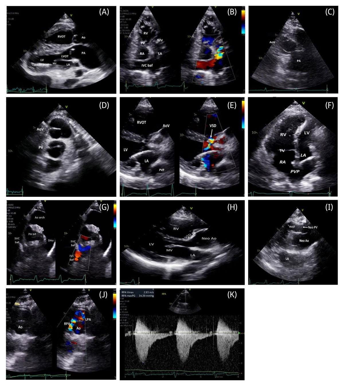
Figure 10
(A) Parasternal long-axis 2-dimensional (2D) transthoracic echocardiography (TTE) demonstrating parallel great arteries in d-transposition of the great arteries (d-TGA) post–atrial switch, with ventriculoarterial discordance and pacer lead in the subpulmonary LV. (B) Intermediate apical TTE color-compare image demonstrating IVC baffle flow to the subpulmonary MV. (C) Parasternal short-axis 2D TTE demonstrating great artery relationship in d-TGA post–atrial switch with the Ao anterior and rightward to the pulmonary trunk. (D) Parasternal short-axis 2D TTE again demonstrating the Ao directly anterior to the pulmonary trunk. (E) Intermediate parasternal long-axis color-compare TTE demonstrating d-TGA with VSD, showing systemic-to-pulmonary shunt across the VSD and pacer lead in the subpulmonary left chambers. (F) Apical 4-chamber 2D TTE demonstrating a dilated, hypertrophied systemic RV and PVP to the RA post–Mustard procedure. (G) Intermediate suprasternal notch color TTE demonstrating SVC and IVC baffle flows post–atrial switch. (H) Parasternal long-axis 2D TTE demonstrating VSD patch and Neo Ao post–arterial switch. (I) High parasternal long-axis 2D TTE demonstrating parallel great arteries post–arterial switch with Neo PV. (J) Intermediate parasternal short-axis color-compare TTE demonstrating the LeCompte maneuver with pulmonary arteries anterior to the ascending Ao and associated RPA stenosis. (K) Color Doppler waveform demonstrating RPA stenosis. Ao: aorta; Ao arch: aortic arch; AoV: aortic valve; DAo: descending aorta; IVC baf: inferior vena cava baffle; LA: left atrium; LIV: left innominate vein; LV: left ventricle; LVOT: left ventricular outflow tract; MV: mitral valve; Neo Ao: neoaorta; Neo PV: neo pulmonary valve (arrow); PA: pulmonary artery; PA bif: pulmonary artery bifurcation; PVP: pulmonary venous pathway; RA: right atrium; RPA: right pulmonary artery; RV: right ventricle; RVOT: right ventricular outflow tract; SVC baf: superior vena cava baffle; TV: tricuspid valve; VSD: ventricular septal defect (arrow/dash line); VSD p: ventricular septal defect patch
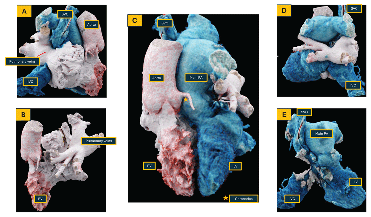
Figure 11
Patient with a history of dextro-transposition of the great arteries post atrial switch operation; coronary computed tomography angiography was performed to assess for coronary and right atrial appendage thrombus. 3D reconstruction. LV: left ventricle; RV: right ventricle; LA: left atrium; RA: right atrium; PA: pulmonary artery; SVC: superior vena cava; IVC: inferior vena cava
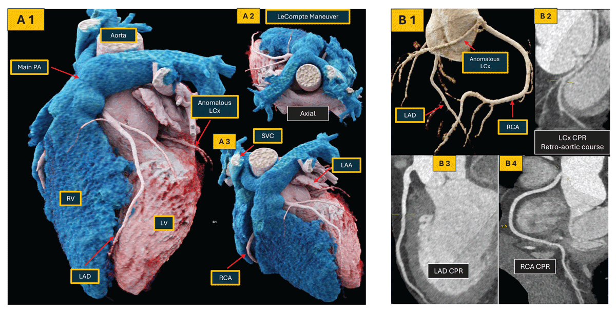
Figure 12
Computed tomography dextro-transposition of the great arteries (d-TGA) after arterial switch procedure with anomalous LCx with a retroaortic course. (A 1-3) 3-dimensional reconstruction of a patient with d-TGA after an arterial switch and LeCompte Maneuver; (B 1-4) curved multiplanar reconstructions of the coronary tree. Note that LCx has an anomalous retro-aortic course after arising from the RCA. LV: left ventricle; RV: right ventricle; PA: pulmonary artery; SVC: superior vena cava; LAA: left atrial appendage; LAD: left anterior descending artery; LCx: left circumflex artery; RCA: right coronary artery.
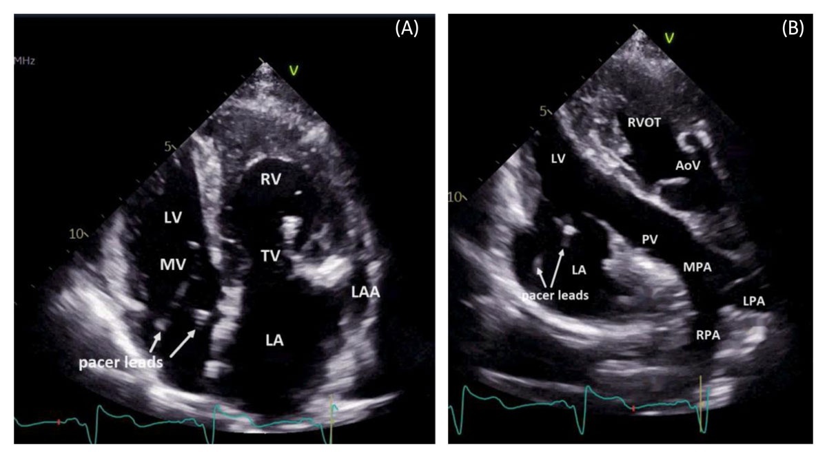
Figure 13
(A) Apical 4-chamber 2-dimensional (2D) transthoracic echocardiography (TTE) demonstrating situs solitus congenitally corrected transposition of the great arteries. (B) Apical LV 3-chamber 2D TTE demonstrating RA-LV, LV-PA, and RV-Ao connections in congenitally corrected transposition of the great arteries. AoV: aortic valve; RA: right atrium; LA: left atrium; LAA: left atrial appendage; LV: left ventricle; MV: mitral valve; MPA: main pulmonary artery; LPA: left pulmonary artery; PV: pulmonary valve; RPA: right pulmonary artery; RV: right ventricle; RVOT: right ventricular outflow tract; TV: tricuspid valve
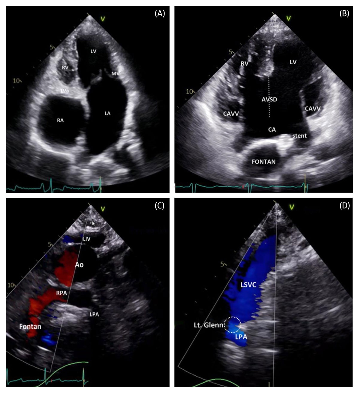
Figure 14
(A) Apical 4-chamber 2-dimensional (2D) transthoracic echocardiography (TTE) demonstrating tricuspid atresia palliated with atriopulmonary Fontan, with plate-like TV atresia and hypoplastic RV. (B) 2D TTE demonstrating an atrioventricular septal defect palliated with Fontan, including a Fontan fenestration stent. (C) Intermediate suprasternal notch color TTE demonstrating an unobstructed Fontan–RPA anastomosis. (D) Left supraclavicular color TTE demonstrating a patent left bidirectional Glenn anastomosis (LSVC–LPA connection). Ao: aorta; CA: common atrium; CAVV: common atrioventricular valve; LA: left atrium; LV: left ventricle; LIV: left innominate vein; Lt. Glenn: left bidirectional Glenn anastomosis (dash circle); RA: right atrium; RPA: right pulmonary artery; RV: right ventricle; MV: mitral valve; TVa: tricuspid valve atresia; LSVC-LPA: left superior vena cava-left pulmonary artery
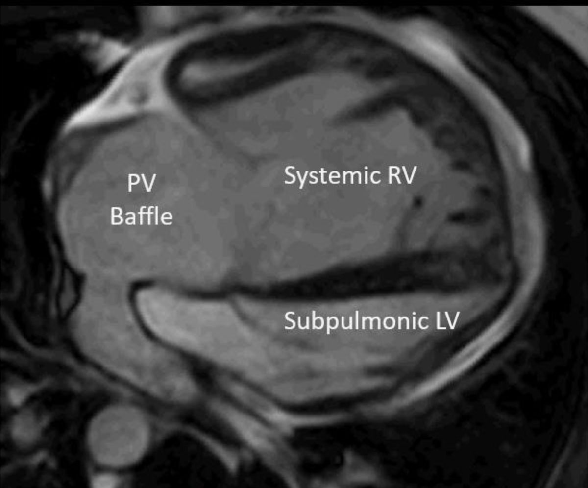
Figure 15
Dilated systemic RV in a patient with d-transposition of the great arteries and atrial switch. PV Baffle: pulmonary venous baffle; RV: right ventricle; LV: left ventricle
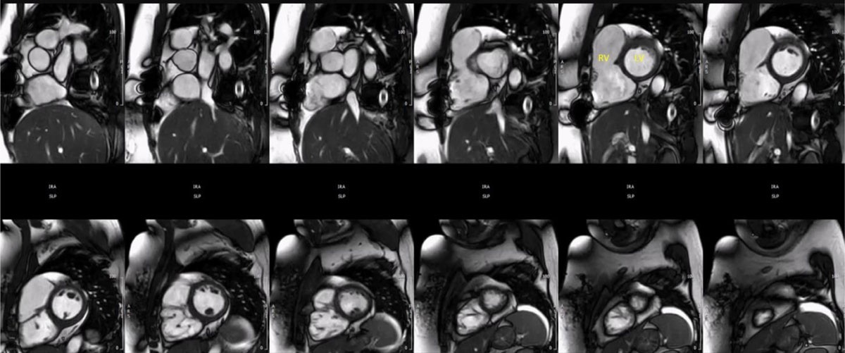
Figure 16
Cardiac magnetic resonance short axis cine imaging stack demonstrating a severely dilated RV in a patient with tetralogy of Fallot with a transannular patch repair and severe pulmonary regurgitation. RV: right ventricle; LV: left ventricle
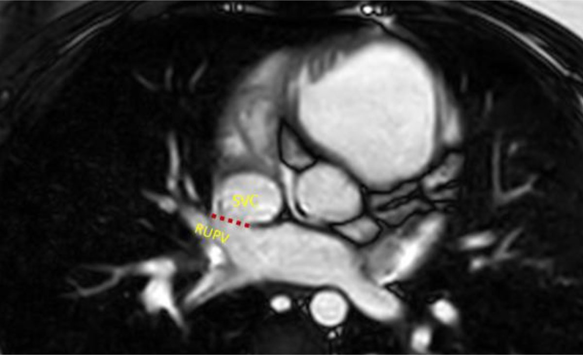
Figure 17
Cardiac magnetic resonance cine imaging in the axial plane demonstrating a sinus venosus defect (red dotted line). SVC: superior vena cava; RUPV: right upper pulmonary vein
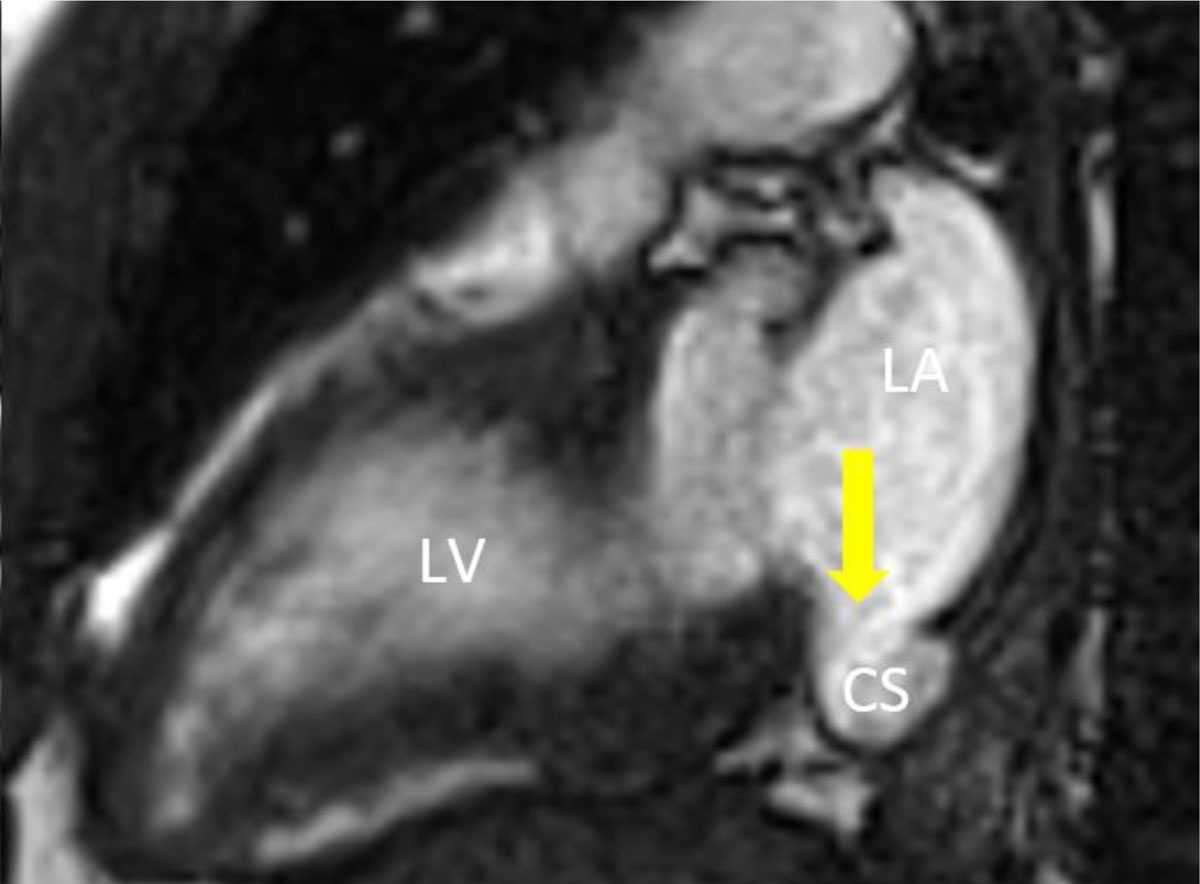
Figure 18
Cardiac magnetic resonance cine imaging of an unroofed coronary sinus defect (arrow) in the two-chamber view. LA: left atrium; CS: coronary sinus; LV: left ventricle
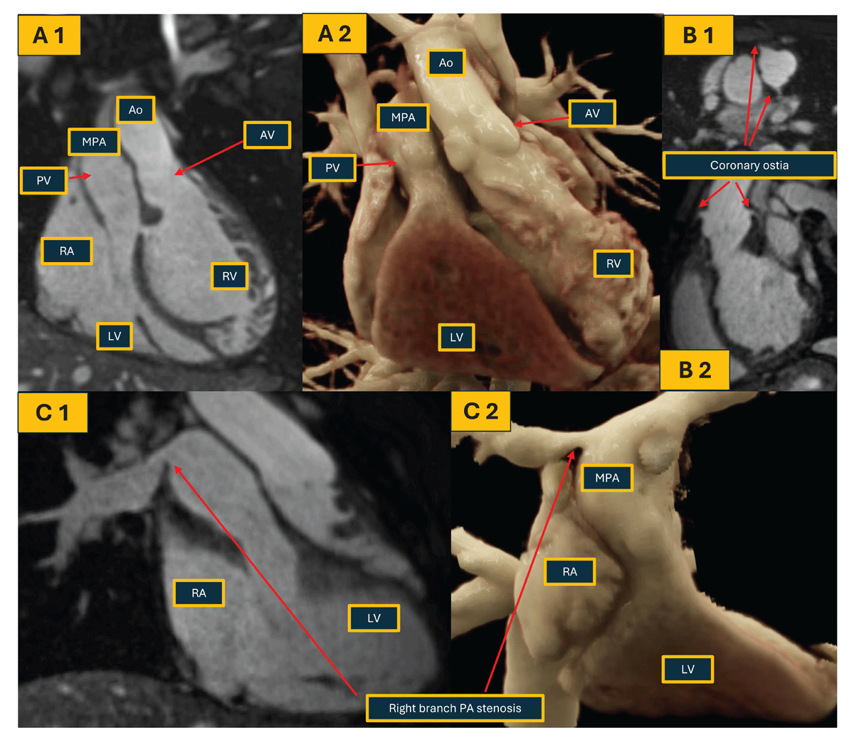
Figure 19
Non-contrast magnetic resonance imaging in a patient with congenitally corrected transposition of the great arteries (CCTGA). (A 1,2) Coronal view of heart and great vessels in 2D and 3D reconstruction demonstrating levo-TGA; (B 1,2) visualization of coronary ostia due to improved spatial resolution (1.6 × 1.6 × 1.6 mm); (C 1,2) identification of right branch pulmonary artery stenosis and post stenotic dilation. RA: right atrium; LA: left atrium; RV: right ventricle; LV: left ventricle; MPA: main pulmonary artery; AV: aortic valve; PV: pulmonic valve; Ao: aorta
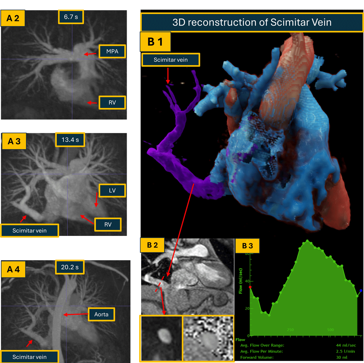
Figure 20
Utilization of time resolved MRA and direct measurement of anomalous pulmonary venous return flow in a patient with Scimitar syndrome (anomalous right lower pulmonary vein). (A 1 Video) Maximal intensity projection demonstrating filling of the Scimitar vein. (A 2-4) Maximal intensity projection of time resolved MRA demonstrating filling of the Scimitar vein. Last few frames also demonstrate persistent left superior vena cava draining into coronary sinus. (B 1) 3D reconstruction of the Scimitar vein; (B 2) GRE image of the Scimitar vein draining into the IVC; (B 3) Flow quantification of the Scimitar vein, direct anomalous pulmonary return flow is 30 mL/beat, 2.5 L/min. MRA: magnetic resonance angiography RV: right ventricle; LV: left ventricle; MPA: main pulmonary artery