
Introduction
Adult congenital heart disease (ACHD) encompasses a broad clinical spectrum, ranging from isolated defects such as atrial/ventricular septal defects (ASD/VSD) and anomalous pulmonary venous return (PAPVR) to complex anatomies including congenitally corrected transposition of the great arteries (CCTGA) and single-ventricle physiology palliated with Fontan circulation. Surgical advances over the past several decades dramatically improved survival for patients with complex congenital heart disease, but the long-term anatomic and physiological sequelae of these repairs have become increasingly apparent as this population ages into adulthood.1,2
Cardiovascular imaging is central to every stage of ACHD care, informing diagnosis, guiding intervention, and supporting longitudinal surveillance.3,4 Transthoracic echocardiography (TTE) remains foundational, yet the complexity of repaired anatomy, altered hemodynamics, and extracardiac pathways frequently necessitates complementary cross-sectional imaging. Cardiac magnetic resonance (CMR) and cardiovascular computed tomography (CCT) provide comprehensive anatomic and functional assessment, flow quantification, and evaluation of surgical repairs.5
This review for general cardiologists and cardiac imagers provides a practical, structured imaging pathway for adults with suspected or known congenital heart disease, with emphasis on modality selection, protocol standardization, and the diagnostic challenges commonly encountered in contemporary ACHD practice. We propose a standardized, efficient imaging sequence starting with (1) anatomy and history, (2) protocolized ACHD TTE, (3) focused CMR with flows, and (4) targeted CT when needed. Each step is designed to answer specific clinical questions and to inform whether and how subsequent modalities should be employed.
Foundational Anatomical and Physiological Assessment for Imaging Prior to Protocol Development
Initial assessment of adults with CHD can be challenging, particularly when native anatomy has been substantially altered by prior surgical repair. Whenever possible, clinicians should obtain previous clinical notes, operative reports, catheterization reports, echocardiograms, and cross-sectional imaging studies such as CCT or magnetic resonance imaging (MRI). These records are invaluable in clarifying native anatomy, post-surgical anatomy, and associated anomalies such as interrupted systemic venous return, aortic arch anomalies, heterotaxy, or situs abnormalities.
When reliable prior documentation is unavailable, the initial evaluation should be deliberately comprehensive, with the explicit goal of reestablishing the patient’s underlying anatomy and physiology. A useful framework for navigating complex congenital anatomy is to anchor the assessment to a fundamental physiological constant: blood flow is continuous. Deoxygenated blood must reach the pulmonary circulation, oxygenated blood must return to the heart, and systemic cardiac output must deliver oxygenated blood to the end organs. In practice, sequentially “following the circulation” through the cardiovascular system provides a methodical, consistent, and reproducible diagnostic framework that is particularly useful in complex CHD (Figure 1).
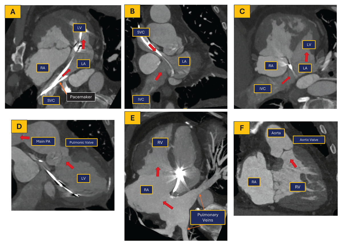
Figure 1
Patient with a history of dextro-transposition of the great arteries post atrial switch operation; coronary computed tomography angiography was performed to assess for coronary assessment and right atrial appendage thrombus assessment. The red arrow shows blood flow through the heart. LV: left ventricle, RV: right ventricle, LA: left atrium, RA: right atrium, PA: pulmonary artery, SVC: superior vena cava, IVC: inferior vena cava
A structured, stepwise approach should therefore define the following structures in addition to intracardiac anatomy: (1) systemic venous return to the heart (or the pulmonary arteries in patients with cavopulmonary anastomosis), (2) pulmonary arterial outflow, (3) pulmonary venous return, and (4) systemic arterial outflow via the aorta and branches. TTE with color Doppler can delineate many of these connections when performed using a comprehensive, anatomy-directed protocol. In adults with limited acoustic windows or complex anatomy, CMR can further refine anatomic definition and physiological characterization with important advantages, including unrestricted imaging plane selection, robust biventricular volumetric assessment, and qualitative and quantitative flow evaluation using phase-contrast sequences for the evaluation of valves, conduits, baffles, and vascular structures.6
Once baseline cardiovascular anatomy has been established, TTE becomes the cornerstone of longitudinal surveillance in ACHD, enabling serial assessment of ventricular size and function; valvular morphology and hemodynamics; right ventricle (RV) and pulmonary artery systolic pressure; baffle patency and function; cardiac masses; and residual or recurrent lesions within native and surgically modified pathways. Abnormal or clinically significant findings should prompt complementary evaluation with CMR, CCT, or cardiac catheterization, as clinically indicated.3,4,7,8
Adult Congenital Transthoracic Echocardiography
Transthoracic echocardiography is an essential component of the multimodality imaging approach to ACHD, playing a central role in both initial assessment and lifelong clinical surveillance.3,4,9
Historically, TTE in patients with ACHD was performed primarily by pediatric echocardiographers in children’s hospitals. However, as survival has improved and acquired comorbidities have become more prevalent, these patients are increasingly receiving care in adult centers. Reflecting the specialized imaging needs of this population, the Intersocietal Accreditation Commission (IAC) introduced the Adult Congenital Transthoracic Echocardiography accreditation in 2023 as a distinct examination, separate from standard adult and pediatric echocardiography. Facilities performing ACHD TTE must also maintain adult TTE accreditation, and interpreting physicians must hold board certification in ACHD, be board eligible, or demonstrate substantial expertise in complex ACHD echocardiography. Sonographers must maintain active credentials in both adult and pediatric echocardiography, making ACHD TTE the only echocardiographic accreditation that requires dual registry.
ACHD TTE is a highly operator-dependent examination in which meticulous image optimization is essential for accurate diagnosis. Comprehensive two-dimensional (2D) and color Doppler sweeps are necessary to define cardiac connections, residual lesions, and postoperative anatomy.
ACHD TTE examinations commonly require 60 to 90 minutes, particularly during initial evaluations and in patients with complex congenital anatomy or technically challenging studies. Effective communication between the sonographer and the interpreting ACHD echocardiographer is essential, particularly when new or clinically significant findings are identified, and joint image review is often necessary. Referral of patients with ACHD to centers with specialized expertise is recommended to optimize clinical outcomes.9 When this is not feasible, studies should be performed and interpreted by the most experienced available sonographer and echocardiographer.
ACHD TTE Protocol
The standard adult TTE protocol, while comprehensive for acquired heart disease, can be limited for the evaluation of adults with CHD.10 A comprehensive ACHD TTE protocol must systematically assess unrepaired and repaired congenital lesions, including associated defects, residual lesions, and postoperative complications. Specialized surgical repairs require targeted protocol modifications and tailored imaging approaches. The protocol should include cardiac situs, morphology, and sequential segmental analysis, together with structural and functional assessment from additional echocardiographic windows and imaging planes beyond those used in standard adult or pediatric examinations.11,12 Protocol implementation should remain consistent with the conventions and workflow of the individual laboratory, including apex orientation and preferred initial imaging window. Before image acquisition, the sonographer should review the patient’s clinical history, prior imaging, and operative reports to understand baseline anatomy, prior repairs, residual lesions, and potential complications, all of which should be documented in the echocardiographic report.
ACHD echocardiographic standards for imaging, interpretation, and reporting continue to evolve.7,8,12,13,14,15,16,17 A suggested ACHD echocardiographic assessment checklist is provided in Table 1. Intermediate windows and nonstandard planes are often necessary for comprehensive evaluation. A comprehensive ACHD TTE examination should include 2D imaging, M-mode, Doppler interrogation (color, pulsed-wave, continuous-wave, and tissue Doppler), and longitudinal strain assessment. Three-dimensional echocardiography provides additional information on valvular anatomy and mechanism of dysfunction, defect size and morphology, chamber volumes, and spatial relationships between cardiac structures that may aid procedural planning.
Table 1
Suggested adult congenital heart disease transthoracic echocardiography protocol checklist by windows, views, and structural assessment. 2C: two-chamber; 2D: two-dimensional; 3C: three-chamber; 3D: three-dimensional; 4C: four-chamber; 5C: five-chamber; Ao: aorta; Abd Ao: abdominal aorta; AoV: aortic valve; AR: aortic regurgitation; ASD: atrial septal defect; Asc Ao: ascending aorta; CS: coronary sinus; CW: continuous-wave Doppler; DAo: descending aorta; FR: frame rate; GA: great arteries; IAS: interatrial septum; IVC: inferior vena cava; IVRT: isovolumic relaxation time; IVS: interventricular septum; LA: left atrium; LAA: left atrial appendage; LAX: long axis; LIV: left innominate vein; LPA: left pulmonary artery; LSVC: left superior vena cava; LV: left ventricle; LVOT: left ventricular outflow tract; MPA: main pulmonary artery; MR: mitral regurgitation; MV: mitral valve; PA: pulmonary artery; Pulm veins: pulmonary veins; Peric: pericardium; PLAX: parasternal long-axis view; PM: papillary muscle; PR: pulmonary regurgitation; PSAX: parasternal short-axis view; PV: pulmonary valve; PW: pulsed-wave Doppler; RA: right atrium; RAA: right atrial appendage; RCA: right coronary artery; RPA: right pulmonary artery; RV: right ventricle; RVIT: right ventricular inflow tract; RVOT: right ventricular outflow tract; SAX: short axis; SSN: suprasternal notch; SVC: superior vena cava; TAPSE: tricuspid annular plane systolic excursion; TDI: tissue Doppler imaging; TV: tricuspid valve; TR: tricuspid regurgitation; VSD: ventricular septal defect.
| ACHD VIEWS [2D, COLOR, PW, CW SEQUENCE]. ZOOM AND 3D AS NEEDED. COLOR FR > 20 | ✓ | |
|---|---|---|
| Left PLAX | ||
| 1 | PLAX: Panoramic PLAX view, zoom as needed (MV, LVOT-AoV) | |
| 2 | High PLAX: VSD patch, AoV, AR, Asc Ao, peric | |
| 3 | 2D/Color Sweep PLAX to RVIT: VSD. ASD, CS, eccentric jets | |
| 4 | PLAX RVIT: RV, TV & TR, RA, CS, IVC | |
| 5 | 2D/Color Sweep PLAX-RVOT: RVOT, PR, outlet VSD | |
| 6 | PLAX RVOT: RVOT, PV, MPA, LPA | |
| Left PSAX | ||
| 7 | 2D/Color Sweep apex to GA: LV, PM, RV IVS, MV, TV, IAS, LA, RA, AoV, RVOT, PV | |
| 8 | PSAX apex: LV, RV, IVS | |
| 9 | PSAX Mid level: LV, PM. RV, IVS | |
| 10 | PSAX MV: LV, RV, IVS, MV, TV, sweep MR | |
| 11 | PSAX GA: panoramic view | |
| 12 | PSAX GA: Zoom AoV | |
| 13 | PSAX GA: focus on LAA | |
| 14 | PSAX GA: focus on Pulm veins (ideally all) | |
| 15 | PSAX GA: focus IAS sweep | |
| 16 | PSAX GA: focus on TV, TR & IVC | |
| 17 | PSAX GA: focus on RVOT and PV, sweep PR | |
| 19 | PSAX GA: MPA. PA branches | |
| 20 | PSAX GA: coronary arteries | |
| APICAL | ||
| 21 | Apical 4C: panoramic view | |
| 22 | 2D/Color Sweep from CS to PV | |
| 23 | Apical 4C: Pulm veins, LA, MV, LV, RA, TV, RV, IAS, IVS, LV strain, TDI | |
| 24 | Apical SC: Pulm veins, LA, MV, LVOT, AoV, Asc Ao, RVOT, IVRT | |
| 25 | Apical 2C: Pulm veins, LA, LAA, MV, LV, CS, LV strain | |
| 26 27 | Apical 3C: Pulm veins, LA, MV, LV, LVOT, AoV, Asc Ao, IVS, RVOT, LV strain Apical 4C RV focused: RA, TV, RV, TDI, TAPSE, RV strain | |
| 28 | Apical RVOT @3: RVOT, PV | |
| 29 | Apical RV Oblique @1-2: SVC, RA, TV, RV, IVS, RVOT, PV, MPA, AoV | |
| 30 | Apical RV2C-@11: RA, TV, RV, IVC, CS | |
| SUBCOSTAL | ||
| 31 | Subcostal transverse @3: abominal Ao, IVC (visceroatrial situs), liver, stomach | |
| 32 | 2D/Color Sweep @3 from IVC/CS to PA | |
| 33 | Subcostal 4C @3: pulm veins, LA, LAA, MV, LV, IAS, IVS, RA, TV, RV | |
| 34 | Subcostal 5C @3: p veins, LA, LAA, MV, LV, LVOT, AoV | |
| 35 | Subcostal RVOT@3: RVOT, PV | |
| 36 | Subcostal RV oblique ~@1-2: RA, TV, RV. RVOT, PV, MPA, AoV | |
| 37 | 2D/Cotor Sweep SAX ~@1 apex to base | |
| 38 | Subcostal SAX GA ~@1: AoV, Pulm veins. LA, RA, IAS, IVS, TV, RVOT, PV, PAs | |
| 39 | Subcostal bicaval -@11: IVC, RA, RAA, SVC, LA, IAS | |
| 40 | Subcostal long-axis of IVC & hepatic veins | |
| 41 | Subcostal Abd Ao: LAX & SAX as needed | |
| SSN | ||
| 42 | 2D/Color Sweep SSN: arch sidedness and branching (SAX/LAX) | |
| 43 | SSN candy cane view ~@1 (left-sided Ao arch): LIV, Asc Ao, arch, DAo, RPA, LA | |
| 44 | SSN left tilt from @1: LPA, Dao, PDA, left pulm veins, LSVC, vertical vein | |
| 45 | SSN @3: LIV, Asc Ao, RPA, Pulm veins (crab view if possible), SVC | |
| SUPRACLAVICULAR | ||
| 46 | LT Supraclavicular ~@12: LSVC/Vertical Vein, Dao | |
| 47 | Rt Supraclavicular ~@12: RSVC, Asc Ao, AoV, RPA | |
| RIGHT PARASTERNAL–Right Lateral Decubitus position | ||
| 48 | Right Parasternal Bicaval ~@11: RAA, RA, IAS, LA, right pulm veins, SVC, IVC | |
| 49 | High Right Parasternal: AoV, asc Ao, RPA | |
| 50 | Right Parasternal @1 - 3: RAA, RA, IAS, LA, Ao. right pulm veins | |
The examination should systematically assess:
Cardiac situs, cardiac position, apex orientation, and sequential segmental anatomy,
Systemic venous anatomy and flow, including the superior and inferior vena cava, innominate vein, hepatic veins, and coronary sinus,
Pulmonary venous connections and flow, ideally including all pulmonary veins,
Atrial morphology, size and function, and appendages,
Interatrial septum,
Atrioventricular valves morphology and function,
Ventricular morphology, size, and function,
Interventricular septum,
Ventricular outflow tracts size and patency,
Semilunar valves morphology and function,
Coronary artery origins and proximal patency,
Arch sidedness, aortic dimensions, and flow, including the aortic root, ascending, transverse, descending, and abdominal aorta,
Main and branch pulmonary artery anatomy and flow, and
Pericardial anatomy and effusion assessment.
Common congenital lesions encountered in adult echocardiography laboratories include bicuspid aortic valve, ASDs, ventricular septal defects, and pulmonary stenosis. However, patients with more complex disease—such as tetralogy of Fallot, TGA, multilevel left-sided obstructive lesions, SVP or complex repairs and palliations including cavopulmonary anastomosis, Ross procedure, Rastelli, conduits, and baffle repairs—should be evaluated at specialized ACHD centers whenever possible.
Imaging-specific Congenital Lesions
Left-Sided Obstructive Lesions
Bicuspid aortic valve and congenital aortic stenosis are among the most common forms of congenital heart disease and may coexist with aortopathy and multilevel left-sided obstruction such as congenital mitral stenosis, subaortic stenosis, and coarctation of the aorta, as seen in Shone’s complex. Echocardiographic assessment should define left ventricular (LV) size and function, left ventricular outflow tract (LVOT) size and patency, aortic valve morphology and hemodynamics, stenosis jet from multiple windows, aortic regurgitation, aortic dimensions, and associated lesions (Figure 2 A, B).
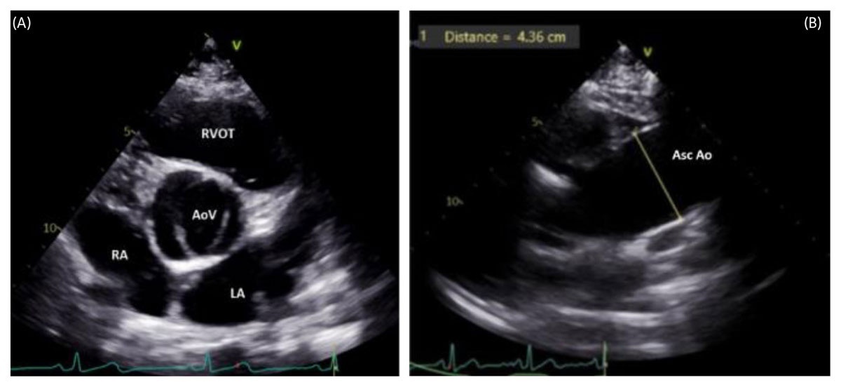
Figure 2
(A) Parasternal short-axis 2-dimensional transthoracic echocardiography (2D TTE) image in systole demonstrating a bicuspid AoV with two cusps/commissures and a characteristic “fish-mouth” appearance. (B) High parasternal long-axis 2D TTE view showing dilatation of the Asc Ao, commonly associated with bicuspid AoV. AoV: aortic valve; LA: left atrium; RA: right atrium; RVOT: right ventricular outflow tract; Asc Ao: ascending aorta
Management is guided by the level and severity of obstruction. Surgical options range from isolated aortic valve replacement to the Ross operation, which requires lifelong surveillance for right ventricle to pulmonary artery conduit degeneration, neoaortic valve dysfunction, and progressive neoaortic root dilation. Percutaneous interventions may include aortic balloon valvuloplasty, transcatheter aortic valve replacement, and stenting for coarctation of the aorta.
Interatrial Communications
The interatrial septum should be systematically evaluated using comprehensive 2D and color Doppler sweeps from multiple imaging windows.18 Optimization of color Doppler settings is particularly important in the presence of pulmonary arterial hypertension. Evaluation should define defect location, size, number, morphology, shunt direction and magnitude, right atrial and RV size and function, RV systolic pressure (RVSP) and pulmonary artery systolic pressure (PASP), LV diastolic function, associated lesions, and residual defects.
Patent foramen ovale and secundum ASD are the most common interatrial communications (Figure 3 A-D). Transcatheter closure is standard for suitable defects. Postprocedural ACHD TTE should assess residual shunt, pulmonary or systemic venous obstruction, device impingement on the atrioventricular valves, device embolization, and device erosion.
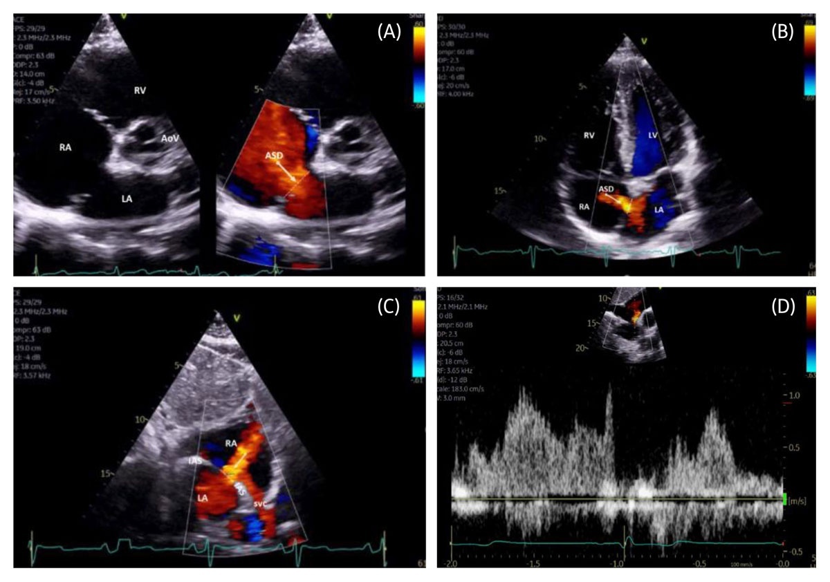
Figure 3
(A) Parasternal short-axis 2-dimensional (2D) color compare transthoracic echocardiography (TTE) image demonstrating a large ostium secundum ASD with left-to-right shunt. (B) Apical 4.chamber color Doppler TTE view showing an ostium secundum ASD with left-to-right shunt. (C) Subcostal bicaval color Doppler TTE view demonstrating an ostium secundum ASD with left-to-right shunt. (D) Subcostal short.axis pulsed.wave Doppler TTE of an ostium secundum ASD with left-to-right shunt. AoV: aortic valve; ASD: atrial septal defect (arrow/dash line); LA: left atrium; LV: left ventricle; RA: right atrium; RV: right ventricle; SVC: superior vena cava
Surgical repair includes primary closure of small defects and patch closure of larger ASDs with deficient rims. In patients with pulmonary hypertension and older adults at risk for heart failure with preserved ejection fraction, a fenestrated patch may reduce postoperative RV failure or diastolic dysfunction, respectively.
Sinus Venosus Defects
Sinus venosus defects are often underrecognized on standard adult TTE, which may delay diagnosis and intervention. Pulmonary venous anomalies are challenging to evaluate by TTE due to their marked anatomic variability. However, trained and experienced ACHD sonographers can detect the most common patterns with high diagnostic yield, including drainage of the right upper pulmonary vein to the superior vena cava (SVC), anomalous connection of the left upper pulmonary vein to the left innominate vein, and anomalous right pulmonary venous connection to the inferior vena cava (Scimitar syndrome)19 (Figure 4 A-D). CMR remains the modality of choice for comprehensive anatomic definition.
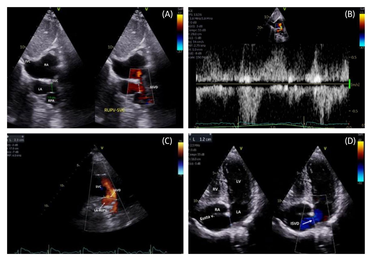
Figure 4
(A) Subcostal bicaval transthoracic echocardiography (TTE) color compare image demonstrating a large SSVD with bidirectional shunt, including a frame showing left-to-right flow from the RUPV ostium to the SVC. (B) Pulsed-wave Doppler demonstrating bidirectional shunt across the SSVD. (C) High right parasternal long-axis color Doppler TTE view of the SVC illustrating large SSVD size with left-to-right shunt from the RUPV to the SVC. (D) Apical 4-chamber view with inferior tilt, color Doppler TTE demonstrating a large inferior sinus venosus defect with left-to-right shunt below the Eustachian valve and tendon of Todaro. IAS: interatrial septum; IVC: inferior vena cava; LA: left atrium; LV: left ventricle; RA: right atrium; RV: right ventricle; RPA: right pulmonary artery; RUPV: right upper pulmonary vein ostium; SSVD: superior sinus venosus defect (green line/arrowed dash line); SVC: superior vena cava
Postoperative anatomy may be challenging to assess without ACHD training. Key residual lesions include pulmonary venous stenosis and SVC obstruction.
Coronary Sinus Defects
Coronary sinus defects are uncommon and may be challenging to diagnose by TTE, requiring a high index of suspicion and careful imaging assessment (Figure 5). A persistent left superior vena cava draining into the coronary sinus is a common associated anomaly and often results in coronary sinus dilation.
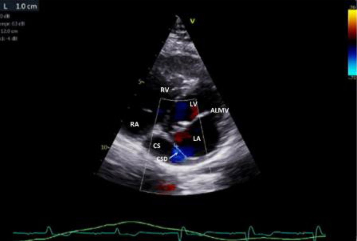
Figure 5
Intermediate parasternal short-axis view, color Doppler transthoracic echocardiography demonstrating an unrepaired coronary sinus defect with left-to-right shunt from the left atrium to the coronary sinus. ALMV: anterior leaflet mitral valve; CS: coronary sinus; CSD: coronary sinus defect; LA: left atrium; LV: left ventricle; RA: right atrium; RV: right ventricle.
Atrioventricular Septal Defects
Atrioventricular septal defect (AVSD) commonly includes an ostium primum atrial septal defect (Figure 6 A, B), an inlet VSD, and a cleft left atrioventricular valve, often with trifoliate valve morphology. Postoperative complications include residual patch leak and persistent or recurrent atrioventricular valve dysfunction.
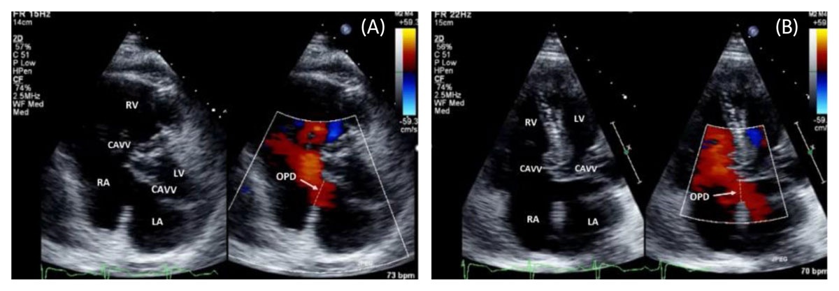
Figure 6
(A) Parasternal short-axis color Doppler transthoracic echocardiography (TTE) demonstrating an OPD with left-to-right shunt in a patient with atrioventricular septal defect. (B) Apical 4-chamber color compare TTE view demonstrating the OPD component of an atrioventricular septal defect with left-to-right shunt. CAVV: common atrioventricular valve; LA: left atrium; LV: left ventricle; RA: right atrium; RV: right ventricle; OPD: ostium primum defect (arrow, dash line)
Ventricular Septal Defects
Ventricular septal defects may occur in isolation or with more complex CHD. Systematic interrogation of the interventricular septum with 2D and color Doppler sweeps from apex to base is essential. Assessment should define VSD type (Figure 7 A), number, location, size, shunt direction and magnitude, VSD gradient (Figure 7 B, D), left heart size and function, RVSP, PASP, and aortic regurgitation. Associated lesions should also be assessed, including double-chambered RV, subaortic obstruction or ridge, aneurysmal tricuspid tissue, sinus of Valsalva rupture, aortic cusp prolapse, overriding or straddling atrioventricular valves (Figure 7 C), Gerbode defect, and coarctation.
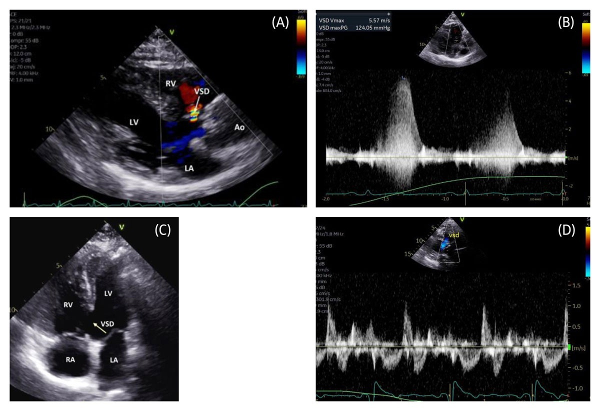
Figure 7
(A) Parasternal long-axis color Doppler transthoracic echocardiography (TTE) demonstrating a small perimembranous VSD with left-to-right shunt. (B) Color Doppler demonstrating a pressure-restrictive perimembranous VSD with left-to-right shunt. (C) Apical 4-chamber 2-dimensional TTE view demonstrating a large inlet VSD. (D) Pulsed-wave Doppler demonstrating a large bidirectional unrestrictive VSD. Ao: aorta; LA: left atrium; LV: left ventricle; RA: right atrium; RV: right ventricle; VSD: ventricular septal defect (arrow, dash line)
Overlap of the VSD or Gerbode jet with tricuspid regurgitation may overestimate the RVSP and falsely suggest pulmonary hypertension. Similarly, double-chambered right ventricle may be missed when overlapping Doppler signals obscure RV outflow tract (RVOT) obstruction, leading to inaccurate PASP estimation if the RVOT gradient is not considered.
Patch closure remains the most common repair. Percutaneous closure may be feasible for selected muscular and perimembranous VSDs. Postprocedural evaluation should assess residual shunt and device impingement on adjacent structures.
Pulmonary Stenosis and Right-Sided Obstructive Lesions
Comprehensive TTE evaluation of the RV, RVOT, pulmonary infundibulum, pulmonary valve, and pulmonary arteries from multiple acoustic windows is required for accurate localization and grading of right-sided obstructive lesions and for optimal clinical management.
In the presence of a high-velocity tricuspid regurgitation jet, pulmonary stenosis (Figure 8 A-C) or RVOT obstruction should be considered before attributing the gradient to pulmonary hypertension. Peak RVOT and pulmonary arterial gradients should be interrogated from multiple imaging planes. The RV inflow-outflow view from the apical and subcostal windows is particularly useful in this assessment (Figure 8 D-F).
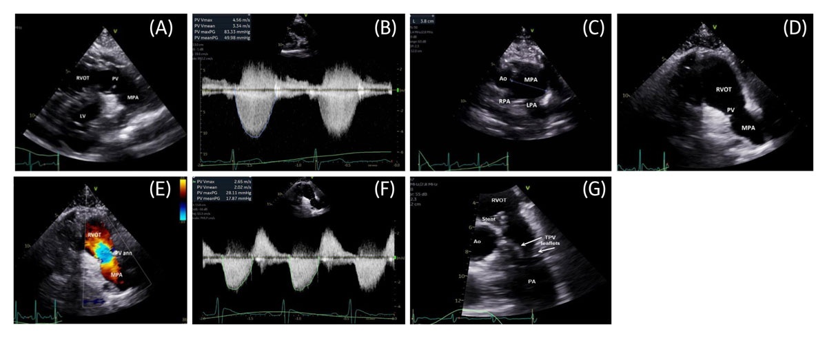
Figure 8
(A) Parasternal short-axis 2-dimensional (2D) transthoracic echocardiography (TTE) demonstrating a stenotic PV with tethered, doming leaflets. (B) Color Doppler waveform demonstrating severe congenital PV stenosis. (C) High parasternal short-axis 2D TTE demonstrating post-stenotic dilatation of the pulmonary trunk. (D) Parasternal short-axis 2D TTE demonstrating a surgically disrupted PV with near absence of leaflet tissue. (E) Parasternal short-axis color Doppler TTE demonstrating severe PV regurgitation with some degree of stenosis following valvectomy. (F) Color Doppler waveform demonstrating mild residual stenosis and severe pulmonary regurgitation. (G) 2D TTE zoomed parasternal short-axis view demonstrating a transcatheter PV replacement with stent in the RVOT and valve leaflets in systole. Ao: aorta; LV: left ventricle; LPA: left pulmonary artery; MPA: main pulmonary artery; PA: pulmonary artery; PV: pulmonary valve; PV ann: pulmonary valve annulus; RPA: right pulmonary artery; RVOT: right ventricular outflow tract; TVP: transcatheter pulmonary valve
Surgical management may include pulmonary valvotomy or valvectomy (Figure 8 E), pulmonary valve replacement, RV to pulmonary artery conduit placement, RV muscle bundle resection, and pulmonary artery augmentation. Transcatheter therapies include pulmonary balloon valvuloplasty, transcatheter pulmonary valve replacement (Figure 8 G), and pulmonary artery stent implantation. Follow-up TTE should assess prosthetic valve function as well as residual or recurrent obstructive and regurgitant lesions.
Tetralogy of Fallot
Patients with tetralogy of Fallot (TOF) typically require closer TTE surveillance. Variants include TOF with pulmonary stenosis, pulmonary atresia, or absent pulmonary valve syndrome (Figure 9 A). Associated anomalies may include right aortic arch, persistent left SVC, ASD, patent ductus arteriosus, retroaortic innominate vein, anomalous coronary artery origin, AVSD, and major aortopulmonary collateral arteries. Palliation may include Blalock–Taussig–Thomas or central shunts, although primary neonatal repair is now standard. Patients should ideally be followed at specialized ACHD centers, and comprehensive TTE is essential.8,10 Assessment should focus on residual pulmonary regurgitation or stenosis, RVOT obstruction, pulmonary artery trunk (Figure 9 B) and branch pulmonary artery dimensions and gradients, residual VSD, tricuspid regurgitation, biventricular size and function, atrial size, RVSP and PASP, proximal aortic dimensions, and associated lesions (Figure 9 C).
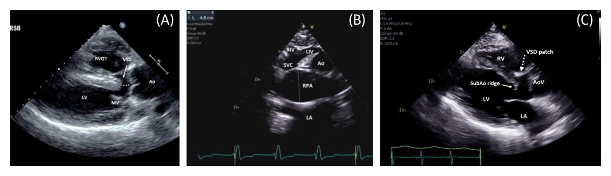
Figure 9
(A) Parasternal long-axis 2-dimensional (2D) transthoracic echocardiography (TTE) demonstrating unrepaired tetralogy of Fallot with overriding Ao and associated VSD. (B) Suprasternal notch 2D TTE demonstrating an aneurysmal RPA in tetralogy of Fallot with absent pulmonary valve. (C) Parasternal long-axis 2D TTE demonstrating repaired tetralogy of Fallot with a large VSD patch and severely dilated RV. Ao: aorta; AoV: aortic valve; LA: left atrium; LV: left ventricle; MV: mitral valve; RV: right ventricle; RVOT: right ventricular outflow tract; RPA: right pulmonary artery; SVC: superior vena cava; LIV: left innominate vein; RIV: right innominate vein; SubAo: subaortic; VSD: ventricular septal defect (overriding the aorta in dash line)
Transposition of the Great Arteries
In dextro-transposition of the great arteries (D-TGA), the aorta arises from the morphologic RV and the pulmonary artery from the morphologic LV, resulting in ventriculoarterial discordance. The aorta is anterior and to the right of the pulmonary trunk. Repair strategies include atrial switch (Mustard or Senning) (Figure 1, Figure 10 A) and arterial switch (Jatene) operations. Associated lesions include VSD, ASD, LVOT obstruction, patent ductus arteriosus, and coronary artery anomalies.
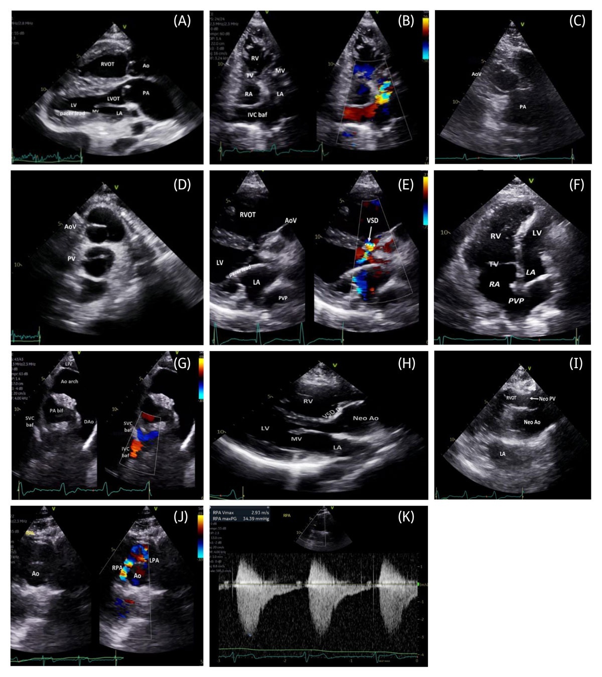
Figure 10
(A) Parasternal long-axis 2-dimensional (2D) transthoracic echocardiography (TTE) demonstrating parallel great arteries in d-transposition of the great arteries (d-TGA) post–atrial switch, with ventriculoarterial discordance and pacer lead in the subpulmonary LV. (B) Intermediate apical TTE color-compare image demonstrating IVC baffle flow to the subpulmonary MV. (C) Parasternal short-axis 2D TTE demonstrating great artery relationship in d-TGA post–atrial switch with the Ao anterior and rightward to the pulmonary trunk. (D) Parasternal short-axis 2D TTE again demonstrating the Ao directly anterior to the pulmonary trunk. (E) Intermediate parasternal long-axis color-compare TTE demonstrating d-TGA with VSD, showing systemic-to-pulmonary shunt across the VSD and pacer lead in the subpulmonary left chambers. (F) Apical 4-chamber 2D TTE demonstrating a dilated, hypertrophied systemic RV and PVP to the RA post–Mustard procedure. (G) Intermediate suprasternal notch color TTE demonstrating SVC and IVC baffle flows post–atrial switch. (H) Parasternal long-axis 2D TTE demonstrating VSD patch and Neo Ao post–arterial switch. (I) High parasternal long-axis 2D TTE demonstrating parallel great arteries post–arterial switch with Neo PV. (J) Intermediate parasternal short-axis color-compare TTE demonstrating the LeCompte maneuver with pulmonary arteries anterior to the ascending Ao and associated RPA stenosis. (K) Color Doppler waveform demonstrating RPA stenosis. Ao: aorta; Ao arch: aortic arch; AoV: aortic valve; DAo: descending aorta; IVC baf: inferior vena cava baffle; LA: left atrium; LIV: left innominate vein; LV: left ventricle; LVOT: left ventricular outflow tract; MV: mitral valve; Neo Ao: neoaorta; Neo PV: neo pulmonary valve (arrow); PA: pulmonary artery; PA bif: pulmonary artery bifurcation; PVP: pulmonary venous pathway; RA: right atrium; RPA: right pulmonary artery; RV: right ventricle; RVOT: right ventricular outflow tract; SVC baf: superior vena cava baffle; TV: tricuspid valve; VSD: ventricular septal defect (arrow/dash line); VSD p: ventricular septal defect patch
After atrial switch repair, systemic venous return is directed to the subpulmonary LV, whereas pulmonary venous return is baffled to the systemic RV (Figure 10 B-G, Figure 11). Echocardiographic assessment should focus on systemic and pulmonary venous baffle patency and leaks, systemic RV size and function, systemic tricuspid valve regurgitation, and PASP estimation and LV systolic pressure from the subpulmonary mitral regurgitation jet (Figure 10 B-G).
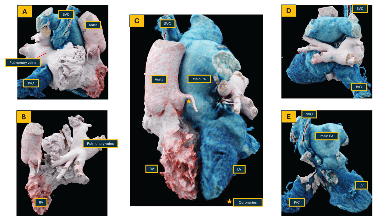
Figure 11
Patient with a history of dextro-transposition of the great arteries post atrial switch operation; coronary computed tomography angiography was performed to assess for coronary and right atrial appendage thrombus. 3D reconstruction. LV: left ventricle; RV: right ventricle; LA: left atrium; RA: right atrium; PA: pulmonary artery; SVC: superior vena cava; IVC: inferior vena cava
Published reference ranges for systemic RV size and function are limited, and guideline-based parameters for the subpulmonary RV are not validated for the systemic RV. Echocardiographic measurements should therefore be used primarily for serial comparison. Useful parameters include RV areas in diastole and systole from a focused apical four-chamber view, fractional area change, tissue Doppler S′ velocity, triplane or 3D estimates of RV volumes and ejection fraction, and RV longitudinal strain. Assessment should include both longitudinal and radial components of contraction. When more accurate quantification is required, CMR should be performed because it provides more reliable RV volumes and ejection fraction.
The arterial switch operation includes great artery translocation, coronary reimplantation, and the LeCompte maneuver, which may predispose to branch pulmonary artery stenosis. After repair, the left ventricle is systemic; the native aortic valve serves as the neopulmonary valve, and the native pulmonary valve as the neoaortic valve (Figure 10 H-J, Figure 12). Echocardiographic assessment should focus on neopulmonary valve or distal pulmonary artery obstruction (Figure 10 K), neoaortic root dilation and regurgitation, branch pulmonary artery stenosis, and ventricular size and function. Tricuspid regurgitation velocity may provide indirect evidence of RVOT obstruction or branch PA stenosis when the pulmonary arteries are not well visualized.
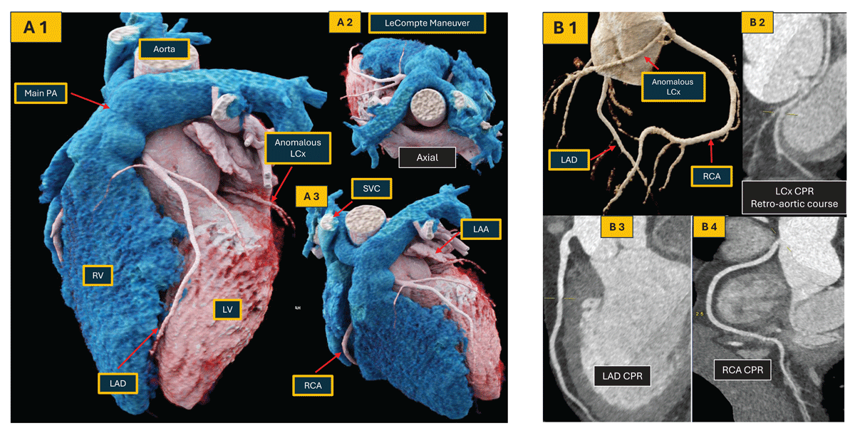
Figure 12
Computed tomography dextro-transposition of the great arteries (d-TGA) after arterial switch procedure with anomalous LCx with a retroaortic course. (A 1-3) 3-dimensional reconstruction of a patient with d-TGA after an arterial switch and LeCompte Maneuver; (B 1-4) curved multiplanar reconstructions of the coronary tree. Note that LCx has an anomalous retro-aortic course after arising from the RCA. LV: left ventricle; RV: right ventricle; PA: pulmonary artery; SVC: superior vena cava; LAA: left atrial appendage; LAD: left anterior descending artery; LCx: left circumflex artery; RCA: right coronary artery.
Congenitally Corrected Transposition of the Great Arteries
Congenitally corrected TGA (CCTGA) is characterized by atrioventricular and ventriculoarterial discordance, resulting in physiologically corrected circulation. The morphologic left atrium connects to the morphologic RV, which gives rise to the aorta and functions as the systemic ventricle, while the morphologic right atrium connects to the morphologic LV, which ejects into the pulmonary artery (Figure 13 A, B). In situs solitus, CCTGA is also referred to as L-transposition of the great arteries because the aorta arises anterior and leftward relative to the pulmonary artery.
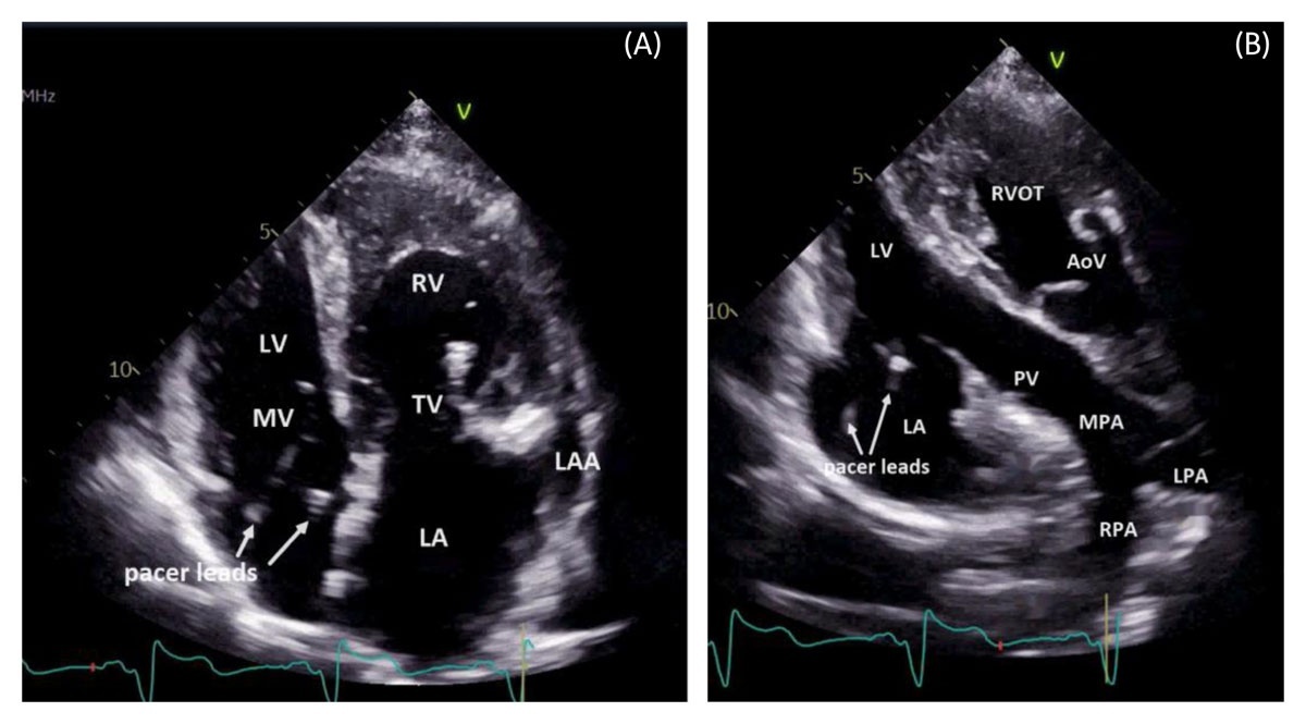
Figure 13
(A) Apical 4-chamber 2-dimensional (2D) transthoracic echocardiography (TTE) demonstrating situs solitus congenitally corrected transposition of the great arteries. (B) Apical LV 3-chamber 2D TTE demonstrating RA-LV, LV-PA, and RV-Ao connections in congenitally corrected transposition of the great arteries. AoV: aortic valve; RA: right atrium; LA: left atrium; LAA: left atrial appendage; LV: left ventricle; MV: mitral valve; MPA: main pulmonary artery; LPA: left pulmonary artery; PV: pulmonary valve; RPA: right pulmonary artery; RV: right ventricle; RVOT: right ventricular outflow tract; TV: tricuspid valve
Echocardiographic assessment is technically challenging and should focus on systemic RV size and function, systemic tricuspid valve regurgitation, pulmonary pressures, and associated lesions. Tricuspid regurgitation is common and may result from RV dilatation occasionally and/or Ebsteinoid tricuspid valve, requiring mechanical valve replacement in unrepaired patients.
Single Ventricle Physiology and Fontan Palliation
Functional univentricular hearts not amenable to biventricular repair are managed with Fontan palliation. Lesions include hypoplastic left heart syndrome, tricuspid atresia (Figure 14 A), severe Ebstein anomaly, TGA with large malaligned VSD and overriding or straddling atrioventricular valves, unbalanced AV canal/AVSD (Figure 14 B), and complex heterotaxy syndromes.
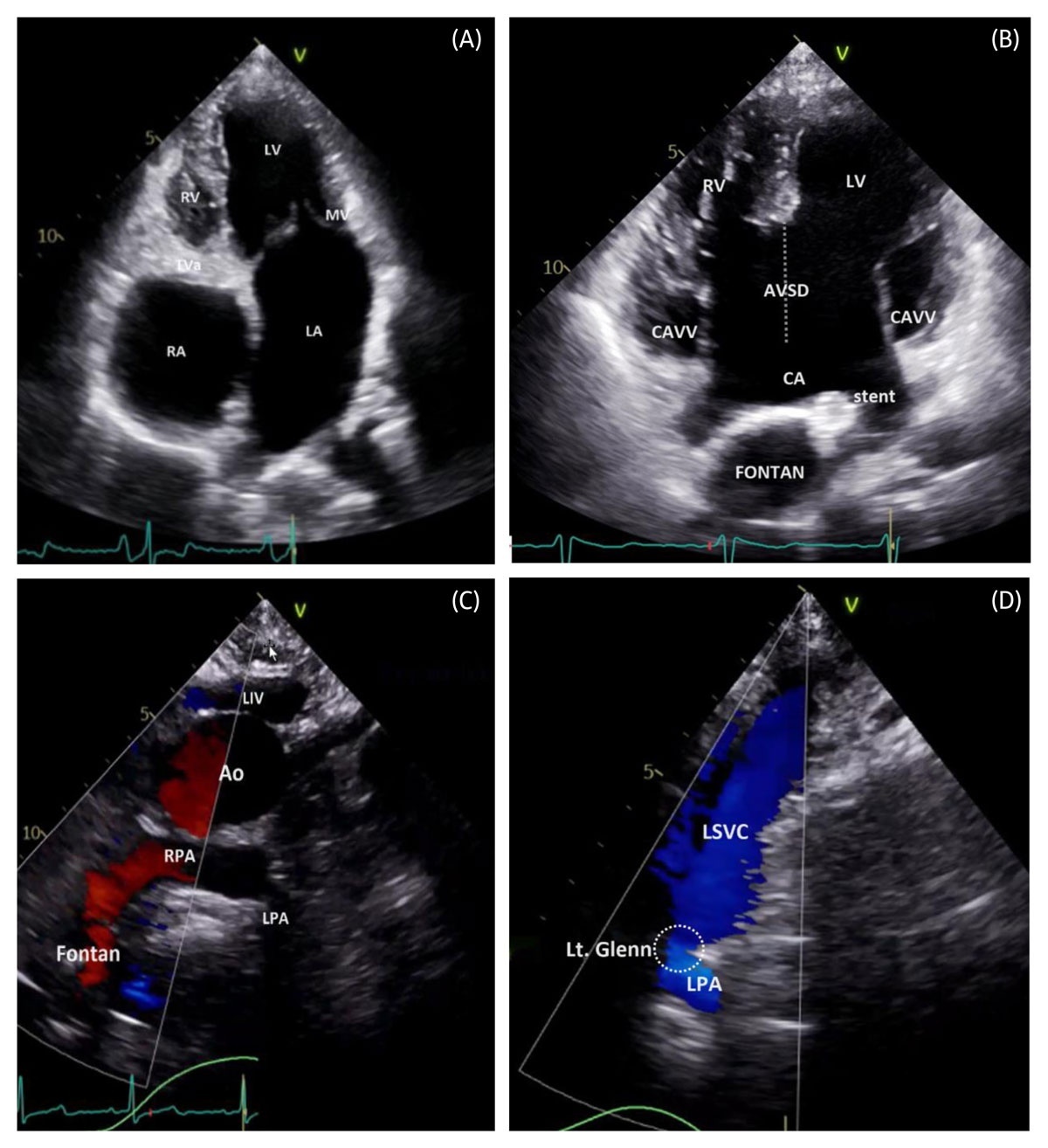
Figure 14
(A) Apical 4-chamber 2-dimensional (2D) transthoracic echocardiography (TTE) demonstrating tricuspid atresia palliated with atriopulmonary Fontan, with plate-like TV atresia and hypoplastic RV. (B) 2D TTE demonstrating an atrioventricular septal defect palliated with Fontan, including a Fontan fenestration stent. (C) Intermediate suprasternal notch color TTE demonstrating an unobstructed Fontan–RPA anastomosis. (D) Left supraclavicular color TTE demonstrating a patent left bidirectional Glenn anastomosis (LSVC–LPA connection). Ao: aorta; CA: common atrium; CAVV: common atrioventricular valve; LA: left atrium; LV: left ventricle; LIV: left innominate vein; Lt. Glenn: left bidirectional Glenn anastomosis (dash circle); RA: right atrium; RPA: right pulmonary artery; RV: right ventricle; MV: mitral valve; TVa: tricuspid valve atresia; LSVC-LPA: left superior vena cava-left pulmonary artery
These patients should be managed at specialized ACHD centers with expertise in multimodality imaging and advanced therapies, including Fontan revision and heart transplantation. Assessment should focus on Fontan pathway patency (Figure 14 C, D), fenestration or leak, Glenn anastomosis patency, systemic ventricular size and function, atrioventricular and semilunar valve function, pulmonary venous flow, ASD patency, pulmonary artery flow, and thrombus.
Echocardiographic protocol must be individualized according to the underlying anatomy and surgical palliation. Key elements of assessment include cavopulmonary pathway patency, fenestration or leak, systemic ventricular size and function, atrioventricular and semilunar valve function, pulmonary venous flow, atrial septal communication patency, pulmonary artery flow, and the presence of thrombus or obstruction of the Fontan pathway.
Transesophageal Echocardiography
Transesophageal echocardiography (TEE) provides important complementary information in ACHD when TTE is suboptimal or incomplete. It is particularly effective for characterization of small intracardiac shunts, sinus venosus and coronary sinus defects, anomalous pulmonary venous connections, Fontan fenestrations, and complex or eccentric regurgitant jets. TEE is especially valuable for detailed evaluation of the systemic atrioventricular valve in patients with a systemic RV, including those with congenitally corrected transposition of the great arteries, prior atrial switch repair, and Fontan physiology. It may also serve as an alternative imaging strategy for ventricular assessment when CMR or CT are contraindicated, unavailable, or limited by device-related artifact or non-MRI-conditional hardware. TEE remains the primary modality for intraprocedural guidance during transcatheter structural and valvular interventions.20
Role of a Focused CMR
Cardiac magnetic resonance occupies a central role in the contemporary evaluation of ACHD patients, complementing TTE by confirming key findings while providing comprehensive cross-sectional definition of cardiovascular anatomy and physiology. Because image acquisition is inherently more demanding in this population, first-time studies typically warrant a 90-minute slot to establish anatomic and physiological baselines, while follow-up examinations can often be completed in 60 minutes using focused, question-driven protocols in experienced programs. These extended time allocations should be regarded as an expected requirement for high-quality imaging rather than a workflow inefficiency given that ACHD management relies heavily on CMR-derived anatomy and quantitative measures for meaningful longitudinal comparison. Although comprehensive ACHD CMR is currently concentrated in quaternary cardiovascular centers, the approach is generalizable across a broader range of CMR laboratories with standardized protocols, focused training, and close physician-technologist collaboration.
Planning the ACHD CMR Study
Protocol selection should begin with a focused review of available echocardiographic data and the specific unanswered clinical questions. The initial objective is an accurate anatomic roadmap, typically achieved with an axial, interleaved 2D stack covering the entire chest using a bright-blood balanced steady-state free precession (bSSFP) sequence, followed by a double inversion recovery-prepared dark-blood T2-weighted single-shot fast spin-echo sequence (HASTE-type), acquired with ECG-gating during free-breathing. Coronal and sagittal reformats are optional and generally unnecessary when a high-quality MR angiographic dataset has been obtained.6 Cine imaging stacks can be valuable in complex congenital anatomies, such as atrial switch physiology and Fontan palliation, where they facilitate assessment for conduit or baffle obstruction, baffle leak, and fenestrations.
Ventricular Size and Function
Quantification of ventricular volumes and systolic function is among the most frequent indications for ACHD CMR. A standard contiguous short-axis cine stack is usually sufficient for this purpose. Long-axis cine views are often helpful to improve basal slice definition and reduce segmentation error near the atrioventricular junction. Breath-held, ECG-gated cine bSSFP remains the preferred approach when feasible. However, contemporary acceleration strategies including parallel imaging and compressed sensing have expanded the applicability of real-time, free-breathing cine acquisitions, which can provide reliable assessment of ventricular size and function in patients with limited breath-hold capacity or significant arrhythmia.21 Figure 15 shows a dilated systemic RV in a patient with D-TGA and atrial switch and Figure 16 shows a severely dilated RV in a patient with TOF and severe pulmonary regurgitation in the setting of a transannular patch repair.
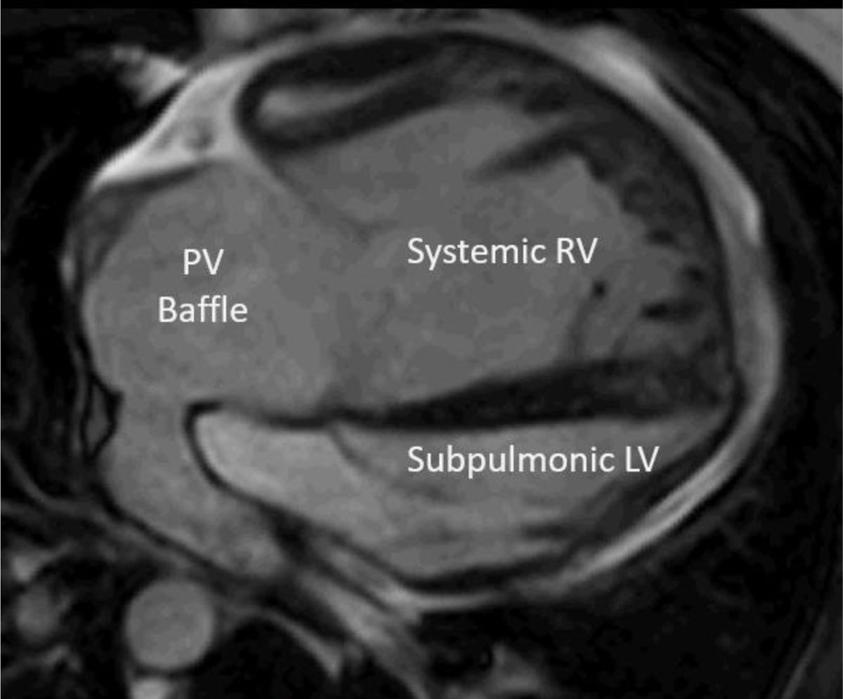
Figure 15
Dilated systemic RV in a patient with d-transposition of the great arteries and atrial switch. PV Baffle: pulmonary venous baffle; RV: right ventricle; LV: left ventricle
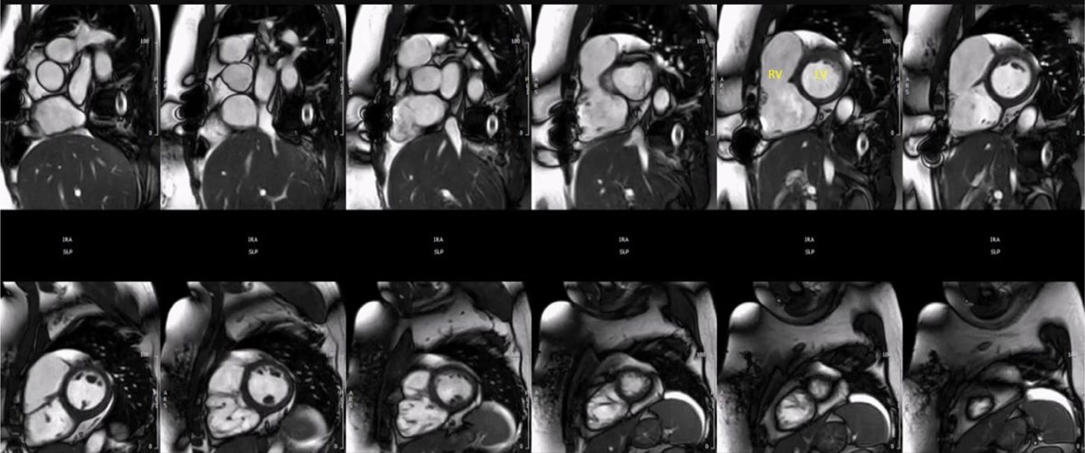
Figure 16
Cardiac magnetic resonance short axis cine imaging stack demonstrating a severely dilated RV in a patient with tetralogy of Fallot with a transannular patch repair and severe pulmonary regurgitation. RV: right ventricle; LV: left ventricle
Anatomical Assessment of Intracardiac Shunts
When atrial morphology or atrial septal evaluation is clinically relevant, a dedicated atrial short-axis stack should be obtained rather than simply extending the ventricular stack. Coverage should be contiguous, without an interslice gap, and should include both atria in their entirety. Spatial resolution should be optimized while maintaining adequate signal-to-noise ratio and breath-hold feasibility, as partial-volume effects can obscure small atrial septal defects. As a practical benchmark, many experienced laboratories target approximately 1.6 × 1.6 mm in-plane resolution with 4-mm slice thickness at 1.5T, with adjustments based on patient size and scanner performance. In good breath-holders, interpolation can be considered to improve apparent image sharpness and may assist assessment of atrial baffles, venous pathways, and fine valvular detail. However, interpolation improves reconstructed rather than acquired spatial resolution and may require phase oversampling, prolonging breath-hold time; its use should therefore be individualized. Similar principles apply when ventricular septal morphology or VSD assessment is clinically important, with imaging plane and coverage tailored to the suspected defect location, adjacent valves, outflow tracts, and prior patch or device material.23 CMR can be particularly helpful to better define anatomy (including pulmonary veins) and plan surgery in patients with sinus venosus defects (Figure 17) and unroofed coronary sinus (Figure 18). When detailed septal anatomy is needed for transcatheter ASD or VSD closure planning, cardiac CT is often helpful, providing reproducible 0.5 mm to 0.6 mm isotropic voxels and excellent delineation of septal rims, defect margins, venous connections, adjacent valves, outflow tracts, and neighboring vascular structures.22
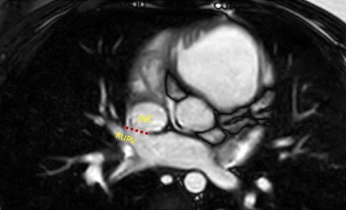
Figure 17
Cardiac magnetic resonance cine imaging in the axial plane demonstrating a sinus venosus defect (red dotted line). SVC: superior vena cava; RUPV: right upper pulmonary vein
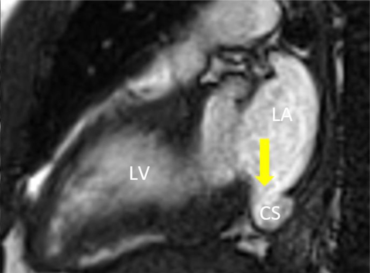
Figure 18
Cardiac magnetic resonance cine imaging of an unroofed coronary sinus defect (arrow) in the two-chamber view. LA: left atrium; CS: coronary sinus; LV: left ventricle
Intracardiac Shunt Assessment with Phase Contrast
For intracardiac shunt evaluation, imaging plane selection is critical. As in echocardiography, a shunt may be missed entirely if the acquisition plane does not intersect the jet. A practical approach is to review prior Doppler data to characterize jet direction, then reproduce an analogous CMR plane using cine bSSFP to identify flow-related signal heterogeneity followed by targeted phase-contrast interrogation. When in-plane phase-contrast imaging is used to localize shunt flow direction, the velocity-encoding axis must be oriented along the expected direction of shunt flow, as an incorrect orientation may underestimate or obscure the jet. Because many atrial-level shunts generate relatively low-velocity flow, velocity encoding should be set low enough to maintain sensitivity, typically around 100 cm/s initially, then increased incrementally if aliasing is observed.24 Once localized, through-plane phase-contrast imaging prescribed perpendicular to the jet can directly quantify shunt flow volume, and this measurement should be correlated with Qp:Qs quantification as a measure of internal validity.
Role of Magnetic Resonance Angiography
Given the frequency of anomalous arterial and venous connections in ACHD—and the need to evaluate stenosis, aneurysmal dilatation, and surgical pathways including conduits, baffles, and anastomoses—a 3D MR angiography (MRA) dataset is often essential and is typically obtained prior to detailed phase-contrast interrogation. MRA should balance spatial resolution, motion control, and acquisition efficiency. Breath-held, ECG-gated approaches reduce motion artifact and improve vessel edge definition, though free-breathing methods may be required in patients with limited compliance. Contrast-enhanced MRA remains widely used and highly reliable for vascular mapping. Noncontrast, free-breathing MRA techniques are increasingly available and may obviate gadolinium in straightforward cases (Figure 19), although bSSFP-based readouts can be susceptible to off-resonance effects, particularly at 3T.25,26,27
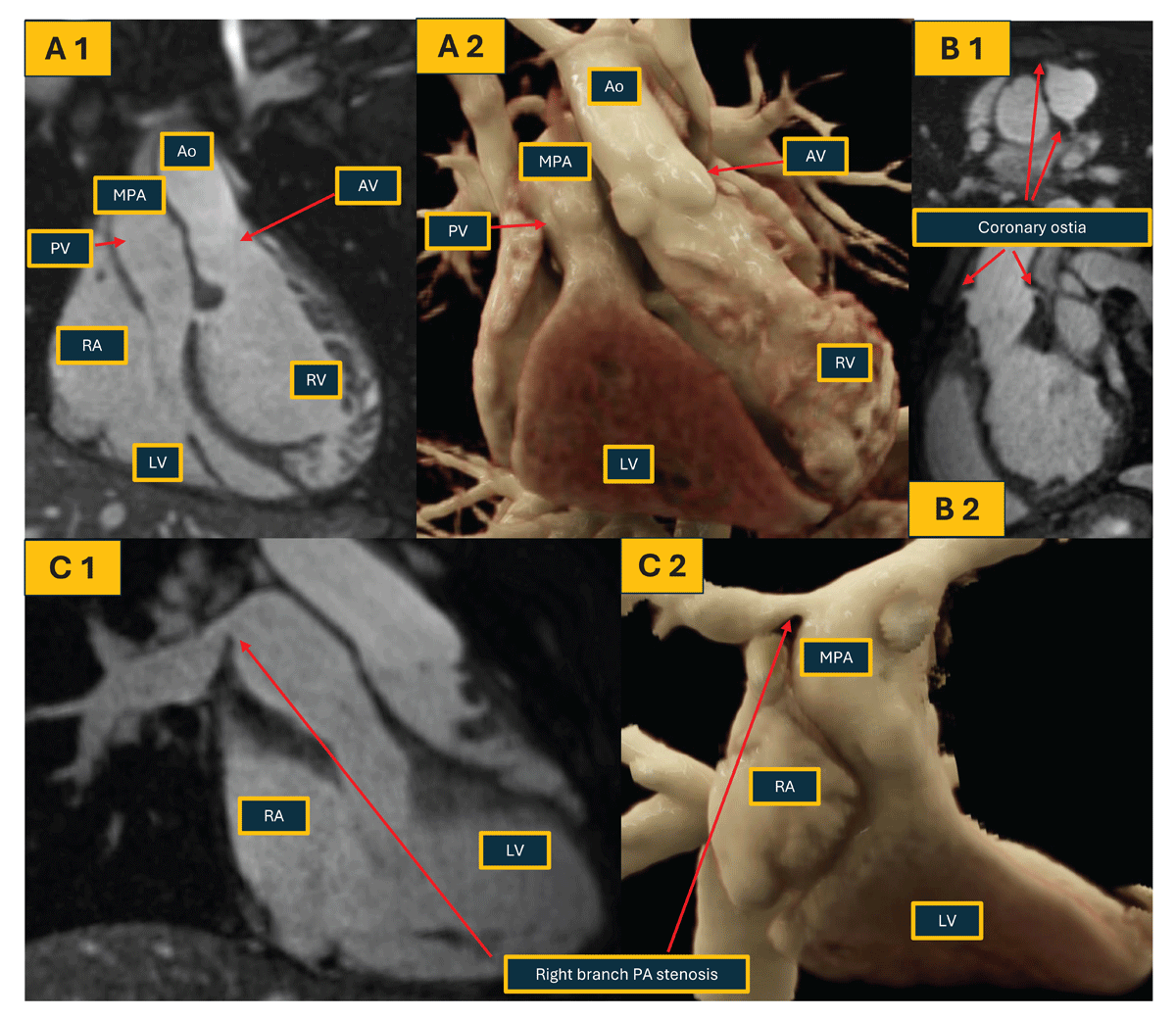
Figure 19
Non-contrast magnetic resonance imaging in a patient with congenitally corrected transposition of the great arteries (CCTGA). (A 1,2) Coronal view of heart and great vessels in 2D and 3D reconstruction demonstrating levo-TGA; (B 1,2) visualization of coronary ostia due to improved spatial resolution (1.6 × 1.6 × 1.6 mm); (C 1,2) identification of right branch pulmonary artery stenosis and post stenotic dilation. RA: right atrium; LA: left atrium; RV: right ventricle; LV: left ventricle; MPA: main pulmonary artery; AV: aortic valve; PV: pulmonic valve; Ao: aorta
Time-resolved contrast-enhanced MRA is particularly valuable for pulmonary venous evaluation in ACHD because it depicts the temporal sequence of contrast transit through the circulation. This dynamic information can help distinguish anomalous pulmonary venous drainage from systemic venous pathways, complex collateral flow, or postoperative venous obstruction, especially when anatomy is ambiguous on static imaging. Although spatial resolution is lower than single-phase high-resolution MRA, modern accelerated acquisitions can improve voxel size in selected implementations 28 (Figure 20).
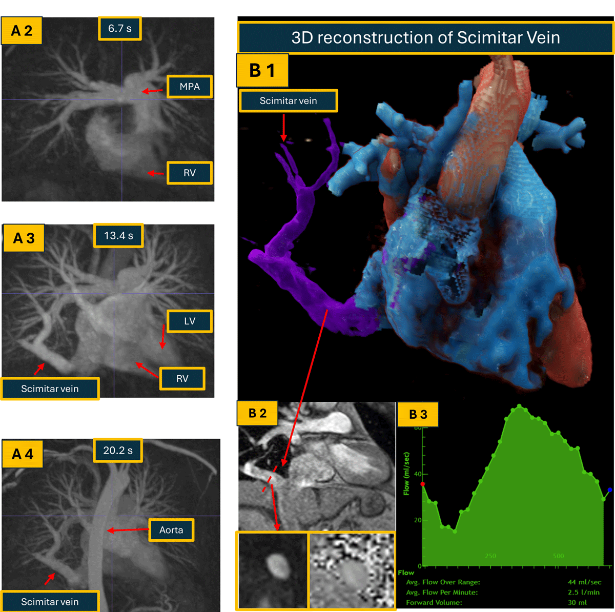
Figure 20
Utilization of time resolved MRA and direct measurement of anomalous pulmonary venous return flow in a patient with Scimitar syndrome (anomalous right lower pulmonary vein). (A 1 Video) Maximal intensity projection demonstrating filling of the Scimitar vein. (A 2-4) Maximal intensity projection of time resolved MRA demonstrating filling of the Scimitar vein. Last few frames also demonstrate persistent left superior vena cava draining into coronary sinus. (B 1) 3D reconstruction of the Scimitar vein; (B 2) GRE image of the Scimitar vein draining into the IVC; (B 3) Flow quantification of the Scimitar vein, direct anomalous pulmonary return flow is 30 mL/beat, 2.5 L/min. MRA: magnetic resonance angiography RV: right ventricle; LV: left ventricle; MPA: main pulmonary artery
This should be integrated into a deliberate multimodality strategy. Echocardiography remains first-line and may show indirect findings such as right-sided chamber dilation, elevated pulmonary pressures, or abnormal Doppler flow, but pulmonary venous anatomy may be incompletely visualized because of acoustic windows, posterior vessel location, and complex postoperative anatomy. CMR and CT should therefore be used when pulmonary venous disease is suspected or right-sided dilation is unexplained. CMR can define anomalous pulmonary venous connections, quantify shunt burden, and assess ventricular remodeling without ionizing radiation, whereas CT provides higher spatial resolution for small pulmonary veins, pulmonary venous stenosis, scimitar anatomy, airway or lung relationships, and procedural planning.
Ferumoxytol-enhanced CMR is increasingly recognized as a valuable alternative contrast strategy, particularly in patients with renal dysfunction. An additional advantage is its prolonged intravascular residence time exceeding 12 hours, attributable to its large molecular size and carbohydrate shell coating.29,30
Optimization of Phase Contrast Flow Imaging for Accurate Qp:Qs Assessment
The MRA dataset serves as the roadmap for planning phase-contrast acquisitions and enables precise prescription of imaging planes perpendicular to the target vessel, improving accuracy and reproducibility of flow quantification in the main pulmonary artery, branch pulmonary arteries, great veins, aorta, and major branches, supporting reliable calculation of Qp:Qs and assessment of differential pulmonary blood flow.
In markedly dilated pulmonary arteries, turbulence and partial-volume effects can underestimate flow at the main pulmonary artery level, making branch pulmonary artery measurements often more reliable. When prescribing branch pulmonary artery imaging planes, slices should be positioned as proximally as feasible while avoiding regions of early branching, as distal placement may underestimate total pulmonary flow. In patients with partial anomalous pulmonary venous return, MRA identifies the anomalous vein or veins and guides targeted phase-contrast measurements for quantification of anomalous venous return and shunt magnitude (Figure 20).
Accurate Qp:Qs assessment is a fundamental component of ACHD CMR. Because phase-contrast flow measurements are susceptible to artifacts related to motion, arrhythmia, through-plane motion, and background phase offset, repeated or supplementary acquisitions are often necessary. In many patients, paired measurements across the aorta and main pulmonary artery are sufficient for routine Qp:Qs quantification. However, in complex anatomies—particularly with multilevel shunts or single ventricle physiology—additional acquisitions involving the SVC, inferior vena cava, descending aorta, and pulmonary veins are essential for comprehensive hemodynamic assessment and internal validation of flow consistency.6
When breath-holding is reliable and initial flow data are technically robust, free-breathing acquisitions may be omitted or limited to repeat measurements of the main pulmonary artery and ascending aorta. In patients with limited breath-hold capacity, arrhythmia, or discrepant measurements, free-breathing or repeated acquisitions help confirm results.
In patients with patent ductus arteriosus, the level of shunt must be considered when interpreting Qp:Qs. Pulmonary arterial flow measured proximal to the ductal insertion reflects systemic rather than total pulmonary flow; in this setting, ascending aortic forward flow includes ductal runoff and may serve as the Qp.31
4D Flow Cardiac Magnetic Resonance Imaging in ACHD
4D flow CMR is an emerging technique providing time-resolved, 3D velocity encoding across a prescribed volume, enabling retrospective visualization and quantification of complex cardiovascular flow.32 This is particularly appealing in ACHD, where abnormal anatomy, repaired outflow tracts, conduits, baffles, Fontan pathways, branch pulmonary arteries, aortopathy, collaterals, and shunts can make conventional 2D phase-contrast plane prescription challenging.33 Potential advantages include retrospective flow quantification, visualization of eccentric jets, and assessment of advanced hemodynamic markers such as vortices, helicity, wall shear stress, kinetic energy, and energy loss.33 Limitations include long acquisition times, lower spatial and temporal resolution compared with targeted 2D phase-contrast imaging, sensitivity to arrhythmia and respiratory motion, and substantial post-processing time and expertise.34 Broader adoption will require faster acquisitions, standardized post-processing workflows, multicenter validation, and evidence that 4D flow-derived metrics meaningfully improve clinical decision-making and patient outcomes.35
Safety and Logistical Considerations in ACHD CMR
CMR is a practical and increasingly accessible modality for ACHD patients. Most adult examinations can be performed without sedation, and anesthesia is almost never required. Pre-scan planning should identify limited breath-hold capacity, arrhythmia, claustrophobia, renal dysfunction, and implanted devices so the protocol can be tailored appropriately.
Most contemporary pacemakers, defibrillators, and intracardiac devices are MRI-conditional or can be scanned under established institutional protocols.36,37 MRI-nonconditional devices and abandoned leads require review by the MRI technologist, supervising physician, and device team before scanning.38 Insufficient data exist to definitively comment on MRI safety in the presence of permanent epicardial leads.39 Device-related artifact should not automatically preclude CMR, as newer sequences and protocol adjustments can often preserve diagnostic image quality.40
Gadolinium use should be individualized. With contemporary macrocyclic agents, the risk of nephrogenic systemic fibrosis is very low when renal function is preserved.41,42 Improved noncontrast cine, phase-contrast, and MRA techniques allow many ACHD questions to be answered without contrast. Advances in device safety, artifact reduction, and noncontrast imaging have made CMR substantially more feasible for ACHD care over the past decade.
Role of Cardiac CT in ACHD
Cardiac CT is a useful complementary tool in ACHD, providing excellent spatial resolution for evaluating small vessels, coronary arteries, calcification, stents, conduits, baffles, surgical anastomoses, airway relationships, and extracardiac vascular anatomy. However, CCT requires iodinated contrast and ionizing radiation and is best reserved for targeted clinical questions rather than routine surveillance.43
CCT is especially valuable when high-resolution anatomy is needed for surgical, sternal reentry, transplant, mechanical support, or transcatheter intervention planning.20,44 After the arterial switch operation, CCT can define coronary anatomy and accurately measure neo-aortic root dilation.45,46 It is also invaluable for transcatheter pulmonary valve replacement planning, including assessment of RVOT dimensions, conduit calcification, coronary proximity, and adjacent mediastinal structures.47
Contrast timing should be tailored to the anatomy and clinical question.48 In patients with suspected baffle obstruction, venous pathway stenosis, thrombosis, or Fontan pathway complications, delayed imaging should be considered, as slow flow and incomplete contrast mixing can mimic thrombus or exaggerate stenosis. Delayed-phase imaging helps distinguish true filling defects from slow contrast transit.49 Fontan CT protocols require particular care because contrast enters the pulmonary arteries passively and may opacify unevenly. In complex cases, bilateral upper-extremity IVs with multiple low-dose, high-pitch acquisitions 30 to 60 seconds apart can capture contrast progression through the Fontan pathway, pulmonary arteries, pulmonary veins, atrium, and ventricle, reducing the risk of nondiagnostic opacification.49
When CMR cannot be performed and ventricular function is needed, retrospective ECG-gated CCT can provide ventricular volumes and systolic function, though at the cost of higher radiation exposure. Overall, cardiac CCT should be used selectively in ACHD, with protocols designed to answer specific clinical questions while minimizing contrast and radiation burden.50
Team Development, Specialized Training, and Quality Frameworks in ACHD Imaging
ACHD-trained sonographers and CMR and CT technologists are an essential but underrecognized component of high-quality ACHD imaging. A central limitation in expanding ACHD echocardiography, CMR, and CCT is not scanner availability but specialized human expertise. ACHD-trained sonographers and technologists are rare because complex congenital imaging remains concentrated in high-volume quaternary centers, leaving regional programs with limited exposure to repaired anatomy, atypical physiology, and lesion-specific protocol adaptation. This gap is increasingly important to address as the ACHD population grows and CMR and CCT hardware becomes more widely available in regional and community settings.
Building sonographer and technologist competency is therefore essential to expanding equitable access. ACHD-trained imaging personnel are not interchangeable with general cardiac imagers because congenital imaging requires expertise in acquiring appropriate planes, recognizing native and postsurgical anatomy, and adapting protocols in real time.48 They function as integral members of the multidisciplinary ACHD team, working closely with cardiologists to ensure examinations are comprehensive and diagnostically complete. This expertise is particularly important when key diagnoses may be missed without congenital experience, such as partial anomalous pulmonary venous return in a patient with an atrial septal defect, baffle leak or obstruction after atrial switch repair, Fontan pathway complications, or de novo presentations with undiagnosed congenital heart disease.
Developing and sustaining this competency requires intentional training, continued case exposure, and close collaboration with ACHD cardiologists and imagers. Investment in ACHD sonographer and CMR/CCT technologist education is a critical strategy to improve diagnostic quality, reduce geographic disparities, and expand meaningful access to advanced imaging for ACHD patients.
Conclusion
Comprehensive multimodality imaging is fundamental to the contemporary care of adults with congenital heart disease. ACHD TTE remains the cornerstone of longitudinal surveillance and requires lesion-specific protocols, advanced technical expertise, and systematic evaluation of native and postsurgical anatomy. CMR and CCT provide complementary strengths for detailed anatomic definition, hemodynamic assessment, flow quantification, and procedural planning, particularly in patients with complex anatomy and limited echocardiographic windows.
Given the heterogeneity and complexity of ACHD, accurate imaging depends not only on advanced technology but also on specialized multidisciplinary expertise. Standardized protocols, dedicated sonographer and technologist training, and close collaboration between congenital imagers and clinicians are essential to ensure accurate diagnosis, longitudinal consistency, and optimal patient outcomes across the growing ACHD population.
Key Points
Advances in pediatric cardiology and surgery have led to a growing population of adults with congenital heart disease, often with complex anatomy and prior repairs, making lifelong, specialized imaging essential for diagnosis and management.
A structured, stepwise multimodality imaging pathway includes a practical 4-stage approach: (1) review clinical history and prior imaging, (2) perform protocolized transthoracic echocardiography (TTE), (3) use targeted cardiac magnetic resonance (CMR), and (4) apply cardiac computed tomography (CT) selectively for detailed anatomy or procedural planning.
TTE remains the first-line tool for initial evaluation and surveillance, but due to complex anatomy and limitations in visualization, CMR and CT are frequently needed for comprehensive anatomical and functional assessment.
Accurate ACHD imaging depends not only on technology but also on specialized training of sonographers, technologists, and cardiologists, along with standardized imaging protocols and collaborative, team-based care.