
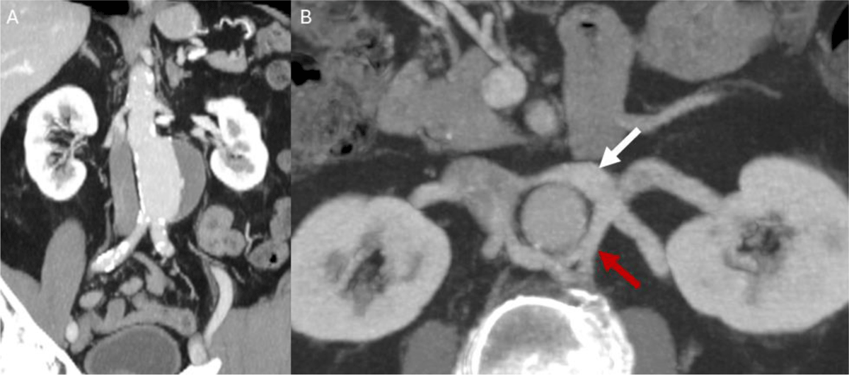
Figure 1
Preoperative computed tomography angiography of the patient. (A) Coronal maximum intensity projection (MIP) image shows a 6 cm infrarenal abdominal aortic aneurysm; (B) axial MIP image shows preaortic (white arrow) and retroaortic branches (red arrow) on the left renal vein.
Video 1
Cinematic-rendered computed tomography angiography of the patient shows the preaortic and retroaortic branches of the circumaortic left renal vein around the abdominal aorta; see also at https://vimeo.com/1208141314.
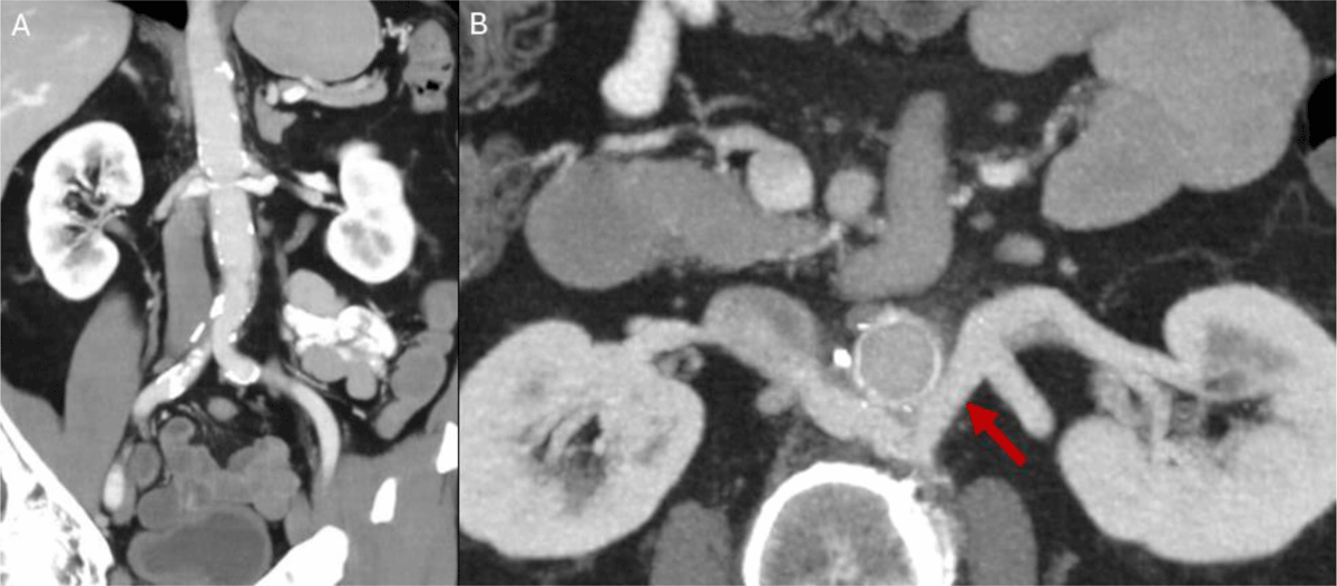
Figure 2
Eight-month follow-up computed tomography angiography of the patient. (A) Coronal abdominal aortic aneurysm image shows an 18 × 9 cm Gelweave bifurcated graft (Terumo) anastomosed from the infrarenal aorta to both iliac arteries; (B) axial maximum intensity projection image reveals the ligated preaortic branch and hypertrophied retroaortic branch of the left renal vein.