
A 72-year-old man presented with left lower quadrant abdominal pain and was found to have a 6 cm infrarenal abdominal aortic aneurysm (AAA) (Figure 1 A). Computed tomographic angiography (CTA) of the patient revealed a type II variant of the circumaortic left renal vein (LRV), with two distinct preaortic and retroaortic branches (Figure 1 B, Video 1).3 The patient underwent an open repair of the AAA with an aorto-bi-iliac graft. Suprarenal control was required due to the poor quality of the infrarenal neck. The preaortic LRV was ligated with 5-0 Prolene sutures for better visualization of the suprarenal aorta and to gain control of the renal arteries. The suprarenal aorta was clamped after identifying the retroaortic LRV to prevent inadvertent clamp injury to the vein. An end-to-end anastomosis of the infrarenal aorta was performed with the 18 × 9 mm Gelweave bifurcated graft (Terumo), followed by distal anastomosis of the graft with the iliac arteries. The patient had an uneventful postoperative course and was discharged on postoperative day 19. Eight-month follow-up CTA imaging revealed a satisfactory aneurysm repair and compensatory hypertrophy of the retroaortic LRV (Figure 2).
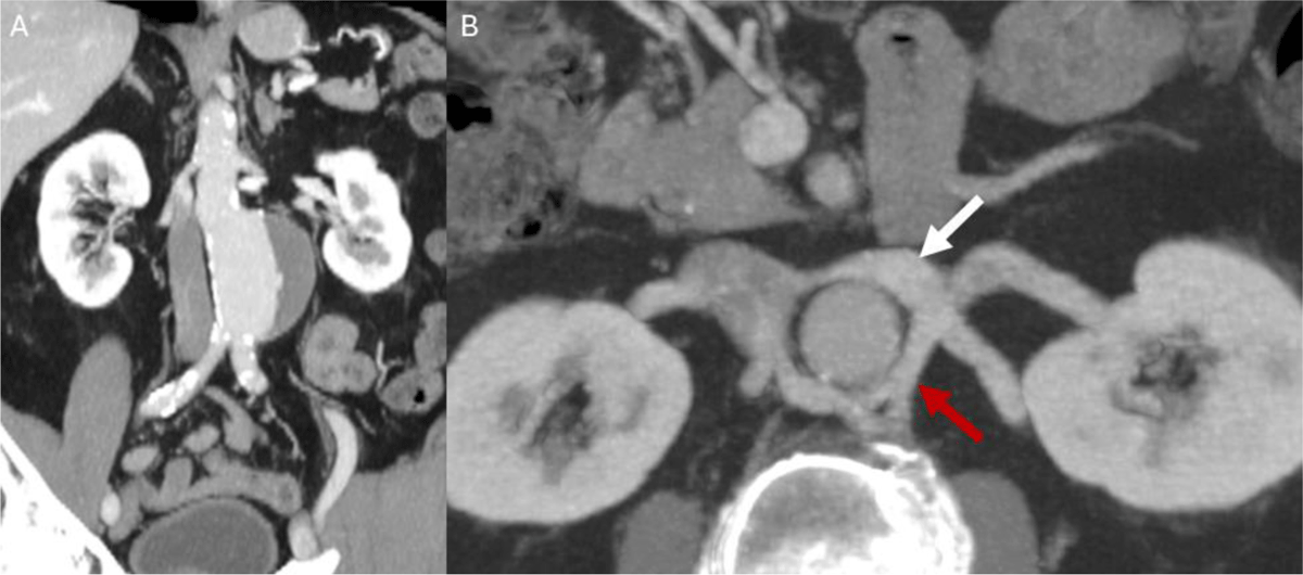
Figure 1
Preoperative computed tomography angiography of the patient. (A) Coronal maximum intensity projection (MIP) image shows a 6 cm infrarenal abdominal aortic aneurysm; (B) axial MIP image shows preaortic (white arrow) and retroaortic branches (red arrow) on the left renal vein.
Video 1
Cinematic-rendered computed tomography angiography of the patient shows the preaortic and retroaortic branches of the circumaortic left renal vein around the abdominal aorta; see also at https://vimeo.com/1208141314.
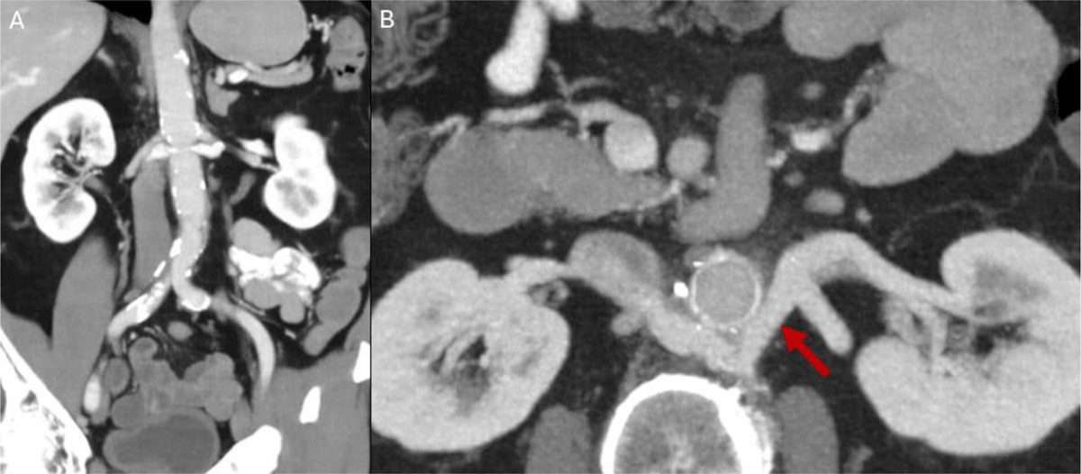
Figure 2
Eight-month follow-up computed tomography angiography of the patient. (A) Coronal abdominal aortic aneurysm image shows an 18 × 9 cm Gelweave bifurcated graft (Terumo) anastomosed from the infrarenal aorta to both iliac arteries; (B) axial maximum intensity projection image reveals the ligated preaortic branch and hypertrophied retroaortic branch of the left renal vein.
This case highlights the importance of preoperative identification of the circumaortic renal vein before open aortic surgery to prevent iatrogenic vascular injury. It also underscores the natural history of a partially ligated circumaortic LRV.