
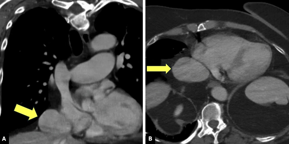
Figure 1
Axial (A) and coronal (B) views of preoperative computerized tomographic venography showing suprahepatic inferior vena cava aneurysm.
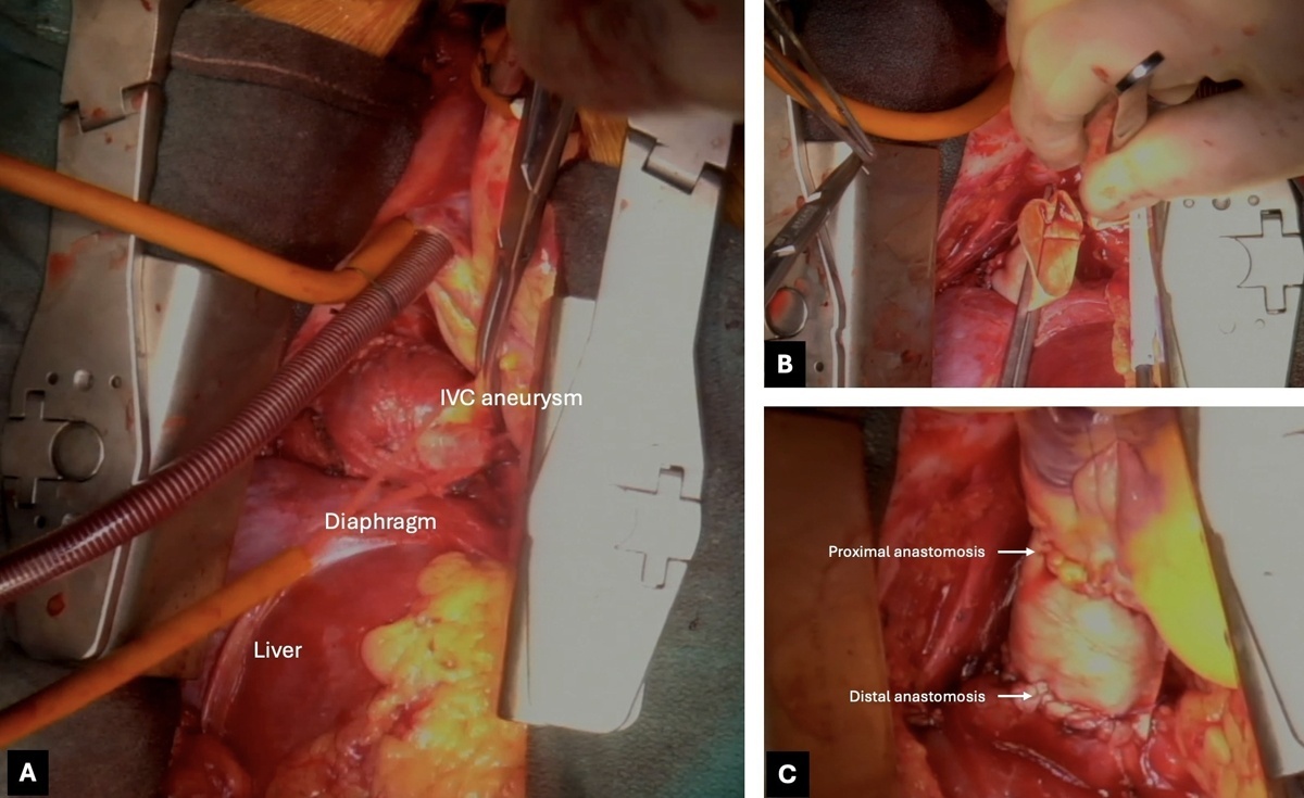
Figure 2
Intraoperative images showing the surgical management of a type I inferior vena cava (IVC) aneurysm. (A) Suprahepatic IVC exposure via median sternotomy and laparotomy IVC aneurysm dissection. (B) Replacement of resected IVC aneurysm with a tubularized bovine pericardial patch. (C) Completed IVC reconstruction.
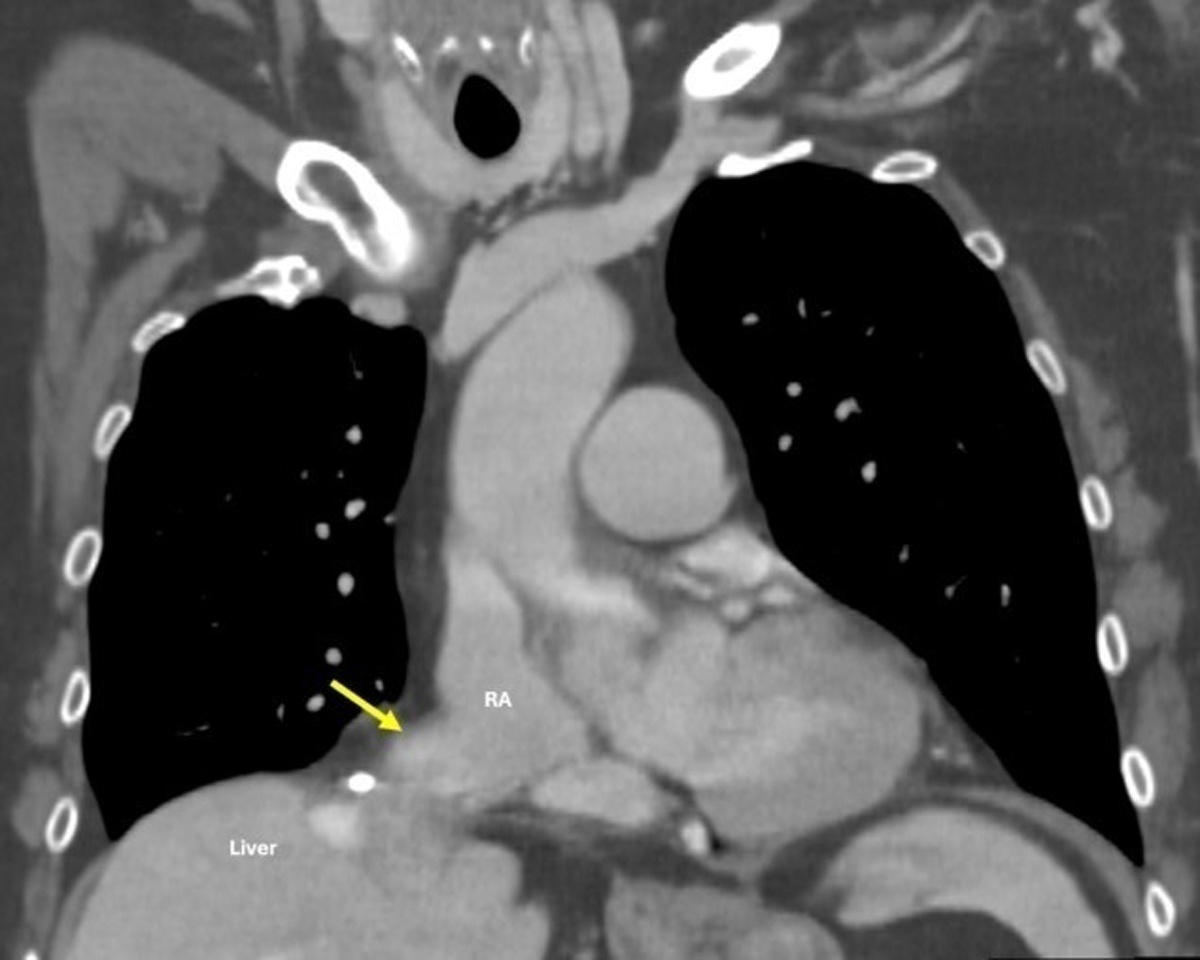
Figure 3
Postoperative computerized tomographic venography showing patent inferior vena cava reconstruction with no residual or recurrent aneurysm. RA: right atrium