
Introduction
Inferior vena cava (IVC) aneurysms are rare vascular dilations that carry the potential for serious complications, including hemorrhage, pulmonary embolism, and other thromboembolic events.1 As of 2026, only 76 cases have been reported in the literature.2 Given the rarity of this condition, standardized management guidelines have not been established. Here, we present a case of a symptomatic type I IVC aneurysm managed surgically, with an uncomplicated postoperative recovery, contributing to the limited body of evidence guiding treatment decisions.
Case Report
A 68-year-old female with a 2.5-year history of a suprahepatic IVC aneurysm presented to the clinic after imaging showed an increase in aneurysm size from 4.8 cm initially to 6 cm on serial axial imaging (Figure 1). She presented with intermittent chest discomfort and anxiety regarding her aneurysm. Her past medical history included gastroesophageal reflux disease, obesity, obstructive sleep apnea, asthma, and hypothyroidism. Current medications included albuterol, levothyroxine, and amitriptyline. Preoperative workup displayed normal heart function with an ejection fraction of 70%. A complete blood count and chemistry panel were within normal limits. This case was reviewed by a multidisciplinary cardiovascular team, which determined that surgical management was necessary due to the symptomatic presentation and enlarged aneurysm size.
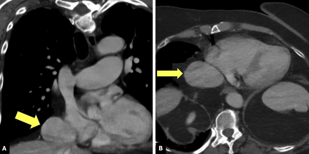
Figure 1
Axial (A) and coronal (B) views of preoperative computerized tomographic venography showing suprahepatic inferior vena cava aneurysm.
The patient was brought to the operating room and placed under general anesthesia. A median sternotomy and upper midline laparotomy were performed to provide exposure of the thoracic and abdominal components of the IVC. The pericardium was opened, and the superior vena cava, right atrium, and IVC were mobilized. The falciform ligament was divided, and the diaphragm was incised to allow exposure of the retrohepatic IVC behind the liver. After systemic heparinization, cardiopulmonary bypass was initiated via cannulation of the distal ascending aorta, superior vena cava, and right femoral vein. Following aortic cross-clamping and cardioplegic arrest, the IVC aneurysm was dissected and resected. Reconstruction of the retrohepatic IVC was performed using an interposition tube graft fashioned from a tubularized bovine pericardial patch, which was anastomosed to the IVC and right atrial junction with running 5-0 Prolene sutures (Figure 2). The patient was weaned from cardiopulmonary bypass without difficulty, and chest tubes were placed prior to closure.
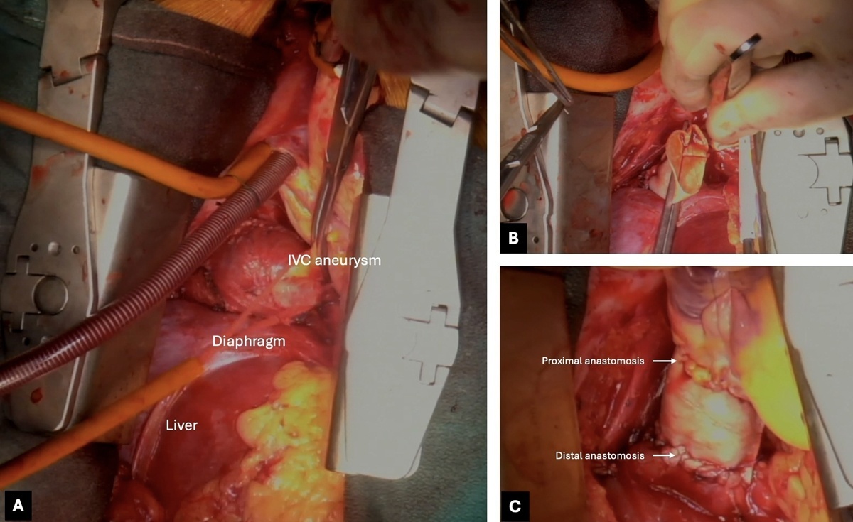
Figure 2
Intraoperative images showing the surgical management of a type I inferior vena cava (IVC) aneurysm. (A) Suprahepatic IVC exposure via median sternotomy and laparotomy IVC aneurysm dissection. (B) Replacement of resected IVC aneurysm with a tubularized bovine pericardial patch. (C) Completed IVC reconstruction.
Postoperatively, the patient was transferred to the cardiovascular intensive care unit, intubated, and successfully extubated that same day. Pain was managed with patient-controlled analgesia, and she was started on aggressive incentive spirometry. A clear liquid diet was initiated on postoperative day 1, and she was transferred to the floor the next day. At 1-month follow-up, the patient had recovered well with full activity and no chest pain or pertinent symptoms. A 6-month surveillance CT venogram demonstrated a patent IVC graft with no residual or recurrent aneurysm (Figure 3).
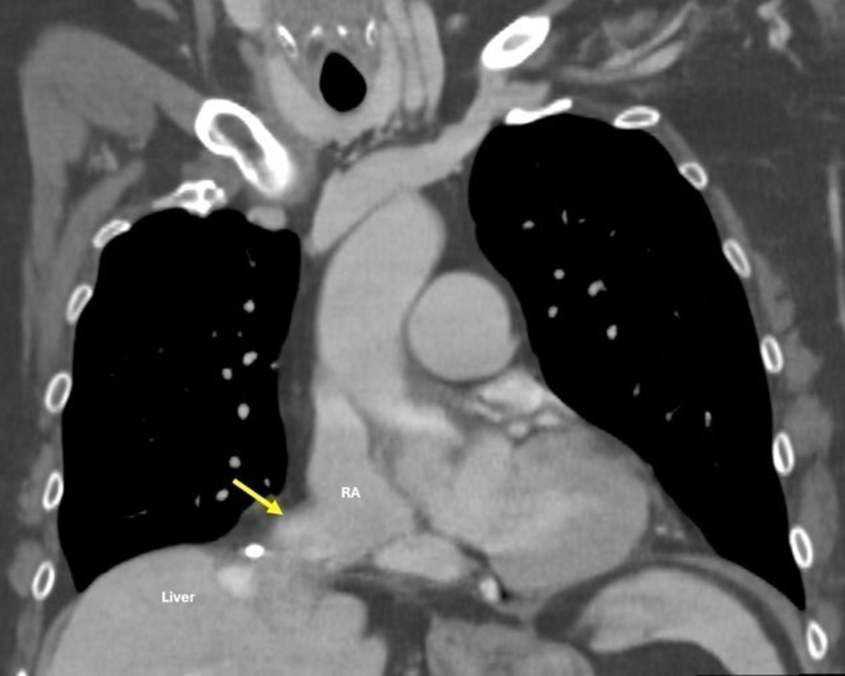
Figure 3
Postoperative computerized tomographic venography showing patent inferior vena cava reconstruction with no residual or recurrent aneurysm. RA: right atrium
Discussion
In this case, a type I IVC aneurysm was treated surgically without postoperative complications. To the best of our knowledge, this is the second reported case of surgically managed type I IVC aneurysm in the literature to date. IVC aneurysms have been classified into four types based on anatomic location, as described by Gradman and Steinberg: type I involves the suprahepatic IVC without interruption; type II is associated with supra- or infrahepatic IVC interruption; type III includes infrarenal aneurysms without an associated venous anomaly; and type IV includes miscellaneous presentations.1 Thompson and Lindenauer proposed an alternative classification based on etiology, categorizing IVC aneurysms as congenital (I), acquired (II), or secondary to an arteriovenous fistula (III).3 For the purposes of this report, we apply the Gradman and Steinberg anatomic classification system.
Type I inferior vena cava (IVC) aneurysms are technically challenging to manage surgically due to proximity to the heart and diaphragm and the need for a combined sternotomy and median laparotomy with cardiopulmonary bypass. Most reported cases have therefore been treated conservatively.4 In the most recent comprehensive review of IVC aneurysms published in 2021, only one case of surgically managed type I IVC aneurysm was identified.1,5 In that case, the patient presented with abdominal pain and a right hemothorax suspected to be secondary to bleeding from the aneurysm, and she survived with an uncomplicated postoperative course.5 Four additional reports of symptomatic type I IVC aneurysms were also managed conservatively, typically with serial imaging surveillance.4,5
Overall, there remains no clear consensus on the management of IVC aneurysms. In contrast to type I aneurysms, type II and type III aneurysms are more frequently considered for active intervention rather than surveillance due to the increased risk of thrombosis and other complications.2 Reported surgical intervention rates reflect this tendency, with 4 of 13 type II aneurysms and 16 of 31 type III aneurysms undergoing resection.6,7 In these cases, indications for surgery most commonly included symptomatic presentation (including hemodynamic instability) and large aneurysm size, both of which increase the risk of rupture.
In the previously reported surgically managed type I IVC aneurysm, the operation was performed through a right thoracotomy with rib retraction, revealing an aneurysm covered by the pericardium overlying the diaphragm. Pericardial and diaphragmatic dissection was performed to expose the lesion, followed by ligation of collateral veins, excision of the aneurysmal mass, and primary closure of the IVC supported with Teflon pledgets.5 Similar operative principles have been described in the surgical management of other IVC aneurysm types. For example, type III IVC aneurysms have been treated with open resection via midline or rooftop laparotomy, followed by excision of the aneurysm and closure of the neck through lateral venorrhaphy or primary repair with 3-0 Prolene sutures.8,9 In another report, a hemodynamically unstable patient with a type II IVC aneurysm underwent emergent surgical excision with reconstruction of the IVC using a vascular graft.7 In our case, surgical management consisted of a sternotomy with aneurysm resection and IVC reconstruction using an interposition graft made from a tubularized bovine pericardial patch. Overall, our approach incorporated principles described in prior surgical cases while adapting the repair to the patient’s anatomy, particularly the need for interposition graft reconstruction in the retrohepatic segment of the IVC.
Although type I IVC aneurysms are more often asymptomatic and therefore managed conservatively, prior literature supports surgical intervention in patients who are hemodynamically unstable, symptomatic, or demonstrate aneurysm progression on serial imaging.10 Our patient met several of these criteria, including progressive enlargement on interval imaging and persistent chest discomfort. Therefore, despite the technical challenges associated with the suprahepatic location typical of type I aneurysms, surgical management was justified.
Importantly, management decisions should not be determined solely by aneurysm classification. Rather, they should be guided by individualized assessment of rupture risk and clinical trajectory. We also deem a multidisciplinary approach to be important given the rare nature of this disease and absence of clear guidelines for management. When high-risk features are present, the benefits of surgical intervention may outweigh the inherent operative risks, even in type I IVC aneurysms.
Conclusion
IVC aneurysms vary in presentation based on their anatomic classification, with management strategies ranging from surveillance with serial imaging to surgical intervention. Operative management is generally reserved for symptomatic cases or those at high risk of rupture and thromboembolic complications, most commonly in type II and III aneurysms, while type I lesions are often managed conservatively due to surgical complexity. This case highlights the feasibility and safety of surgical repair to mitigate the risk of rupture of a symptomatic, enlarging type I IVC aneurysm.
Competing Interests
Drs. Michael J. Reardon and Alan B Lumsden are members of the editorial board of the Methodist DeBakey Cardiovascular Journal, serving on a voluntary basis. The other authors have no competing interests to declare.