
Introduction
Progressive leaflet calcification is the primary driver of aortic stenosis (AS) in the majority of elderly individuals.1,2 Importantly, a subset of patients manifests severe AS caused by extensive aortic leaflet thickening with little or no associated calcification. Several pathological processes, including rheumatic heart disease, systemic lupus erythematosus, medications, and prior chest radiation, have been associated with the development of AS characterized by leaflet thickening with minimal calcification.3 We present an older adult with severe AS despite an almost complete absence of aortic valve calcification, highlighting a rare and diagnostically challenging presentation.
Case Presentation
A 79-year-old man with a past medical history significant for hypertension, hyperlipidemia, small-vessel ischemic disease, and AS was referred to the valve clinic for preoperative cardiovascular risk stratification prior to shoulder surgery. He denied chest pain, dyspnea, palpitations, or syncope and reported no significant change in functional capacity. He endorsed occasional orthostatic symptoms but otherwise felt well from a cardiovascular standpoint.
Transthoracic echocardiography (TTE) performed as part of the preoperative evaluation demonstrated progression of AS severity compared with a study performed 3 years earlier, which had shown moderate AS. The preoperative examination revealed an aortic valve area of 0.9 cm2 and mean transaortic gradient of 32 mm Hg. The TTE findings are provided in Videos 1, 2, 3 and 4.
Video 1
Parasternal long-axis view focused on the aortic valve demonstrating marked leaflet thickening and restricted systolic opening of a trileaflet aortic valve without overt valvular calcification; see also at https://vimeo.com/1207815788.
Video 2
Apical three-chamber view of simultaneous two-dimensional imaging (left panel) and color Doppler interrogation (right panel) demonstrating turbulent systolic flow across the aortic valve consistent with hemodynamically significant aortic stenosis. Mild aortic regurgitation is also present; see also at https://vimeo.com/1207819149.
Video 3
Parasternal short-axis view demonstrating a trileaflet aortic valve with markedly restricted leaflet excursion and asymmetric leaflet thickening despite minimal visible valvular calcification; see also at https://vimeo.com/1207820076.
Video 4
Parasternal long-axis view demonstrating preserved left ventricular systolic function and severe restriction of aortic valve opening. Despite a suboptimal acoustic window, progressive severe aortic stenosis is evident; see also at https://vimeo.com/1207821147.
Following the TTE findings, transesophageal echo-cardiography (TEE) was recommended for further aortic valve evaluation given the asymmetrical thickening of the leaflets. A follow-up TEE performed approximately 2 months later demonstrated moderate aortic valve stenosis, with an aortic valve area of 1.2 cm² by 3-dimensional planimetry and a mean transvalvular gradient of 24 mm Hg. Moderate aortic regurgitation was also noted.
Over the subsequent follow-up period, the patient underwent serial TTE at approximately 6-month intervals. These studies demonstrated persistent and progressive AS, with aortic valve area decreasing from 1 cm² down to 0.79 cm² and systolic mean Doppler gradients increasing from 34 mm Hg to 41 mm Hg. During this interval, the patient remained clinically asymptomatic with respect to AS and had a normal N-terminal pro-B-type natriuretic peptide (NT-proBNP) level. Six months later, the patient underwent repeat TTE, which demonstrated severe aortic valve stenosis, with a mean systolic Doppler gradient of 40 mm Hg and an aortic valve area of 0.85 cm² by Doppler. Moderate aortic regurgitation was also present. At this time, the patient had reported some decreased exercise tolerance despite a normal NT-proBNP level. After discussion of treatment options, he agreed to proceed with intervention. Serial assessments of AS severity over time are summarized in Table 1.
Table 1
Serial echocardiographic assessment of aortic stenosis severity. TEE: transesophageal echocardiography; TTE: transthoracic echocardiography
| TIME POINT | MODALITY | AORTIC VALVE AREA (CM2) | MEAN GRADIENT (MM HG) |
|---|---|---|---|
| First visit | TTE | 0.9 | 32 |
| Subsequent 2-months visit | TEE | 1.2 | 24 |
| Subsequent 6-months visit | TTE | 1 | 34 |
| Subsequent 12-months visit | TTE | 0.79 | 41 |
| Subsequent 6-months visit | TTE | 0.85 | 40 |
| Intraoperative | TEE | 0.92 | 43 |
| Subsequent 6-months visit | TTE | - | 6 |
To further assess the functional significance of his aortic valve disease and confirm a cardiac etiology for his symptoms, cardiopulmonary exercise testing was performed using a treadmill Naughton protocol, which demonstrated mild cardiac output limitation during exercise, with a peak oxygen consumption of 1.56 L/min (83% predicted) and O2 pulse of 78% predicted. The patient achieved a peak workload of 5.4 measured metabolic equivalents (6.6 predicted) with a normal chronotropic response, reaching a peak heart rate of 148 beats per minute (106% predicted).
One month later, a contrast-enhanced cardiac computed tomography (CT) angiogram was performed, demonstrating a low aortic valve calcium (AVC) burden, with an aortic valve calcium score of 76.5 AU as shown in Figure 1. His cardiac catheterization revealed nonobstructive coronary artery disease.
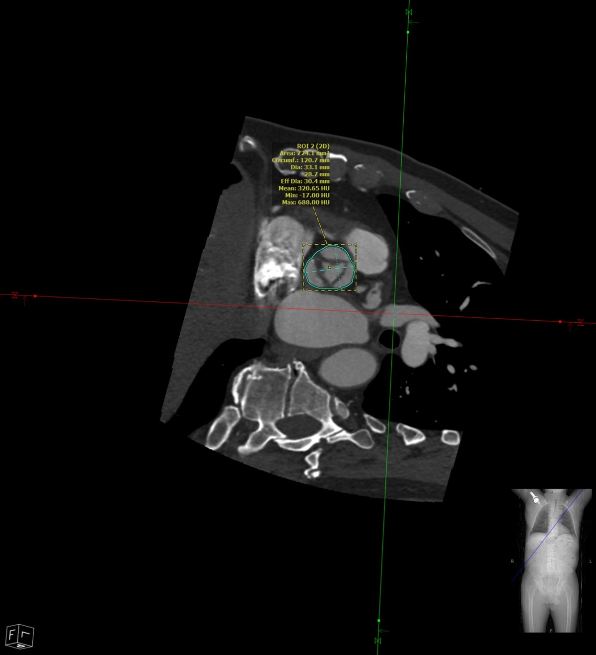
Figure 1
Cardiac computed tomography angiogram cross-sectional image demonstrating focal minimal calcification of the aortic valve (circled).
Following a multidisciplinary heart team discussion, the patient was deemed a high-risk candidate for transcatheter aortic valve replacement (TAVR) given the fibrotic nature of the aortic stenosis; this was despite a Society of Thoracic Surgeons Predicted Risk of Mortality score of 2.4% (intermediate risk), a tricuspid aortic valve morphology, annular dimensions of 21.1 mm × 25.6 mm, and absence of left ventricular outflow tract calcification. Although TAVR has been performed in selected patients with noncalcific aortic stenosis, traditionally the absence of calcification remains a major limitation because of impaired valve anchoring and sealing, rendering this anatomy relatively unfavorable for transcatheter treatment and increasing the risk of prosthesis embolization.4,5 As a result, the patient subsequently underwent open surgical bioprosthetic aortic valve replacement with concomitant left atrial appendage ligation. Intraoperative TEE demonstrated severe AS with a trileaflet aortic valve and a mean systolic Doppler gradient of 43 mm Hg (Figure 2). Post-procedural echocardiography showed a well-functioning bioprosthetic aortic valve with a mean systolic Doppler gradient of 6 mm Hg, without prosthetic aortic regurgitation or paravalvular leak. Histopathologic examination of the surgically explanted aortic valve demonstrated degenerative fibrous changes with minimal calcification, consistent with the clinical diagnosis of significant AS (Figure 3).
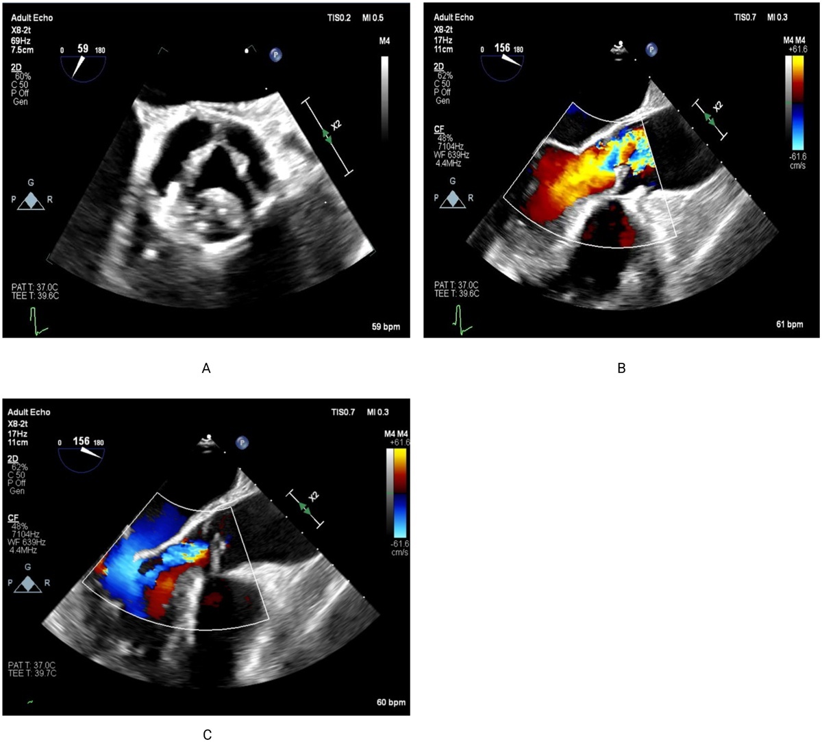
Figure 2
Transesophageal echocardiography findings. (A) Short-axis view of the aortic valve in systole demonstrating restricted leaflet opening with severe leaflet remodeling. (B) Long-axis view demonstrating aliasing at the level of the aortic valve before surgical intervention. (C) Long-axis view showing moderate aortic regurgitation.
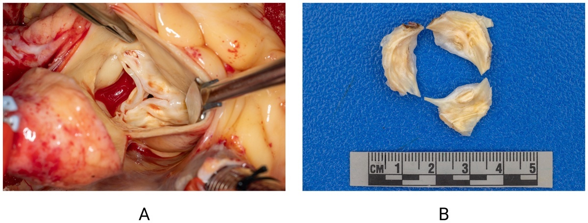
Figure 3
Surgical and pathological evaluation of the native aortic valve. (A) Intraoperative view of the aortic valve showing severe stenosis characterized by fibrotic thickening and degenerative changes of the leaflets. (B) Histopathological specimen of the aortic valve showing fibrotic changes with minimal calcification.
Discussion
Degenerative calcific disease continues to be the predominant cause of severe AS in adults, and AVC burden is routinely used as a marker of disease severity and prognosis.6 Our case is notable for severe AS in the near absence of macroscopic or CT-detectable calcification, indicating that extensive fibro-inflammatory remodeling of the valve can, in rare instances, produce critical obstruction without substantial calcium deposition. This phenotype challenges the conventional calcium-centric paradigm and suggests that calcification alone may be insufficient to accurately characterize AS severity.
Computed tomography aortic valve calcium (CT-AVC) scoring has been validated as a complementary tool for adjudicating AS severity. Contemporary guidelines recommend sex-specific AVC thresholds to define severe AS, but these thresholds are derived predominantly from populations in whom macroscopic calcification is the principal driver of obstruction.7 As a result, CT-AVC may underestimate disease severity in patients with a fibrosis-predominant phenotype, as demonstrated in the present case with an AVC score of 76.5 AU despite hemodynamic evidence of severity.
In a retrospective cohort study of 563 patients with severe symptomatic AS, Abramowitz et al. demonstrated that 93 patients exhibited low CT-AVC, with a mean AVC score of approximately 1,278 ± 485 within this low-calcification subgroup.8 In contrast, our patient’s AVC score of 76.5 AU indicates an almost complete absence of calcification, representing a markedly more extreme phenotype and further expanding the spectrum of noncalcific severe AS reported in the literature.
In another retrospective cohort of 136 patients who received TAVR, including 21 patients with minimally calcified AS, Xiong et al. found that the noncalcified AS group was significantly younger than our patient, with a mean age of 70.0 years; this suggests that our case represents a more advanced-age presentation of this uncommon phenotype. Additionally, Xiong et al. quantified valvular calcium burden using the volume of aortic root calcification as determined by FluoroCT 3.0 software,4 whereas in our case, valve calcification was assessed using the standard Agatston calcium score, which is the most commonly used metric in clinical practice.
Notably, Nonaka et al. reported a case describing severe AS in the setting of a noncalcified bicuspid aortic valve,5 whereas our patient had a trileaflet aortic valve with an almost complete absence of calcification, highlighting that this rare entity is not limited to bicuspid valve morphology and may also occur in anatomically tricuspid valves.
Conclusion
This case highlights a rare presentation of hemo-dynamically severe AS occurring in an elderly patient in the near absence of aortic valve calcification. Compared with previously reported cohorts, our patient represents a more extreme phenotype, with a markedly lower calcium burden and older age at presentation. These findings emphasize that severe AS can occur through mechanisms other than calcification and that CT-based calcium scoring may underestimate disease severity in selected patients. Careful integration of clinical findings, echocardiographic hemodynamics, and multimodality imaging is therefore essential to accurately diagnose and manage this uncommon but clinically important entity.
Artificial Intelligence Statement
Artificial intelligence was used solely to assist with language editing of the manuscript. All authors reviewed and approved the final version and accept full responsibility for the accuracy, integrity, and content of the work.
Competing Interests
The authors have no competing interests to declare.