
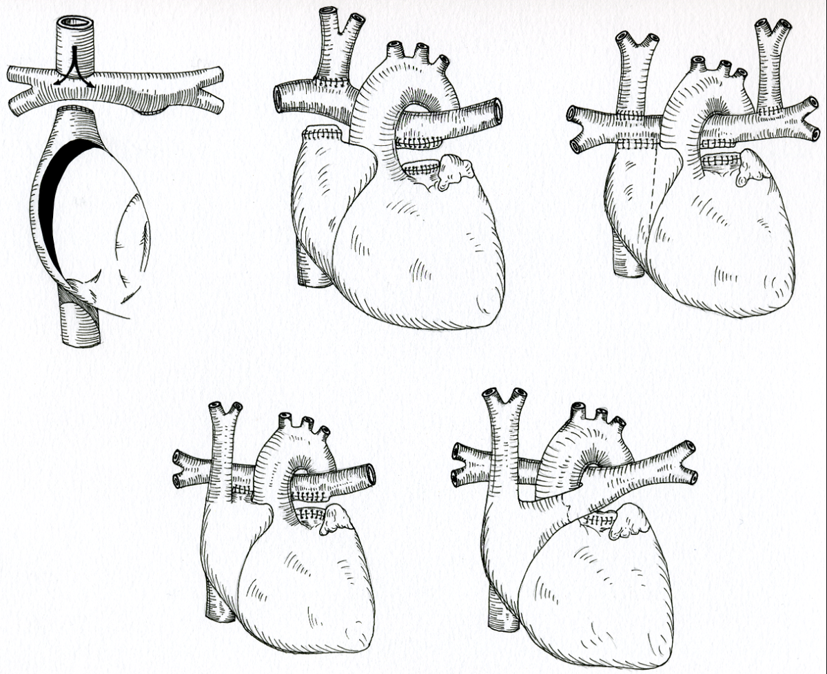
Figure 1
Schematic representation of the various Fontan modifications.
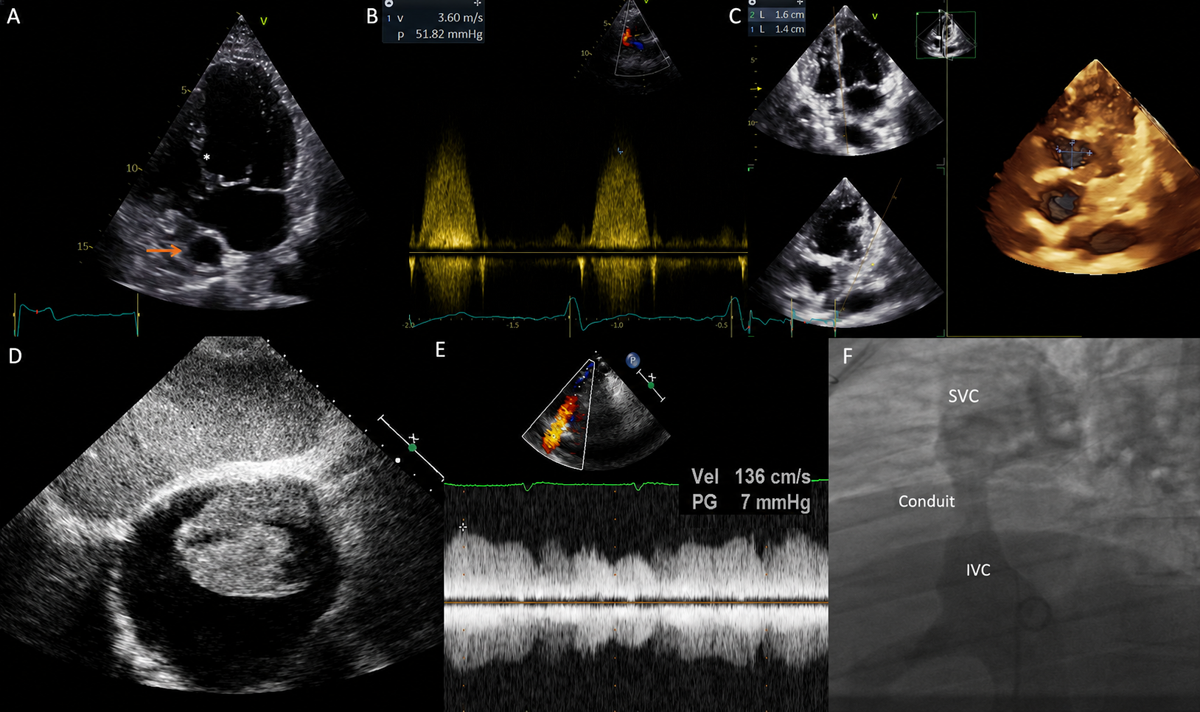
Figure 2
Possible Fontan features on imaging. (A) Transthoracic echocardiographic apical 4-chamber view in a patient with double inlet left ventricle and rudimentary right ventricle. The asterisk indicates the bulboventricular foramen, while the arrow points to the extracardiac Fontan conduit. (B) Continuous Doppler signal of a restrictive bulboventricular foramen in a patient with tricuspid atresia and transposed great vessels resulting in significant subaortic obstruction. (C) Bulboventricular foramen size assessed with 3-dimensional echocardiography and en face visualization with multiplanar reconstruction. (D) Subcostal echocardiographic view showing a bulky thrombus inside the grossly dilated right atrium in a patient with direct atriopulmonary connection. (E) Suprasternal view with pulsed Doppler interrogation revealing increased flow velocity at distal conduit anastomosis with reduced respiratory variations, suggestive of Fontan conduit obstruction. (F) Venous angiography during cardiac catheterization in the same patient as E confirming a discrete stenosis of the conduit, which was successfully treated with conduit stenting. SVC: superior vena cava; IVC: inferior vena cava
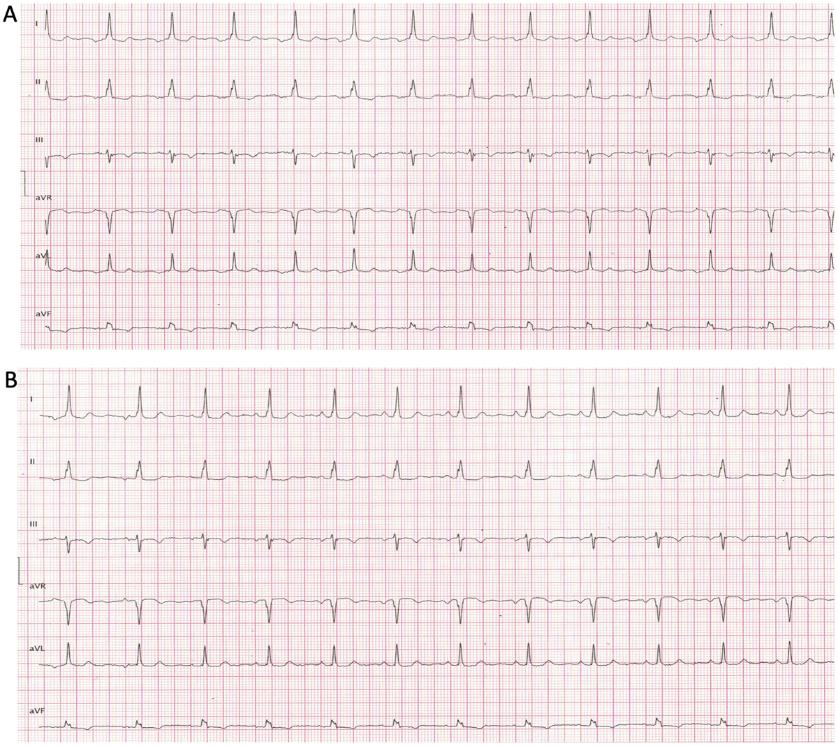
Figure 3
Electrocardiogram of a Fontan patient (A) during atrial tachycardia and (B) in sinus rhythm.