
Introduction
Single-ventricle physiology, or functionally univentricular hearts, refers to a heterogeneous group of complex congenital heart defects in which a single dominant ventricle supports both the systemic and pulmonary circulations.1,2 The dominant ventricle may exhibit right-, left-, mixed-, or indeterminate-sided morphology, whereas any secondary chamber is typically rudimentary and functionally inadequate to support independent circulation. Single-ventricle lesions are overall rare, collectively accounting for approximately 0.5% to 2% of congenital heart defects, with an estimated incidence of 2 to 4 per 10,000 live births depending on inclusion criteria.3 A male predominance has been reported in several subtypes. Familial recurrence occurs in a minority of cases, and genetic associations are multifactorial and incompletely defined. Although syndromic and chromosomal associations have been described, no single chromosomal abnormality has been consistently linked to isolated single-ventricle physiology.4 Over the last five decades, staged surgical palliation culminating in the Fontan procedure (Figure 1)—routing systemic venous return directly to the pulmonary arteries—has transformed survival, and most patients now reach adulthood.5,6 Nevertheless, Fontan circulation remains palliative: pulmonary blood flow is passive, preload reserve is limited, and chronic systemic venous hypertension is intrinsic. With time, patients may develop arrhythmias, thromboembolism, ventricular dysfunction, pathway obstruction, lymphatic disorders, liver disease, and reduced exercise capacity. This review provides a practical, physiology-based overview for general cardiologists, emphasizing early recognition of deterioration and timely referral to adult congenital heart disease (ACHD) centers.
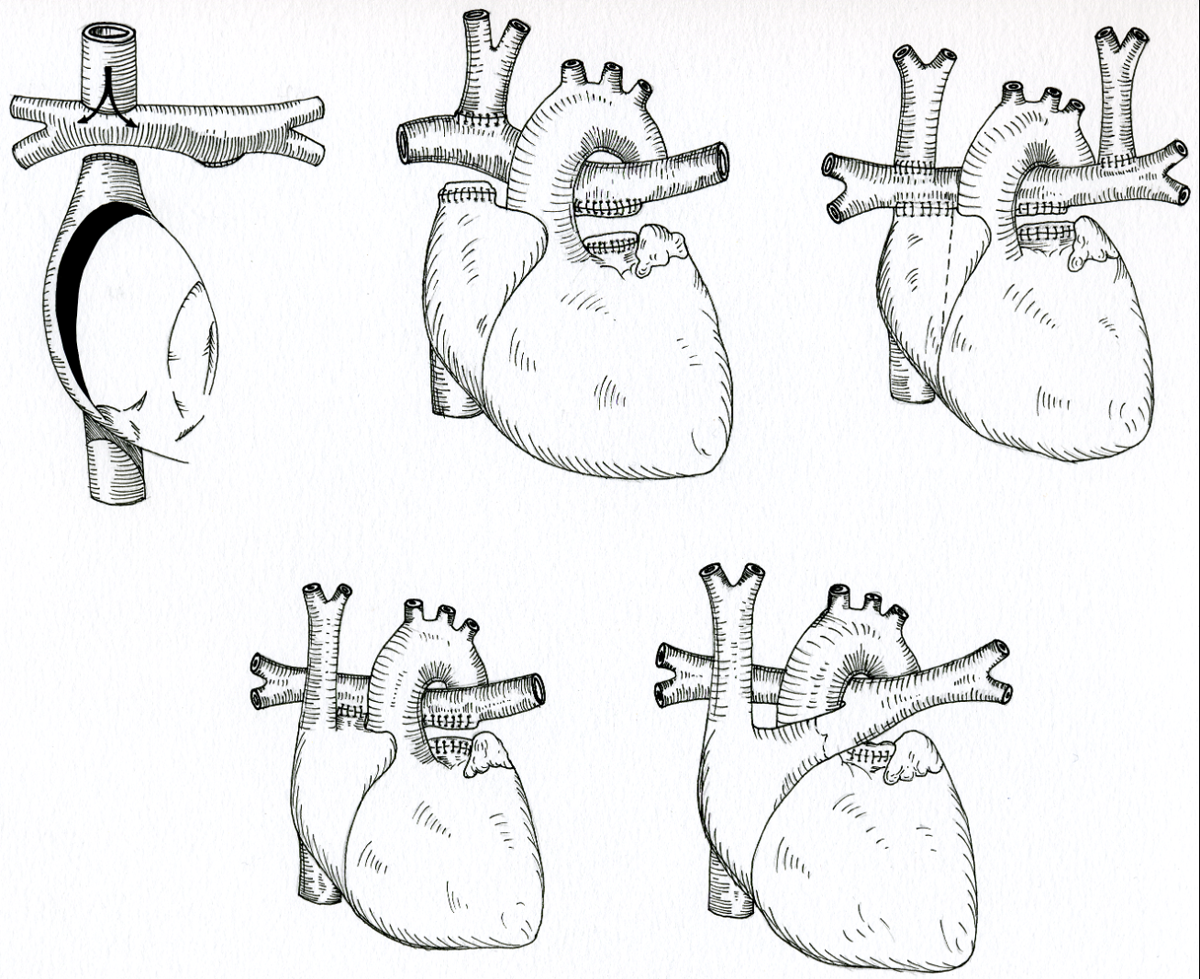
Figure 1
Schematic representation of the various Fontan modifications.
Anatomical Substrate of Single Ventricles
A ventricle is defined by an inlet component (atrioventricular valve to papillary muscle insertions), a trabecular/apical component, and an outlet (infundibular) tract supporting the semilunar valve. The presence of an inlet component is essential for defining a true ventricle. In its absence, a cavity consisting only in an outlet portion is described as a rudimentary chamber. In the heterogenous group of single-ventricle malformations, one chamber is dominant while the other is rudimentary or nonfunctional.
Classic double-inlet variants include double-inlet left ventricle (approximately 80%), double-inlet right ventricle, and the least common forms with indeterminate morphology of the ventricular chamber. For practical orientation, lesions may be grouped according to the morphology of the dominant pumping chamber: conditions resulting in a dominant single left ventricle include double-inlet left ventricle and tricuspid atresia; conditions resulting in a dominant single right ventricle include hypoplastic left heart syndrome, mitral atresia, and selected double-outlet right ventricle variants with severe left-sided hypoplasia; true univentricular or indeterminate forms include rare hearts with a solitary ventricular cavity or complex heterotaxy. Univentricular physiology may also occur when two ventricles are present but cannot be separated safely (eg, straddling/overriding atrioventricular [AV] valves, coronary-dependent right ventricle in pulmonary atresia with intact septum).
Any ventriculo-arterial relationship may coexist, including concordant or discordant connections, double-outlet configurations, or single-outlet physiology. When a rudimentary ventricle is present, the bulboventricular foramen is the communication between the dominant ventricle and the underdeveloped ventricular chamber (Figure 2 A-C). If this communication is restrictive, subaortic or subpulmonary obstruction may occur and may evolve over time, particularly in discordant ventriculo-arterial connections.7 Associated lesions such as AV valve abnormalities (hypoplasia, dysplasia, clefts, straddling/overriding), pulmonary valve stenosis or atresia, subpulmonary obstruction, and aortic arch lesions are frequent and influence palliation strategy and long-term outcomes.
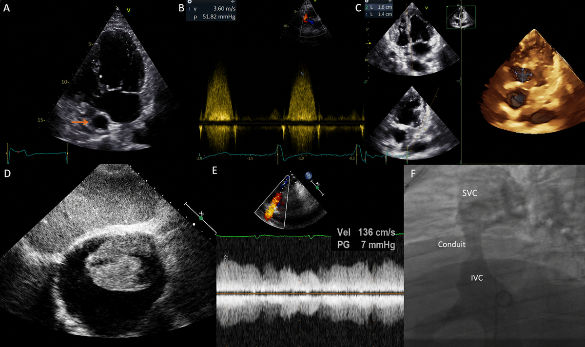
Figure 2
Possible Fontan features on imaging. (A) Transthoracic echocardiographic apical 4-chamber view in a patient with double inlet left ventricle and rudimentary right ventricle. The asterisk indicates the bulboventricular foramen, while the arrow points to the extracardiac Fontan conduit. (B) Continuous Doppler signal of a restrictive bulboventricular foramen in a patient with tricuspid atresia and transposed great vessels resulting in significant subaortic obstruction. (C) Bulboventricular foramen size assessed with 3-dimensional echocardiography and en face visualization with multiplanar reconstruction. (D) Subcostal echocardiographic view showing a bulky thrombus inside the grossly dilated right atrium in a patient with direct atriopulmonary connection. (E) Suprasternal view with pulsed Doppler interrogation revealing increased flow velocity at distal conduit anastomosis with reduced respiratory variations, suggestive of Fontan conduit obstruction. (F) Venous angiography during cardiac catheterization in the same patient as E confirming a discrete stenosis of the conduit, which was successfully treated with conduit stenting. SVC: superior vena cava; IVC: inferior vena cava
Surgical Palliation and Fontan Circulation
Surgical palliation of single-ventricle physiology is typically performed in staged procedures that ultimately culminate in the Fontan circulation, first described in 1971 (Figure 1).8 The goal of this staged approach is to separate the systemic and pulmonary circulations, reserving the dominant ventricle for the systemic circulation. It may involve a systemic-to-pulmonary artery shunt (eg, modified Blalock-Taussig shunt) in patients with reduced pulmonary blood flow or a pulmonary artery banding to balance pulmonary blood flow and protect the pulmonary vasculature. Atrial septectomy may be required when atrial-level restriction impairs mixing. Stage 2, usually performed between 3 and 6 months of age, consists in a bidirectional cavopulmonary connection (Glenn), directing superior vena cava blood to the pulmonary arteries and partially decompressing the ventricle.9 Fontan completion is usually performed between 2 and 4 years and directs inferior vena cava flow to the pulmonary arteries, establishing total cavopulmonary connection. Contemporary techniques include lateral tunnel and extracardiac conduit Fontan. Earlier atriopulmonary connections are largely abandoned due to late atrial dilation and arrhythmias. Some patients undergo creation of a fenestration (typically 4-6 mm) between the Fontan pathway and atrium to decompress the circuit in higher-risk settings at the cost of mild cyanosis and potential paradoxical embolism.
Fontan Circulation Physiology and Long-term Outcome
Fontan palliation establishes a circulation in which systemic venous return is directed to the pulmonary arteries without an interposed subpulmonary ventricle. Pulmonary blood flow becomes entirely passive, driven by the pressure gradient between the systemic venous circulation and the systemic atrium. As a result, cardiac output depends critically on low pulmonary vascular resistance (PVR), adequate preload, and preserved ventricular function.
This circulation is characterized by:
Elevated systemic venous pressure, necessary to maintain pulmonary flow,
Reduced ventricular preload, limiting stroke volume and exercise capacity,
A chronic low cardiac output state, particularly during stress or exertion, and
Absence of pulsatile pulmonary flow, affecting pulmonary vascular development.
Successful Fontan physiology requires an unobstructed pathway of the entire circuit (Fontan conduit, pulmonary arteries, veins, ventricular outflow), low PVR, preserved systolic and diastolic ventricular function, and competent systemic AV valve function. Failure to meet these conditions increases early and late Fontan failure risk. Any cause of increased atrial pressure may be transmitted retrogradely to the pulmonary vasculature, resulting in elevated PVR. As PVR rises, central venous pressure must increase further to sustain passive pulmonary blood flow in the Fontan circulation, thereby exacerbating systemic venous congestion.
Long-term survival has improved and many patients now live well into adulthood.6 Large cohort studies report survival rates of approximately 90% at 30 years and 80% at 40 years of age in contemporary cohorts.10 However, morbidity remains substantial and typically increases with time, driven by arrhythmias, thromboembolism, ventricular dysfunction (especially with a systemic morphologic right ventricle), systemic AV valve regurgitation, elevated PVR, and pathway obstruction. Exercise limitation is common and reflects limited stroke-volume reserve in a circulation constrained by the Fontan pathway and passive pulmonary blood flow; contributing factors include an elevated transpulmonary gradient or PVR, chronotropic incompetence, impaired ventricular filling, pathway obstruction, AV valve regurgitation, and deconditioning. Declining peak oxygen consumption correlates with adverse outcomes. A recently validated clinical score incorporating age at the first visit, history of atrial tachyarrhythmia, heart failure, New York Heart Association class, systolic blood pressure, and resting arterial oxygen saturation was demonstrated to discriminate patients at higher risk of death or transplant.11
Late Complications and Management of Adult Patients
Adult Fontan care aims to identify anatomic lesions and physiologic derangements that may precipitate Fontan failure and to address extracardiac complications early.12 Follow-up is ideally delivered in a multidisciplinary ACHD program with coordinated arrhythmia management, thromboprophylaxis, imaging/hemodynamic surveillance, and liver/lymphatic assessment.
Arrhythmias
Rhythm disturbances are common after Fontan surgery and significantly contribute to hospitalization, Fontan failure, and late mortality. Atrial arrhythmias occur in over 60% of adult Fontan patients, with intra-atrial reentrant tachycardia, atrial flutter, and atrial fibrillation representing the main substrates.13 Mechanisms include atrial scarring, dilation, elevated atrial pressures, ventricular dysfunction, AV valve regurgitation, and Fontan pathway obstruction. In the preload-dependent Fontan circulation, atrial tachyarrhythmias can acutely impair ventricular filling and reduce cardiac output, necessitating prompt rhythm control and anticoagulation when indicated.12 Intra-atrial reentrant tachycardia is particularly important after Fontan palliation and may present with deceptively modest ventricular rates; therefore, a resting heart rate persistently above 100 beats/min in clinic should be considered a red flag for possible intra-atrial reentrant tachycardia until proven otherwise. New or recurrent arrhythmias warrant evaluation for treatable factors and reassessment of Fontan function. Long-term management includes antiarrhythmic drugs, although its use may be limited by proarrhythmic effects and extracardiac organ dysfunction. Chronic antiarrhythmic medications may also complicate recognition of atrial arrhythmia (Figure 3). Catheter ablation should be considered but is technically challenging due to complex anatomy and access limitations and should be performed by an experienced team.13,14 In selected patients with failing atriopulmonary Fontan and refractory atrial arrhythmias, surgical conversion to total cavopulmonary connection combined with arrhythmia surgery (maze/ablation) can reduce arrhythmia burden and improve hemodynamics.12,15 Sinus node dysfunction and AV block are also common, potentially requiring permanent pacing. Epicardial systems are preferred due to venous access limitations, and device programming generally aims to maximize atrial pacing and minimize ventricular pacing. When ventricular pacing is unavoidable, careful site selection is essential because chronic ventricular pacing can lead to dyssynchrony, systemic ventricular dysfunction, and worse long-term outcomes.16,17
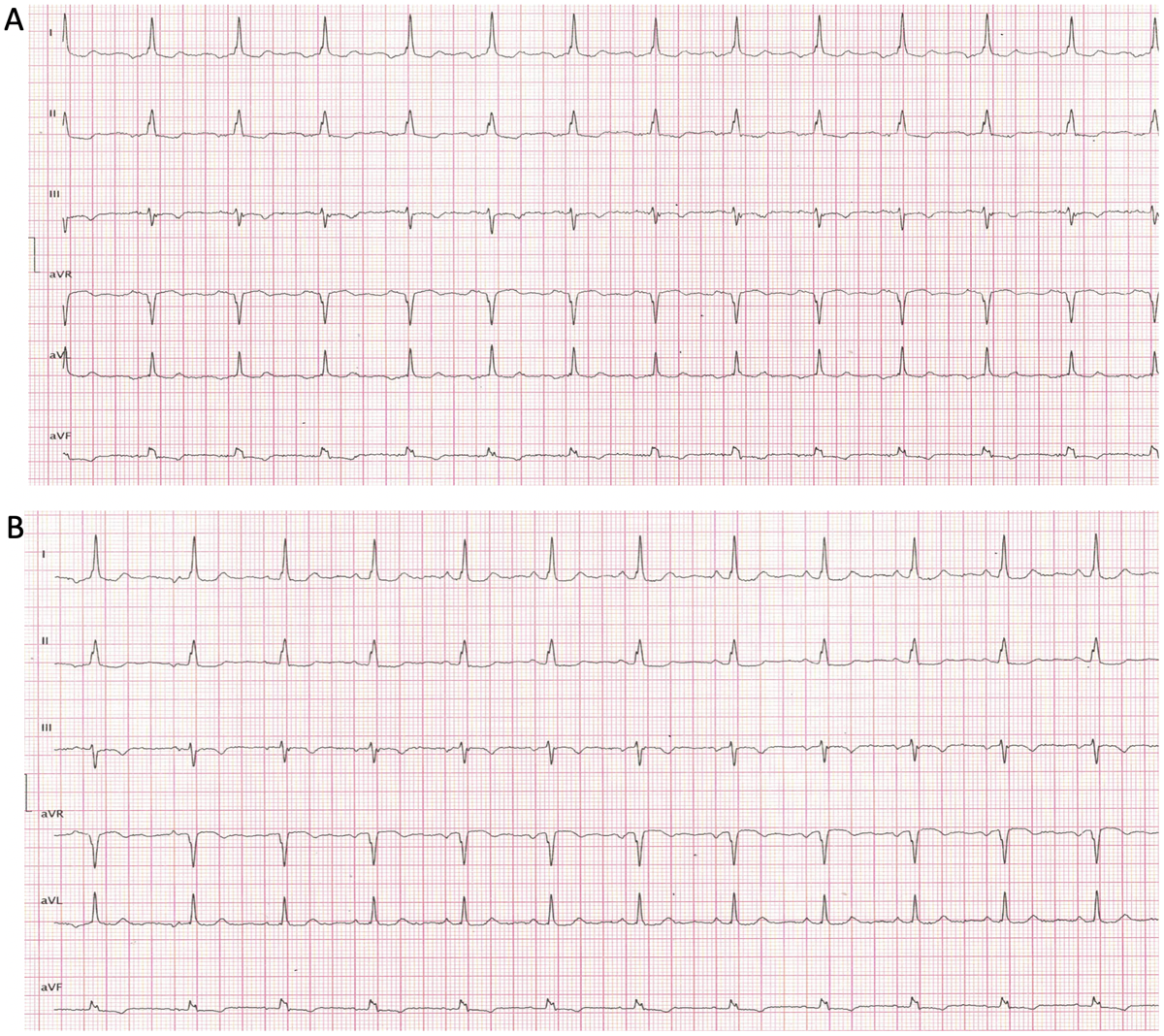
Figure 3
Electrocardiogram of a Fontan patient (A) during atrial tachycardia and (B) in sinus rhythm.
Thromboembolic and Bleeding Risk
Both thromboembolic and bleeding complications are well recognized in Fontan circulation and contribute substantially to mortality and morbidity. Thrombotic events are described in approximately 6% to 25% of patients across series.18,19,20 The Fontan physiology is intrinsically prothrombotic due to chronic systemic venous hypertension, low-flow states, absence of pulsatile venous flow, compounded by atrial dilation, atrial arrhythmias, endothelial dysfunction, prosthetic material, and reduced cardiac output.20 Coagulation abnormalities are common in Fontan patients and are largely attributed to liver dysfunction, low-grade factor consumption, and loss of proteins in conditions such as protein-losing enteropathy (PLE). Reduced levels of vitamin K–dependent clotting factors have been reported, together with increased antithrombin and tissue factor pathway inhibitor levels. However, decreased protein C and elevated factor VIII counterbalance this profile, resulting in a fragile and unstable hemostatic state with concurrent risks of thrombosis and bleeding.20
Patients with older Fontan configurations with an atriopulmonary connection are particularly prone to thrombus formation within the grossly dilated right atrium (Figure 2 D). Events include intracardiac thrombus, pulmonary embolism, stroke, peripheral embolism, and pulmonary embolism. Pulmonary embolism can be catastrophic in the Fontan circulation, leading to an acute increase in PVR that may severely compromise passive pulmonary blood flow and precipitate hemodynamic collapse. In patients with fenestrated Fontan, right-to-left shunting permits paradoxical systemic embolization. At the same time, bleeding risk is increased by Fontan-associated liver disease (FALD), portal hypertension, thrombocytopenia, and acquired coagulopathy, creating a complex balance between thrombosis and bleeding. Current guidelines recommend lifelong thromboprophylaxis with either antiplatelet therapy or anticoagulation, individualized according to patient risk profile, presence of atrial arrhythmias, prior thromboembolism, or prosthetic material.12 Use of non-vitamin K antagonist oral anticoagulants appear well tolerated in this complex population from small cohorts,21,22 but data comparing efficacy across agents are lacking.
Lymphatic Complications
Lymphatic complications are increasingly recognized as a major source of morbidity in patients with Fontan circulation. Chronic systemic venous hypertension and elevated central venous pressure lead to lymphatic congestion, impaired drainage, and abnormal lymphatic remodeling. This may manifest clinically as PLE or, more rarely, as plastic bronchitis. In plastic bronchitis, lymphatic leakage into the airways causes formation of airway casts and severe respiratory compromise.23 PLE is characterized by abnormal loss of serum proteins into the intestinal lumen (albumin and immunoglobulins) and represents one of the most challenging complications encountered by persons living with the Fontan circulation. PLE occurs in approximately 4% to 13% of Fontan patients and is associated with substantial morbidity and poor survival, about 50% at 5 years.24 Pathophysiology is multifactorial and involves chronic systemic venous hypertension, low cardiac output and reduced bowel perfusion, local proinflammatory state,25 intestinal lymphatic congestion, and lymphangiectasia.26 Clinical manifestations include peripheral edema, ascites, pleural effusions, chronic diarrhea, weight loss, and susceptibility to infections. Diagnosis is supported by hypoalbuminemia and hypoproteinemia, lymphopenia, low immunoglobulins, and elevated fecal α1-antitrypsin clearance.27
Once PLE is confirmed, the diagnostic work-up should focus on the research for potentially reversible causes, including Fontan pathway obstruction through cardiac catheterization.12 Management prioritizes optimization of Fontan hemodynamics and reduction of venous pressure, supportive treatment with a dedicated nutritional plan (high-protein, low-fat diet with medium-chain triglycerides), albumin, and immunoglobulin infusion, when necessary.28 Other pharmacological treatment options have recently been proposed, including oral corticosteroids (eg, budesonide), low-molecular-weight heparin, octreotide, and midodrine; however, data are still limited to small series.29,30,31,32 Interventional therapy may include Fontan fenestration creation and, in experienced centers, lymphatic system percutaneous embolization.33,34 Patients with chronic PLE should also be reassessed for Fontan failure and potential candidacy for heart transplant (HTx).
Fontan Pathway Obstruction
Because pulmonary flow is passive, unobstructed venous pathways is pivotal. Obstruction can occur at the cavopulmonary or conduit anastomoses, within lateral tunnels or extracardiac conduits, or in branch pulmonary arteries and may develop acutely or subacutely due to neointimal proliferation, thrombosis, conduit calcification, or distortion. Presentation includes declining exercise tolerance, refractory effusions/ascites, edema, progressive cyanosis, or recurrent atrial arrhythmias. Echocardiography may raise the suspicion of Fontan pathway obstruction by demonstrating high-velocity flow with reduced or absent respiratory variations35 (Figure 2 E,F) and should be complemented by cardiac magnetic resonance (CMR) or computed tomography (CT). However, interpretation of contrast-enhanced imaging is challenging due to the unique, nonpulsatile, and preferential pulmonary blood flow of the Fontan circulation, which may generate streaming artifacts mimicking filling defects. Dual-site contrast injection from the upper and lower extremities can improve diagnostic accuracy.36 Cardiac catheterization remains central when obstruction is suspected, providing direct hemodynamic assessment and enabling angioplasty or stenting.37 A luminal reduction of ≥ 25% or a pullback gradient ≥ 1 mm Hg has been proposed as a threshold for clinically significant stenosis.38
Fontan-Associated Liver Disease
Chronic systemic venous hypertension and reduced cardiac output lead to progressive and inevitable multiorgan failure. Among these, FALD represents a major source of morbidity. Hepatic congestion, progressive fibrosis, and eventual cirrhosis may manifest decades after surgery.39 Histopathologic changes range from centrilobular fibrosis to advanced cirrhosis, and many patients develop complications of portal hypertension, including ascites, variceal bleeding, and hepatic encephalopathy, as well as an increased risk of hepatocellular carcinoma.39
FALD is frequently clinically silent until advanced stages, making structured surveillance essential. Recent ACHD guidelines recommend at least annual liver assessment with multidisciplinary evaluation and involvement of hepatologists experienced in FALD.12 In patients with cirrhosis, hepatology follow-up every 6 months is recommended as early detection of hepatocellular carcinoma and timely treatment may improve outcomes.40 Evaluation may include laboratory tests, noninvasive fibrosis assessment, elastography, and cross-sectional imaging. Liver biopsy remains the gold standard for fibrosis staging but is limited by bleeding risk and sampling variability due to the patchy nature of FALD.41 Severe or decompensated FALD warrants optimization of Fontan hemodynamics and consideration of combined heart-liver transplantation in selected patients.42
Mechanisms and Management of Cyanosis
Most patients without residual shunts maintain resting oxygen saturation of 92% to 95%, while persistent values < 90% warrant evaluation for right-to-left shunting or impaired pulmonary blood flow. Causes include persistent or intentional fenestration, veno-venous collaterals typically connecting the tributaries of superior/inferior vena cava inferior with the pulmonary veins bypassing the lungs, and pulmonary arteriovenous malformations (PAVMs), which may also increase hemoptysis risk during intercurrent infections. Occurring in up to ~30% after Glenn anastomosis, PAVMs are thought to result from absence of hepatic factors that regulate pulmonary microvascular homeostasis.43 Large PAVMs may be occluded percutaneously, but definitive management often requires restoration of hepatic venous flow via Fontan completion. Persistent fenestration can cause mild cyanosis while decompressing the circuit.
Transcatheter closure may be considered if cyanosis is significant and Fontan pressures are acceptable, following careful hemodynamic assessment.44 Veno-venous collaterals can be closed percutaneously, but elevated venous pressures must be addressed to prevent recurrence.45 Aortopulmonary collaterals are also common, and while not associated with cyanosis, they may increase ventricular load and pulmonary pressures while reducing systemic output. Closure may improve systemic flow, especially in patients with progressive ventricular enlargement or elevated end-diastolic pressures,12,46 and is often performed before HTx to minimize intraoperative bleeding risk.47
Ventricular Failure: Current Treatment Options and Transplant Considerations
Systemic ventricular failure is increasingly recognized as a major driver of late Fontan circulatory failure stemming from cumulative myocardial stress, chronic preload limitation, ventricular maladaptation, AV valve regurgitation, cyanosis, and pulmonary vascular and lymphatic abnormalities that evolve over decades. This multifactorial heart failure (HF) phenotype may present as either systolic or diastolic dysfunction and frequently coexists with elevated central venous pressures and extracardiac organ dysfunction. Despite emerging promising data on the use of novel HF medications in ACHD,48,49,50,51,52 the role of guideline-directed medical therapy for Fontan patients remains largely uninvestigated. Although small retrospective series reported potential beneficial effects in Fontan patients after use of sodium-glucose cotransport-2 inhibitors,53,54 validation in larger-scale studies is still needed. Guideline-directed medical therapy is often used off-label in this complex population, especially in the context of chronic complications such as PLE. Use of pulmonary vasodilators to reduce PVR has been shown to improve exercise performance in select adults with Fontan circulation after exclusion of elevated ventricular end-diastolic pressure with cardiac catheterization55,56; however, whether these agents favorably impact non–exercise-related Fontan parameters remain unclear.
Persistent ventricular dysfunction is an important determinant of late morbidity and mortality and a key signal for advanced therapies evaluation. Recent guidelines recommend that patients with signs of refractory ventricular dysfunction or progressive Fontan circulatory failure undergo assessment for advanced HF therapies such as ventricular assist devices or HTx listing in specialized centers.12 HTx is considered a definitive option for selected patients with Fontan failure, including those with PLE, plastic bronchitis, or multiorgan compromise unlikely to respond to conventional therapies. Although historically associated with higher perioperative risk, contemporary transplant outcomes in Fontan patients have shown post-transplant survival comparable to non-Fontan congenital recipients, with 1-year survival rates often exceeding 80%.57,58 Support with a ventricular assist device, both as a bridge to transplant and as a potential standalone therapy in select failing Fontan patients, is increasingly reported.59,60 A comprehensive multidisciplinary evaluation prior to HTx—including assessment of PVR, end-organ function, and immunologic risk—is critical to optimize outcomes in this high-risk population.12 The role of additional, nontransplant cardiac surgical procedures in adults with Fontan circulatory failure (including AV regurgitation correction) remains controversial and may be considered after multidisciplinary discussion and careful review of the risk-benefit ratio.12 Although moderate or greater AV regurgitation is associated with increased mortality, whether AV surgery improves outcomes remains a matter of debate.61,62
Lifelong Surveillance
General cardiologists commonly perform first- and second-line evaluation of adult Fontan patients.
First-line Evaluation
Physical examination often reveals elevated jugular venous pressure that may be nonpulsatile and lacks inspiratory collapse, hepatomegaly, a single second heart sound, and murmurs related to systemic AV valve regurgitation or subaortic obstruction. Peripheral edema, ascites, pleural effusions, or unexplained hypoalbuminemia should prompt evaluation for venous congestion or PLE.
Electrocardiography may show sinus rhythm, junctional rhythm, atrial flutter/fibrillation, or complete AV block; abnormal P-wave morphology reflects atrial enlargement.
Chest radiography may show atrial enlargement in atriopulmonary Fontan and cardiomegaly with ventricular dysfunction or valve regurgitation.
Routine laboratory testing should include renal and hepatic function, complete blood count (attention to thrombocytopenia and erythrocytosis), NT-proBNP, serum albumin and immunoglobulins, iron studies (particularly in cyanotic patients with secondary erythrocytosis), and alpha-fetoprotein as part of Fontan-associated liver disease surveillance. In suspected PLE, 24-hour stool alpha-1 antitrypsin clearance is indicated.
Clinical tools such as the VAST score (varices, ascites, splenomegaly, thrombocytopenia) may help identify clinically significant portal hypertension and stage the severity of Fontan-associated liver disease. Given the commonly reduced skeletal muscle mass in Fontan patients, cystatin C may provide a more reliable estimate of renal function than creatinine.63
Transthoracic echocardiography is the cornerstone of surveillance and should assess ventricular morphology and function, systemic AV valve competence, evidence of outflow obstruction, Fontan pathway patency and flow characteristics and also rule out thrombus.7,35 Use of advanced echocardiographic tools including 3-dimensional echocardiography and global longitudinal strain may allow more precise evaluation of ventricular function, and structure definition (Figure 2 C).35
Transesophageal echocardiography is useful when transthoracic windows are limited and to evaluate extracardiac conduits or suspected thrombus.
Second-line Evaluation
Cardiac CMR provides comprehensive assessment of ventricular volumes and function, Fontan anatomy, pulmonary artery flow distribution, thrombus, and collateral vessels and cardiac CT, which is useful to explore Fontan pathway patency.
Cardiopulmonary exercise testing should assess exercise capacity periodically and also provides important prognostic information.64,65
Cardiac catheterization is indicated when obstruction is suspected, when reintervention is considered, or when hemodynamic clarification is required; it enables angioplasty and stenting of branch pulmonary arteries or pathway stenoses.
Specialized ACHD evaluations should happen every 6 to 12 months, with closer surveillance warranted in the presence of complications. Red flags prompting early ACHD referral include new-onset atrial arrhythmias, progressive oxygen desaturation, refractory effusions or ascites, and features suggestive of Fontan circulatory failure. Before noncardiac procedures, Fontan patients should undergo ACHD-guided pre-procedural risk stratification with early involvement of cardiac anesthesia because anesthesia-induced reductions in preload, increases in PVR, or positive-pressure ventilation may destabilize Fontan hemodynamics.
Lifestyle and Patient Empowerment
Regular physical activity and healthy lifestyle engagement are important components of care for individuals living with a Fontan circulation. Although exercise capacity is often reduced in this population, structured exercise interventions have been shown to be safe and to improve peak oxygen uptake, cardiac function, and quality of life.66,67 Discussion on routine physical activity should be integrated into clinical visits to enhance adherence and patient empowerment, favoring noncompetitive aerobic activity (eg, walking, cycling on level terrain, moderate swimming). Supervised, low-intensity resistance training is often appropriate, guided by cardiopulmonary exercise testing when available, avoiding sustained isometric strain and dehydration or overheating. Patients with Fontan circulation should also be counselled on prevention of infections since even minor infectious illnesses may precipitate hemodynamic instability.68 Routine immunization is usually recommended, including annual influenza69 and COVID-19 vaccination70,71 and pneumococcal vaccination when indicated.69 The risk of infective endocarditis in patients with Fontan circulation is generally lower than in other complex congenital heart defects, but it should nonetheless be acknowledged.72 In patients with PLE, attention to nutritional status is essential. Patients with Fontan circulation may also face significant psychosocial challenges, including anxiety, depression, and difficulties with employment or daily life, and these should be addressed as part of comprehensive care.73 Comprehensive patient education focused on understanding Fontan physiology, recognizing early warning signs, adhering to follow-up schedules, and actively participating in lifestyle decisions is fundamental to fostering awareness, self-management, and long-term empowerment in this complex population.74
Pregnancy and Contraception in Women with Fontan Circulation
Pregnancy in women with a Fontan circulation is considered high-risk and requires careful preconception counseling and multidisciplinary management in specialized ACHD and high-risk obstetric centers.75 Hemodynamic changes during pregnancy may be poorly tolerated in the setting of limited preload reserve, elevated venous pressures, ventricular dysfunction, cyanosis, or arrhythmias. Maternal complications include HF, atrial arrhythmias, thromboembolism, and worsening cyanosis, while fetal risks include miscarriage, prematurity, fetal growth restriction, and neonatal complications. Pregnancy is generally discouraged in women with features of complicated Fontan. Pre-pregnancy assessment should include detailed evaluation of ventricular function, oxygen saturation, arrhythmia burden, liver status, and thrombotic risk. Effective contraception counseling is essential during follow-up to avoid unplanned pregnancies. Estrogen-containing contraceptives are typically avoided because of the increased thromboembolic risk. Progestin-only methods are generally preferred, and long-acting reversible contraception is often recommended given its efficacy and safety profile.76 Barrier methods alone are less reliable and should not be the sole strategy when pregnancy would pose substantial risk. Shared decision-making, early counseling, and individualized risk assessment are central to optimizing maternal and fetal outcomes in this complex population.77
Conclusion
Fontan circulation has transformed single-ventricle physiology from a uniformly fatal condition into a chronic disease compatible with adult survival. However, it remains a palliative physiology leading to progressive cardiac and multisystem complications. Arrhythmias, thromboembolism, ventricular dysfunction, pathway obstruction, lymphatic disorders, and FALD frequently coexist and can amplify one another. General cardiologists play a critical role in early recognition of deterioration, managing acquired cardiovascular conditions, and coordinating multidisciplinary care. Lifelong surveillance and timely referral to specialized ACHD centers—particularly when “red flag” features emerge—are essential to optimizing quality of life and long-term outcomes.
Key Points
Fontan circulation is a palliative physiology; survival into adulthood is common, but progressive morbidity remains expected.
Passive pulmonary blood flow makes Fontan hemodynamics highly sensitive to pulmonary vascular resistance, ventricular compliance, and pathway obstruction.
Atrial tachyarrhythmias are frequent and may precipitate hemodynamic decompensation and thromboembolism; prompt rhythm control and risk-based anticoagulation are essential.
Thromboembolic events occur in 6% to 25% of patients; risk is highest with atrial arrhythmias, ventricular dysfunction, dilated atria, and fenestration.
Protein-losing enteropathy and Fontan-associated liver disease are major multisystem complications that require active surveillance and hemodynamic optimization.
Persistent oxygen saturation < 90% warrants evaluation for fenestration, venovenous collaterals, and pulmonary arteriovenous malformations.
Lifelong follow-up in collaboration with a center specializing in adults with congenital heart disease, with early referral for red flags, improves outcomes and enables timely interventions.