
Introduction
Heart failure (HF) is an increasingly common manifestation of adult congenital heart disease (ACHD) and a major determinant of hospitalization, reduced functional capacity, and premature mortality.1 Contemporary survival into adulthood has transformed congenital heart disease (CHD) into a chronic condition; however, lifelong exposure to abnormal hemodynamics, surgical myocardial injury, arrhythmogenic substrates, and extracardiac sequelae predisposes these patients to progressive ventricular dysfunction.2,3
Traditional definitions describe HF as a clinical syndrome resulting from structural or functional cardiac abnormalities leading to elevated filling pressures or impaired forward flow.2,4 In ACHD, this framework is necessary but insufficient. HF may arise from intrinsic systolic or diastolic dysfunction, chronic pressure or volume overload, residual shunts, systemic or pulmonary vascular disease, arrhythmias, hypoxemia, or consequences of surgical palliation.3,5,6 Importantly, patients may exhibit substantial functional limitation despite preserved conventional ejection fraction (EF), reflecting impaired ventricular reserve, abnormal ventriculoarterial coupling, or chronotropic incompetence rather than overt systolic failure.7
Recognition of HF in ACHD is further complicated by frequent under-reporting of symptoms. Having lived with lifelong physiological limitations, patients may normalize HF symptoms, thus resulting in poor correlation between patient-reported symptoms and objective measurements of disease severity.7,8 Given this complexity and heterogeneity, baseline and longitudinal evaluation is essential. In fact, the updated American College of Cardiology (ACC)/American Heart Association (AHA)/Heart Rhythm Society (HRS)/International Society for Adult Congenital Heart Disease (ISACHD)/Society for Cardiovascular Angiography and Interventions (SCAI) Guideline for the Management of Adults With Congenital Heart Disease recommends that all patients with ACHD, regardless of classification, seek expert care when undergoing a variety of cardiac and noncardiac procedures to incorporate expert opinion, provide guideline-based medical therapy, and improve outcomes.9
Assessment must define ventricular morphology, loading conditions, myocardial substrate, arrhythmia burden, and potentially reversible structural lesions. Multimodal imaging, cardiopulmonary exercise testing (CPET), biomarker trends, rhythm monitoring, and selective invasive hemodynamic assessment together provide a comprehensive physiological profile.6,10,11 In ACHD, CPET interpretation should be lesion-specific, as exercise capacity varies substantially by congenital diagnosis and surgical history, and absolute population norms may underestimate disease burden.7
Early identification of modifiable contributors such as atrioventricular (AV) valve regurgitation, conduit obstruction, pulmonary hypertension (PH), iron deficiency, hepatic dysfunction, or sleep disordered breathing may significantly alter clinical trajectory.3,10,12 Perioperative and anesthetic literature further emphasize the multisystem nature of ACHD-related HF, highlighting the interplay between ventricular morphology, arrhythmia substrates, and extracardiac organ dysfunction.5 A ventricle-based framework facilitates clinical decision-making. In ACHD, the systemic ventricle may be a morphologic left ventricle (LV), morphologic right ventricle (RV), or single ventricle in Fontan circulation, each with distinct pathophysiologic challenges and trajectories of failure.3,13
Systemic Left Ventricle
Physiology
Adults with a systemic LV include those with repaired shunt lesions (including atrial and ventricular septal defects and patent ductus arteriosus); left-sided obstructive lesions such as coarctation of the aorta and congenital aortic valve lesions; and complex conotruncal abnormalities including tetralogy of Fallot (ToF), transposition of the great arteries following arterial switch repair, and double-outlet right ventricle.
Although anatomically suited for systemic afterload, chronic exposure to residual lesions and abnormal loading conditions drives progressive remodeling.13 Pressure overload from residual outflow obstruction or systemic hypertension promotes concentric hypertrophy, impaired relaxation, and eventual diastolic dysfunction. Volume overload from valvular regurgitation or residual shunts leads to eccentric dilation and declining systolic performance. Historical cohort data demonstrate that HF progression in CHD reflects shared pathways of maladaptive remodeling across ventricular morphologies, including those with a systemic LV.14 Surgical myocardial injury, chronic neurohormonal activation, and diffuse fibrosis further contribute to long-term deterioration. Cardiac magnetic resonance (CMR) studies show that myocardial fibrosis correlates with reduced exercise capacity and adverse outcomes even before overt systolic decline.14
Diastolic dysfunction frequently precedes reduced EF, reflecting increased chamber stiffness and impaired filling. Ventricular–ventricular interaction, particularly in the presence of RV pressure or volume overload, may distort septal mechanics and compromise LV filling. Neurohormonal activation is present but heterogeneous, likely contributing to variable response to conventional HF therapies.15
Testing
Evaluation of HF in adults with a systemic LV requires longitudinal multimodal assessment as conventional systolic indices may underestimate disease severity in the presence of concentric remodeling, abnormal loading conditions, or predominant diastolic dysfunction.11 Testing should define ventricular morphology, quantify loading conditions, identify residual structural lesions, and assess functional reserve.
Transthoracic echocardiography (TTE) is the first-line modality and provides assessment of chamber size, wall thickness, systolic performance, diastolic function, and valvular pathology. In pressure-overload states, preserved EF may coexist with impaired relaxation and elevated filling pressures, necessitating careful evaluation of transmitral inflow, tissue Doppler velocities, and left atrial size. Global longitudinal strain (GLS) has emerged as a sensitive marker of early dysfunction and may detect impairment before EF declines; strain abnormalities correlate with reduced exercise capacity and may provide incremental prognostic value in ACHD.6 CMR is the reference standard for ventricular volumes, mass, and EF and is particularly valuable when echocardiographic windows are limited or anatomy is complex.6,11 CMR also permits evaluation of residual shunts and conduit patency. Tissue characterization with late gadolinium enhancement (LGE) and parametric mapping identifies focal and diffuse fibrosis that have been associated with adverse outcomes and diminished functional capacity.13,14
CPET provides integrative assessment of functional reserve and frequently reveals impairment not evident at rest.7,8 Reduced peak oxygen consumption correlates with morbidity and mortality in ACHD, and declining values may signal progressive dysfunction.8 Ventilatory efficiency and oxygen pulse further characterize stroke volume augmentation and cardiopulmonary interaction during exertion. Biomarkers such as B-type natriuretic peptide (BNP) and N-terminal pro-B-type natriuretic peptide (NT-proBNP) support longitudinal monitoring although interpretation must consider lesion type, ventricular morphology, renal function, and arrhythmia burden; serial trends are generally more informative than isolated values.15 Rhythm surveillance is essential as atrial and ventricular arrhythmias may precipitate or exacerbate HF symptoms.16 Invasive hemodynamic assessment is indicated when noninvasive findings are inconclusive or when residual lesions or PH are suspected.10
Pharmacologic Management
Pharmacologic therapy for HF in adults with a systemic LV is largely extrapolated from acquired LV systolic dysfunction trials because patients with ACHD have been underrepresented in randomized studies.3,4,17 Management should be individualized and guided by ventricular function, chronic loading conditions, residual structural lesions, and arrhythmia burden.3,9 Renin–angiotensin–aldosterone system (RAAS) inhibition remains central in patients with systemic LV systolic dysfunction.17 Angiotensin converting enzyme inhibitors and angiotensin receptor blockers are used to reduce afterload and attenuate maladaptive remodeling, although lesion-specific randomized data in ACHD are limited.3,15 Mineralocorticoid receptor antagonists may be considered in symptomatic patients with reduced EF, particularly when progressive remodeling or fibrosis is suspected, with careful monitoring of renal function and electrolytes.17 Betaadrenergic blockers (BBs) are frequently employed in patients with reduced systolic function or coexisting arrhythmias.16 Beyond heart rate control, betablockade may mitigate remodeling and reduce arrhythmia-mediated decompensation. However, tolerance may be limited in patients with sinus node dysfunction or chronotropic incompetence, necessitating cautious titration.16 The role of angiotensin receptor–neprilysin inhibitors in systemic LV ACHD is evolving.15 Although these agents improve outcomes in acquired HF, congenital populations were excluded from pivotal trials.17 Observational data suggests potential improvement in symptoms and natriuretic peptide levels, but robust outcome data remains lacking.5 Sodium–glucose cotransporter2 (SGLT2) inhibitors similarly demonstrate benefit in acquired HF across EFs, yet experience in ACHD remains limited and initiation should consider blood pressure and renal function.15,17 Diuretics remain essential for management of congestion, particularly in patients with valvular regurgitation or elevated filling pressures.3 Excessive preload reduction, however, may compromise forward flow in patients with diastolic dysfunction or fixed stroke volume.4 Pharmacologic therapy should complement, not delay, correction of residual structural lesions, significant valvular disease, or uncontrolled arrhythmias, which often drive systemic LV deterioration.3,9 Expert care within specialized ACHD centers facilitates integration of medical and interventional strategies and is one of several updated recommendations in the ACC/AHA/HRS/ISACHD/SCAI Guideline for the Management of Adults With Congenital Heart Disease.
Subpulmonic Right Ventricle
Physiology
The subpulmonic RV in adults with CHD operates in a low-pressure, high-compliance pulmonary circuit and is uniquely sensitive to chronic perturbations in preload and afterload. Unlike the systemic LV, the RV’s thinner free wall and crescentic geometry confer limited adaptive reserve in the setting of sustained stress.13,18 Chronic volume overload frequently occurs in repaired ToF, most often due to right ventricular outflow tract (RVOT) patch reconstruction and resultant longstanding pulmonary regurgitation.18 Regurgitant flow leads to progressive RV dilation, altered fiber orientation, and increased wall stress; early remodeling may preserve stroke volume via Frank-Starling mechanisms but persistent dilation ultimately results in impaired contractile efficiency, mechanical dyssynchrony, and declining reserve.18,19 Importantly, deterioration often occurs before overt reduction in resting RV EF.19 Chronic pressure overload, typically due to residual RVOT obstruction or PH, produces a distinct maladaptive trajectory with adaptive hypertrophy, fibrosis, and reduced compliance over time.18,19 Adaptive hypertrophy initially maintains output but increases myocardial oxygen demand and reduces compliance.18 As afterload persists, fibrosis, capillary rarefaction, and reduced contractility develop.14,15
Ventricular–ventricular interaction plays a central role. RV dilation shifts the interventricular septum leftward, impairing LV filling and reducing systemic output.13 Increased pericardial constraint in dilated biventricular states further limits diastolic filling. Emerging evidence suggests that inflammatory pathways may contribute to subpulmonic RV dysfunction, with elevated inflammatory markers correlating with clinical HF severity and adverse remodeling.20 This highlights the multifactorial nature of RV failure in ACHD, extending beyond purely mechanical loading conditions. Myocardial fibrosis is increasingly recognized as a key substrate for RV dysfunction; interstitial and replacement fibrosis reduce compliance and contribute to arrhythmogenesis.13,14 Atrial and ventricular arrhythmias are common and may precipitate acute decompensation by shortening diastolic filling time and impairing forward flow.16 Overall, subpulmonic RV failure reflects the interplay of abnormal loading, septal mechanics, myocardial remodeling, inflammation, and rhythm disturbance.2,14,21
Testing
Accurate evaluation of the subpulmonic RV requires multimodal and serial assessment.6,11 TTE remains the initial modality, assessing RV dimensions, fractional area change, tricuspid annular plane systolic excursion, and RVOT pathology6; however, geometric assumptions limit precision. Doppler assessment of pulmonary regurgitation severity and estimation of pulmonary artery pressure are essential but may be unreliable in severe regurgitation.19 CMR is the reference standard for RV volumetrics and regurgitant fraction quantification; in repaired ToF, serial CMR thresholds for RV end-diastolic volume index and EF, interpreted in conjunction with symptoms and/or arrhythmia burden, guide timing of pulmonary valve replacement.19 Tissue characterization with LGE and parametric mapping may detect fibrosis associated with adverse outcomes.13,14
CPET provides integrative physiologic assessment; reduced peak oxygen consumption, elevated VE/VCO2 slope, and abnormal chronotropic response reflect impaired RV contractile reserve or abnormal pulmonary vascular response.7,8 Exercise testing is particularly valuable when resting imaging appears stable but symptoms progress.11 Biomarkers such as BNP may correlate with RV dilation and pulmonary pressures, although interpretation requires caution due to lesion variability. Serial trends are more informative than isolated values.15 Rhythm surveillance via ambulatory monitoring is essential because atrial flutter, atrial fibrillation, and ventricular tachyarrhythmias frequently complicate RV remodeling.16 Invasive hemodynamic assessment clarifies pulmonary vascular resistance, pressure gradients, and operability in patients with suspected PH or residual obstruction and is often necessary before intervention.10,22
Pharmacologic Management
Evidence for disease-modifying pharmacotherapy in isolated subpulmonic RV dysfunction remains limited.3,15 Diuretics are the primary agents for symptomatic congestion, particularly in patients with severe pulmonary or tricuspid regurgitation.3 Careful titration is required to avoid excessive preload reduction, which may compromise forward flow in preload-dependent ventricles.4 RAAS inhibition has theoretical benefit in attenuating maladaptive remodeling, but clinical data in isolated subpulmonic RV failure are sparse and heterogeneous.3,15 These agents may be considered in patients with concomitant LV dysfunction or systemic neurohormonal activation.17 Beta-blockers may assist in arrhythmia control but should be used cautiously given potential chronotropic intolerance and limited evidence for reverse remodeling.16 Pulmonary vasodilators play a defined role when pulmonary vascular resistance is elevated.10,22 By reducing RV afterload, these agents can improve functional capacity and delay progression; however, they are not indicated in isolated pulmonary regurgitation without PH.10 Ultimately, timely correction of residual structural lesions, particularly, PVR, remains the most effective strategy for preserving RV function.9
Systemic Right Ventricle
Physiology
In congenitally corrected transposition of the great arteries or following atrial switch repair for dextro-transposition of the great arteries, the morphologic RV sustains systemic pressures, placing a ventricle designed for low afterload into a chronic high-pressure environment.23 Chronic systemic afterload induces hypertrophy, progressive dilation, and increasing wall stress.23,24 Unlike the LV, the RV’s fiber architecture and thinner free wall confer limited capacity for sustained pressure adaptation over time; ultimately, contractile reserve diminishes and systolic dysfunction develops.25
Systemic AV valve (tricuspid) regurgitation is both a consequence and accelerator of failure; annular dilation and leaflet tethering worsen regurgitation, increasing volume load and exacerbating remodeling.23,26 Advanced CMR feature-tracking studies demonstrate that the subpulmonic LV mechanics significantly influence systemic RV performance, underscoring the importance of biventricular interaction in this physiology.27 Earlier observational work similarly identified systemic RV dilation and progressive dysfunction as major determinants of HF-related morbidity, even before modern imaging techniques were available.25
Myocardial fibrosis is prevalent and correlates with adverse outcomes and arrhythmias; interstitial fibrosis contributes to diastolic dysfunction while replacement fibrosis predisposes to ventricular arrhythmia.14 Ventricular–ventricular interaction further impairs systemic output as septal displacement and altered geometry compromise LV filling.27 Chronotropic incompetence and sinus node dysfunction are common in transposition physiology and are often exacerbated after atrial switch repair, leading to reduced exercise reserve.20 Failure is typically insidious, underscoring the importance of early detection.23,24,28
Testing
Assessment of the systemic RV (sRV) requires a structured, longitudinal, multimodal strategy because conventional systolic indices frequently underestimate disease severity.6,11 Progressive dilation, valvular regurgitation, and impaired contractile reserve may precede overt decline in resting EF.23,24 Evaluation must integrate structural imaging, tissue characterization, functional capacity testing, rhythm surveillance, and invasive hemodynamics when indicated.9,11
TTE provides essential information regarding ventricular size, global systolic performance, and systemic AV valve competence.6 However, geometric complexity limits volumetric accuracy. Parameters such as fractional area change and tricuspid annular plane systolic excursion may not reliably reflect global performance. Deformation imaging has gained importance; reduced GLS correlates with impaired functional capacity and adverse outcomes and may detect early dysfunction before EF declines. CMR is the reference standard for volumetric assessment and is central to risk stratification.6,11 Accurate quantification of indexed volumes allows detection of progressive dilation even when EF remains relatively preserved.23 Phase-contrast imaging quantifies systemic AV valve regurgitant fraction, informing timing of surgical intervention.26 Tissue characterization with LGE identifies focal fibrosis associated with arrhythmias and adverse outcomes, while parametric mapping provides insight into diffuse interstitial fibrosis.13,14
CPET frequently reveals impairment not evident at rest.11 Decline in peak oxygen consumption correlates with worsening ventricular performance and predicts hospitalization and mortality.29 Reduced oxygen pulse suggests limited stroke volume augmentation during exertion, and chronotropic incompetence may substantially limit exercise capacity independent of resting systolic indices.16 Serial CPET offers objective longitudinal data and may detect deterioration before imaging changes appear.11 Biomarkers such as BNP and NT-proBNP support longitudinal monitoring but require contextual interpretation.29 Rising trends may reflect progressive dilation, worsening systemic AV valve regurgitation, or increasing wall stress.23,26
Arrhythmia surveillance is essential since atrial and ventricular arrhythmias are both markers and mediators of sRV decline.16 Supraventricular tachyarrhythmias impair diastolic filling and may precipitate acute decompensation while ventricular arrhythmias confer risk of sudden cardiac death, particularly in the presence of fibrosis or advanced dilation.13,14 Invasive hemodynamic assessment is indicated when noninvasive findings are inconclusive or when evaluating candidacy for surgical intervention or advanced HF therapies.9 Measurement of systemic ventricular end diastolic pressure, pulmonary artery pressures, pulmonary vascular resistance, and cardiac output may reveal hemodynamic compromise not fully captured by imaging.10
Integration of progressive dilation, declining systolic or strain parameters, worsening peak VO2, biomarker trends, arrhythmia burden, and fibrosis provide the most accurate representation of disease trajectory and informs timely intervention.9
Pharmacologic Management
Pharmacologic therapy for HF in patients with a systemic RV remains largely extrapolated from trials in LV systolic dysfunction as ACHD populations have been underrepresented in randomized studies.3,9,15 Management must be individualized, guided by ventricular morphology, degree of systemic AV valve regurgitation, arrhythmia burden, blood pressure tolerance, and overall hemodynamic profile.9 RAAS inhibition is commonly employed to mitigate adverse remodeling, reduce afterload, and attenuate neurohormonal activation.17 However, clinical studies in sRV populations demonstrate heterogeneous results, with variable effects on ventricular size, function, and exercise capacity.23,24 Mineralocorticoid receptor antagonists may be considered in patients with progressive systolic dysfunction or symptomatic HF, although evidence supporting outcome benefit remains limited.17 BBs are frequently used, especially in the presence of arrhythmias or reduced systolic function.16 Their theoretical benefits include reduced myocardial oxygen demand, mitigation of maladaptive remodeling, and improved arrhythmia control.16 However, tolerance may be limited by sinus node dysfunction, chronotropic incompetence, or AV conduction disease, most commonly iatrogenic after atrial switch repair in D-transposition and intrinsic, progressive conduction disease in L-transposition. Angiotensin receptor-neprilysin inhibitors have an evolving role in sRV failure.15 Although beneficial in acquired HF, congenital populations were excluded from pivotal trials.17 Early observational experience suggests potential improvements in symptoms and biomarker profiles, but robust outcome data remain lacking.15 SGLT2 inhibitors have emerged as disease-modifying agents in acquired HF across a spectrum of EFs.17 Limited early experience in ACHD suggests acceptable tolerability, but their impact on remodeling or longterm outcomes in sRV populations remains unknown.15 Diuretics remain essential for management of congestion, particularly in patients with significant systemic AV valve regurgitation or elevated filling pressures.3,26 Judicious use is critical, as excessive preload reduction may impair forward flow in the pressure-loaded sRV.4 Pulmonary vasodilators are generally reserved for patients with concomitant pulmonary vascular disease.10,22 In isolated sRV dysfunction without PH, routine use is not supported.10
Importantly, pharmacologic therapy should not delay timely surgical intervention for significant systemic AV valve regurgitation, which often represents a pivotal driver of progressive remodeling and clinical decline.23,26 Optimization of valvular competence and rhythm control may confer greater long-term benefit than escalation of medical therapy alone.9 Multidisciplinary management within a specialized ACHD center remains essential.
Single Ventricle/Fontan Circulation
Physiology
Fontan physiology represents a unique circulatory model in which systemic venous return flows passively into the pulmonary arteries without a subpulmonic ventricle.3,10 Cardiac output is therefore preload-limited and highly dependent on low pulmonary vascular resistance (PVR). Even modest increases in PVR or reductions in venous return can significantly impair forward flow. Chronic elevation in central venous pressure drives hepatic congestion, fibrosis, lymphatic dysfunction, and systemic inflammation.6,18 Ventricular performance may be morphologic LV or RV, but diastolic dysfunction and reduced contractile reserve frequently develop over time.3,4
Fontan failure is inherently multisystemic. Reduced preload limits exercise capacity while venous congestion contributes to ascites, protein-losing enteropathy, and hepatic dysfunction.11,30 Fibrosis and AV valve regurgitation accelerate decline.13,26 Earlier foundational work demonstrated that patients with single-ventricle physiology, whether morphologic LV or RV, share common pathways of progressive HF, including impaired ventricular reserve, chronic volume/pressure loading, and neurohormonal activation.25,31 These mechanisms are amplified in the Fontan circulation due to the absence of a subpulmonic pump.
Testing
Evaluation of patients with Fontan physiology requires a comprehensive and longitudinal approach because clinical limitation reflects the interplay between ventricular function, elevated systemic venous pressure, pulmonary vascular resistance, arrhythmias, and end-organ involvement.3,10 Resting systolic indices alone often underestimate disease severity given the circulation’s dependence on preload and passive pulmonary flow.4
TTE remains the initial imaging modality and allows assessment of ventricular size, systolic function, AV valve competence, and Fontan pathway flow.6 Even moderate AV valve regurgitation may significantly reduce effective forward flow in this preload-dependent circulation.26 Diastolic function assessment is particularly important because impaired relaxation or increased stiffness can meaningfully limit preload and output despite preserved EF.4 CMR provides reference-standard quantification of ventricular volumes and EF and is especially valuable when echocardiographic windows are limited.11 Accurate measurement of end-diastolic volume is critical since small reductions in preload may translate into significant decreases in cardiac output.10 CMR also permits quantification of AV valve regurgitant fraction, assessment of collateral flow, and evaluation of the Fontan pathway for anatomic obstruction.11,26 Tissue characterization with LGE and parametric mapping can identify myocardial fibrosis associated with reduced exercise capacity and adverse outcomes.13,14
CPET is central to functional assessment.11 Reduced peak oxygen consumption reflects limited preload augmentation, chronotropic incompetence, abnormal pulmonary vascular response, or impaired ventricular reserve.4,29 Serial changes in peak VO2 provide important prognostic information and may precede structural deterioration on imaging. Biomarkers such as BNP and NT-proBNP support longitudinal monitoring but require contextual interpretation, as values may be influenced by ventricular morphology, hepatic congestion, and renal function. Serial trends are generally more informative than isolated measurements.29
Arrhythmia surveillance is essential since atrial tachyarrhythmias can markedly impair ventricular filling and forward flow in the Fontan circulation.16 Even brief episodes may precipitate symptomatic decline, warranting periodic ambulatory monitoring.
Invasive hemodynamic assessment is indicated when noninvasive findings are discordant with clinical status or when considering intervention.10 Direct measurement of central venous pressure, transpulmonary gradient, and PVR clarifies the relative contribution of ventricular dysfunction versus Fontan pathway obstruction or pulmonary vascular disease. Catheterization also permits evaluation of collateral vessels and hepatic venous pressures in patients with advanced Fontan-associated liver disease (FALD).30 Given the multisystem nature of Fontan failure, integration of imaging, exercise capacity, biomarker trends, rhythm status, hemodynamics, and end-organ function provides the most accurate representation of disease trajectory and informs timely referral for advanced therapies.9,10
Pharmacologic Management
Pharmacologic therapy in Fontan patients remains largely supportive because robust evidence for disease-modifying benefit is limited.3,15 The unique preload-dependent and nonpulsatile pulmonary circulation necessitates careful consideration of hemodynamic effects when initiating therapy.9 Treatment should be individualized according to ventricular morphology, degree of AV valve regurgitation, PVR, arrhythmia burden, and end-organ involvement.9
Diuretics are commonly used to manage systemic venous congestion, including peripheral edema, ascites, and pleural effusions.3 However, excessive diuresis may reduce ventricular filling and further limit forward flow.4 Careful titration with close monitoring of renal function and electrolytes is essential.17 RAAS inhibitors are frequently prescribed with the theoretical aim of reducing afterload and mitigating maladaptive remodeling.15,17 However, randomized trials in Fontan populations have not consistently demonstrated improvements in ventricular function or exercise capacity.3,15 Hypotension and renal dysfunction may limit tolerability.17 Beta blockers may be considered in patients with ventricular dysfunction or recurrent tachyarrhythmias.16 Excessive heart rate suppression may impair cardiac output in patients who rely on chronotropic response to augment flow during exertion.
Pulmonary vasodilators, particularly phosphodiesterase-5 inhibitors, have shown potential benefit in selected Fontan patients by reducing PVR and improving preload to the systemic ventricle.22 Small studies demonstrate improvements in exercise capacity and hemodynamic parameters, although long-term outcome data remain limited.22 Use is most appropriate in patients with elevated transpulmonary gradients or evidence of increased PVR.10 Anticoagulation or antiplatelet therapy is often indicated due to elevated thromboembolic risk related to venous stasis and altered coagulation profiles.30 Management must balance bleeding risk, particularly in those with hepatic dysfunction.
Importantly, pharmacologic therapy should not substitute for correction of mechanical contributors to Fontan failure, such as AV valve regurgitation, pathway obstruction, or uncontrolled arrhythmias.9,26 Optimization of Fontan hemodynamics through structural or electrophysiologic intervention frequently yields greater benefit than escalation of medical therapy alone.9 Multidisciplinary management within a specialized ACHD center is essential.
Advanced Heart Failure Therapies Associated With ACHD
Advanced HF therapies—including heart transplantation, durable mechanical circulatory support (MCS), temporary mechanical support, and structured palliative strategies—are increasingly central to the management of patients with ACHD and refractory HF.9,32,33,34 Progression to advanced HF in ACHD is heterogeneous and influenced by anatomic complexity, prior surgical reconstruction, pulmonary vascular disease, arrhythmia burden, and end-organ dysfunction.23,24
Timing and Referral Considerations
Recognition of advanced HF in ACHD requires integration of conventional markers—refractory symptoms, recurrent hospitalizations, declining peak oxygen consumption, and intolerance of medical therapy—with lesion-specific indicators, a process further complicated by under-recognition and under-reporting of symptoms that can delay timely identification of advanced disease.9,29 Progressive systemic AV valve regurgitation in systemic RV physiology, worsening ventricular dilation despite preserved EF, rising natriuretic peptide levels, recurrent arrhythmias, and evidence of FALD or protein-losing enteropathy should prompt early evaluation.26,29,30 Expert consensus emphasizes the importance of early transplant in ACHD, as delayed evaluation may limit candidacy due to progressive end-organ disease, sensitization, or fixed pulmonary vascular resistance.21 This is particularly relevant in patients with systemic RV failure, complex Fontan physiology, or a history of multiple prior sternotomies.
Heart Transplantation
Heart transplantation remains the definitive therapy for end-stage ACHD-related HF when medical and interventional strategies are insufficient,32,34 with adults with Fontan circulation and those with a systemic right ventricle representing the populations at highest lifetime risk for progression to advanced heart failure and eventual need for transplantation.35 Candidate selection requires assessment of pulmonary vascular resistance, ventricular morphology, prior surgical history, and sensitization status.34 Elevated pulmonary vascular resistance must be evaluated for reversibility because fixed elevation may represent a contraindication.10 In Fontan patients, evaluation for hepatic fibrosis is essential, and combined heart-liver transplantation may be considered in selected cases with advanced liver disease.30,36 Operative complexity is frequently greater than in acquired cardiomyopathy due to multiple prior sternotomies, vascular anomalies, and extensive collateralization.34 Sensitization from prior transfusions or homografts increases the risk of antibody-mediated rejection and complicates donor matching. Comprehensive reviews of transplant outcomes in ACHD emphasize the need for multidisciplinary expertise, individualized surgical planning, and careful perioperative management to optimize survival.37 Despite higher early post-transplant mortality, long-term survival after heart transplantation in patients with ACHD is now equal to or even superior to non-ACHD populations, especially when managed at experienced centers,32,34 and physicians should familiarize themselves with the updated HF sections of the ACC/AHA/HRS/ISACHD/SCAI Guideline for the Management of Adults With Congenital Heart Disease.9
Mechanical Circulatory Support
Durable ventricular assist devices (VADs) are increasingly used as bridge to transplant, bridge to candidacy, or destination therapy in ACHD.32,34 Use in systemic LV and systemic RV failure has expanded, although complex anatomy may complicate inflow cannula positioning and outflow graft routing.23,24 In systemic RV physiology, trabeculated anatomy and anterior ventricular orientation require individualized surgical strategies.23 Contemporary series demonstrate expanding use of durable VAD therapy in end-stage ACHD, although anatomic complexity continues to influence device selection, complication profiles, and long-term outcomes.38 Complications include bleeding, thrombosis, infection, and device malposition, with risk heightened in patients with prior surgical reconstruction or hepatic dysfunction.30,34 Nevertheless, outcomes have improved with growing institutional experience, and MCS may successfully stabilize patients awaiting transplantation.9,32,34
Temporary Mechanical Support
Temporary mechanical support modalities, including extracorporeal membrane oxygenation (ECMO), are used in acute decompensation, cardiogenic shock, or perioperative failure.34 ECMO may serve as bridge to recovery, bridge to decision, or bridge to transplant.32 Outcomes depend heavily on underlying anatomy, duration of support, and end-organ function. In Fontan physiology, temporary support requires careful management to avoid exacerbation of systemic venous congestion.10,30
Combined and Multiorgan Transplant
In selected ACHD patients—particularly those with advanced FALD, irreversible pulmonary vascular disease, or complex systemic venous abnormalities—combined organ transplantation may be necessary.30,36 Combined heart-liver transplantation is increasingly considered in patients with cirrhosis or significant portal hypertension, while heart-lung transplantation may be appropriate when pulmonary vascular resistance is not reversible.10,30 Multidisciplinary evaluation is essential in determining candidacy.9
Palliative and Supportive Strategies
Palliative care plays a critical and complementary role in the care of adults with congenital heart disease, particularly given the lifelong nature, clinical complexity, and unpredictability of this population. Dedicated palliative care frameworks tailored to ACHD have been shown to improve symptom management, facilitate complex decision-making, and support patients and families through advanced HF trajectories.39 Management focuses on symptom-directed diuresis, arrhythmia control, and mitigation of end-organ complications, while structured goals-of-care discussions ensure alignment of therapy with patient preferences.9 For patients who are not candidates for transplantation or MCS, early integration of palliative care is essential. Early palliative engagement may improve quality of life and reduce unnecessary hospitalizations, particularly in patients with progressive systemic RV failure, advanced Fontan physiology, or refractory arrhythmias.39
Conclusion
HF in ACHD represents a distinct and highly heterogeneous clinical entity shaped by ventricular morphology, lifelong abnormal loading conditions, surgical myocardial injury, arrhythmia substrates, and extracardiac sequelae. The systemic ventricle—whether morphologic left, morphologic right, or single ventricle—demonstrates phenotype-specific vulnerability to adverse remodeling, myocardial fibrosis, AV valve dysfunction, and progressive functional deterioration. Foundational studies have shown that HF progression across congenital morphologies reflects shared pathophysiologic pathways, including impaired reserve, chronic loading abnormalities, and neurohormonal activation. Importantly, clinically significant limitations often develop despite preserved conventional systolic indices, underscoring the limitations of directly applying acquired HF paradigms without modification (Figure 1). Accurate evaluation requires longitudinal, multimodal assessment integrating TTE, CMR, CPET, biomarkers, rhythm surveillance, and selective invasive hemodynamics. Structural lesions, arrhythmias, pulmonary vascular disease, and end-organ dysfunction frequently act as modifiable contributors and must be systematically identified and addressed. Insights from perioperative and anesthetic perspectives further emphasize the multisystem nature of ACHD-related HF and the importance of comprehensive physiologic assessment.5 Pharmacologic therapy for ACHD-related heart failure remains largely extrapolated from acquired HF and demonstrates variable efficacy across ventricular phenotypes. Management should therefore be individualized, physiology driven, and closely integrated with timely correction of valvular, structural, or electrophysiologic pathology. In advanced disease, early referral for transplantation or MCS improves candidacy and outcomes. Multiorgan transplantation—including heart-liver transplantation—has become increasingly relevant for patients with advanced Fontan-associated liver disease or irreversible end-organ dysfunction. Palliative care plays a critical and complementary role across the HF continuum, supporting symptom management, complex decision-making, and quality of life for patients who are not candidates for advanced therapies or who face progressive multisystem decline. As the ACHD population continues to grow and age, the development of lesion-specific therapeutic strategies, refined risk stratification tools, and integrated multidisciplinary care models will be essential to improve both survival and quality of life in this expanding patient population.
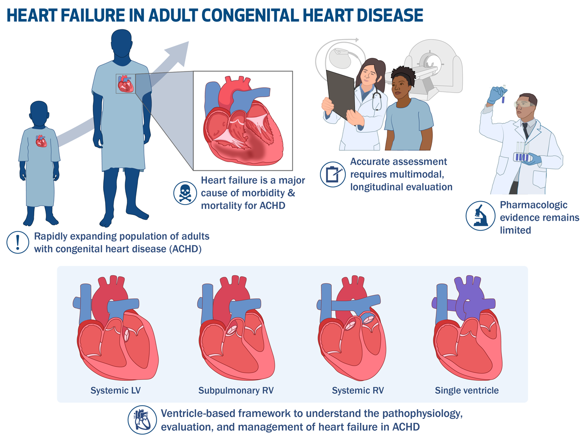
Figure 1
Heart failure in adult congenital heart disease.
Key Points
Heart failure (HF) is a major cause of morbidity and mortality in adults with congenital heart disease, driven by mechanisms distinct from acquired HF.
Accurate assessment requires multimodal, longitudinal evaluation from an expert in adult congenital heart disease since dysfunction often precedes changes in ejection fraction and patients frequently underreport heart failure symptoms. Ventricular morphology shapes pathophysiology and management, necessitate tailored diagnostic and therapeutic strategies across systemic left ventricle, subpulmonic right ventricle (RV), systemic RV, and Fontan circulations.
Pharmacologic evidence remains limited, making individualized care and correction of structural contributors essential.
Transplantation and mechanical circulatory support can offer meaningful benefits but are constrained by anatomical complexity and endorgan disease, underscoring the importance of early referral and integrated palliative care.