
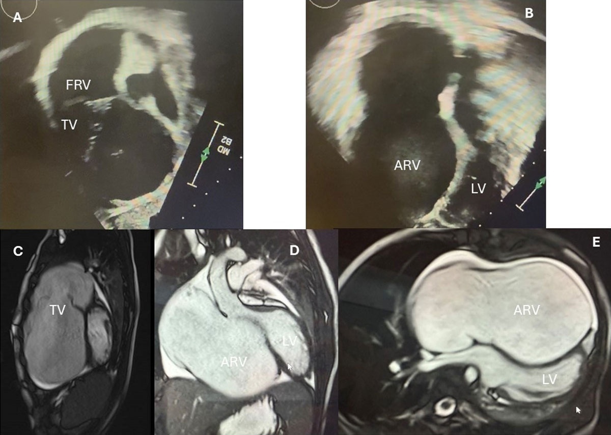
Figure 1
Severely rotated tricuspid valve. (A) Echocardiogram subcostal view exposing the tricuspid leaflets in the right ventricular outflow tract (RVOT), with a very small functional right ventricular (RV) chamber. (B) Echo 4-chamber view showing a large atrialized RV and no tissue in the septal area. The septal leaflet compresses the LV cavity. (C) Magnetic resonance imaging shows images showing the rotated TV in the RVOT, and (D, E) the interventricular septum compressing the left ventricular cavity. FRV: functional right ventricle; TV: tricuspid valve; ARV: atrialized right ventricle; LV: left ventricle
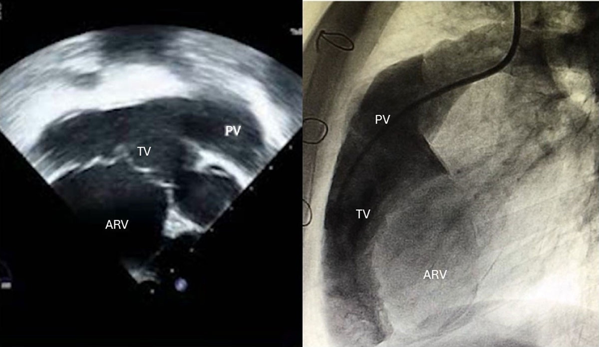
Figure 2
(A) Echocardiogram and (B) heart catheterization images of a cyanotic patient, showing a rotated tricuspid valve into the right ventricular (RV) outflow tract, with a small functional RV. PV: pulmonary valve; TV: tricuspid valve; ARV: atrialized right ventricle
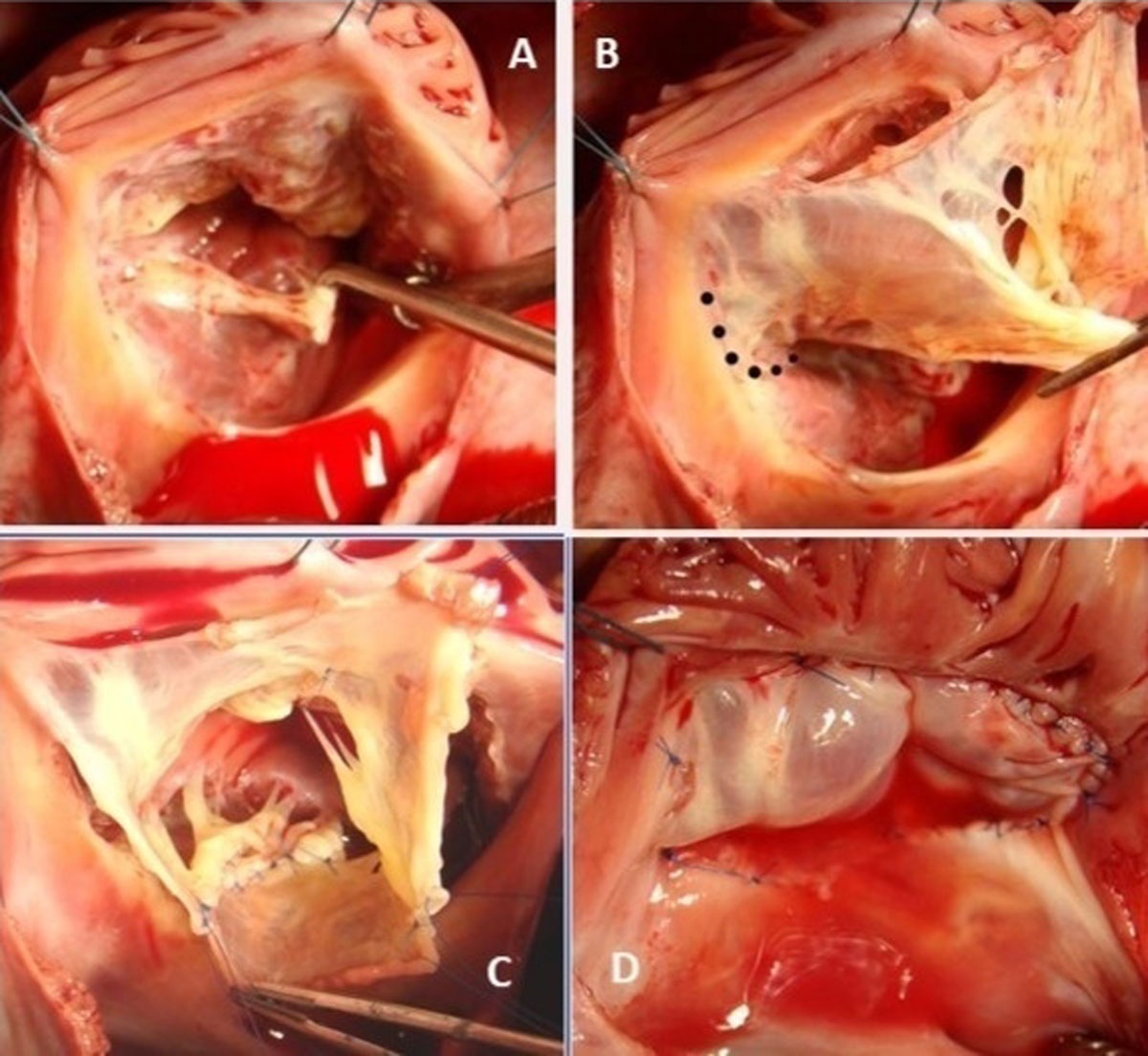
Figure 3
Surgical steps of the Cone repair: (A) Valve inspection shows a small, displaced septal leaflet. (B) The anterior leaflet was detached from the annulus, and the dotted lines show the location to detach the medial portion of the septal leaflet. (C) The Cone was reconstructed by rotating and suturing the valvar leaflets. (D) The base of the Cone was sutured to the truer atrioventricular junction and the saline test shows a competent valve.