
Introduction
Primary tricuspid valve (TV) regurgitation encompasses a group of congenital (Ebstein Anomaly, TV dysplasia) or acquired conditions (myxomatous degeneration of the tricuspid valve, endocarditis, carcinoid syndrome, rheumatic heart disease, or chest irradiation) in which the primary problem is TV dysfunction. Here we focus on Ebstein anomaly (EA) and TV dysplasia; however, the same surgical repair techniques, diagnostic tests, and surgical indications apply to all primary TV regurgitation diseases.
Tricuspid valve regurgitation (TR) causes right ventricle (RV) and right atrium (RA) dilation and dysfunction. Due to the diversity of anatomical presentations, varying degrees of leaflet-valve rotation, and RV muscle involvement, clinical outcomes are heterogeneous; some patients require immediate intervention after birth, whereas others lead near-normal lives into adulthood.
Ebstein anomaly is a recognized RV cardiomyopathy that may also involve the left ventricle (LV). There are factors beyond TR that complicate the disease course, clinical severity and, consequently, the urgency and type of management. Those factors include intrinsic RV myopathy, RV diastolic dysfunction, septal dyskinesis, adverse RV-LV interactions (eg, impaired LV filling due to RA/RV dilation), LV noncompaction cardiomyopathy, heart rhythm abnormalities, RV electromechanical desynchrony, and, in neonates, poor lung development secondary to cardiomegaly.1,2,3
For asymptomatic patients, the rationale for early valve intervention is to prevent irreversible consequences of ventricular volume overload. The results of the Cone procedure—with reverse remodeling of the RV, improvement in biventricular stroke volume, and low procedural risk—are encouraging for proceeding with surgical repair at an earlier age4,5,6,7 and can be applied to any anatomical type of TV.
Diagnostic Testing: Initial Diagnosis
Transthoracic Echocardiogram
Transthoracic echocardiogram (TTE), the standard diagnostic test for TR, demonstrates valve anatomy, the degree of regurgitation, and associated abnormalities including pulmonary valve disease (atresia, stenosis, insufficiency), atrial septal defect (ASD), ventricular septal defect, atrioventricular septal defect, aortic arch obstruction, mitral cleft, and other abnormalities.
Pulmonary systolic pressure and RV systolic pressure can be estimated with the regurgitant jet in the tricuspid valve along with qualitative evaluation of RV size and function. In EA, it can be challenging to precisely define the RV function, delineate the atrialized RV portion, identify the thin RV wall, determine the size of the functional RV, and assess tricuspid valve rotation on echocardiography. Figures 1A and 1B demonstrate a severely rotated valve on the echocardiographic images.
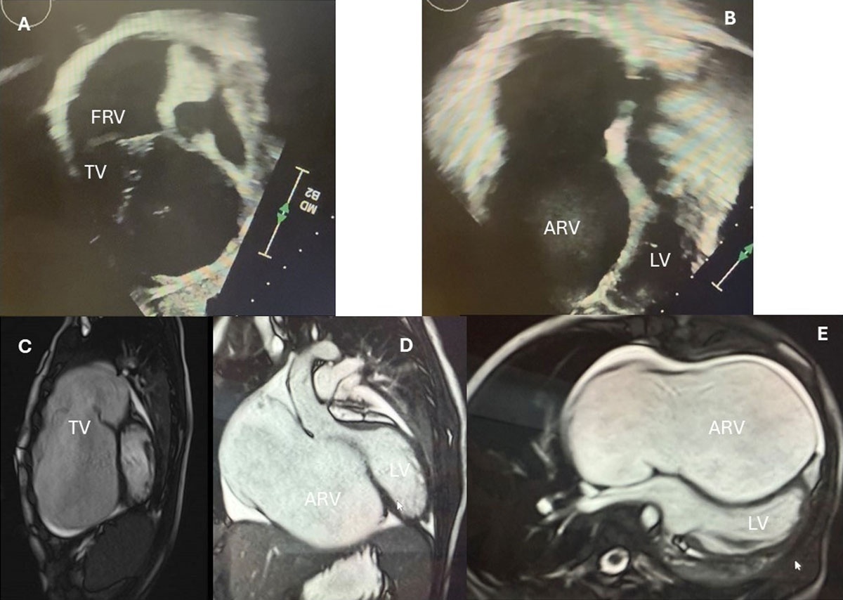
Figure 1
Severely rotated tricuspid valve. (A) Echocardiogram subcostal view exposing the tricuspid leaflets in the right ventricular outflow tract (RVOT), with a very small functional right ventricular (RV) chamber. (B) Echo 4-chamber view showing a large atrialized RV and no tissue in the septal area. The septal leaflet compresses the LV cavity. (C) Magnetic resonance imaging shows images showing the rotated TV in the RVOT, and (D, E) the interventricular septum compressing the left ventricular cavity. FRV: functional right ventricle; TV: tricuspid valve; ARV: atrialized right ventricle; LV: left ventricle
Anatomy and function of the RA and RV are characterized by linear dimensions as well as by 2-dimentional (2D) and 3D volumes and ejection fraction (EF).
Some echocardiographic parameters—such as tricuspid annular plane systolic excursion (TAPSE) and RV fractional area change—are limited by their load dependence, so advanced imaging modalities such as RV global longitudinal strain or free wall strain imaging can be a sensitive index for quantifying RV dysfunction in the presence of severe TR, with prognostic value superior to TAPSE and RV fractional area change.7,8,9,10
Chest X-ray
The cardiac silhouette diameter is usually enlarged. The cardiothoracic ratio > 0.65 correlates with a high risk of sudden death and is considered a parameter for surgical intervention.4,11
Electrocardiography and Holter Monitoring
In the presence of congenital TV disease, it is mandatory to elucidate the presence of pre-excitation (Wolf-Parkinson-White syndrome), heart block, prolonged QT syndrome, and arrhythmias. A history of arrhythmias or pre-excitation on the electrocardiogram indicates the need for Holter monitoring.
A diagnostic electrophysiology study with catheter ablation, if feasible, should be done even in asymptomatic patients with Wolf-Parkinson-White syndrome. One third of EA adults with ventricular pre-excitation present multiple accessory pathways and a high risk for sudden cardiac arrest.4,12,13,14 In adults with EA, most symptomatic arrhythmias are atrial and probably related to RA dilation secondary to long-term TV regurgitation.
Cardiac Magnetic Resonance Imaging
The American Association for Thoracic Surgery (AATS) Expert Consensus on EA in adults and children recommended cardiac magnetic resonance imaging (CMR) for comprehensive assessment of ventricular volumes and evaluation of tricuspid regurgitation fraction and RV stroke volume. Ventricular volumes and the degree of TR are important in surgical decisions and postoperative follow-up. CMR is strongly recommended whenever feasible.4 Protocols for imaging and data post-processing are needed to standardize the diagnosis and reports.
CMR can assess LV involvement in EA, including diffuse and/or focal myocardial fibrosis and strain as well as EF, particularly in the adult population with long-term abnormal interventricular interaction and who can tolerate the exam without sedation.4,15,16,17,18
In Figure 1, echocardiographic and MRI images demonstrate the LV compressed by the enlarged atrialized RV.
Cardiac Catheterization
Valuable clinical information for decision-making regarding valve intervention can be obtained in the catheterization laboratory. Older adult patients usually present with low cardiac index, which can be a sign of poor prognosis and contraindication for surgical TV intervention.4,19,20
Direct measurements of intracavitary pressures, assessment of LV systolic and diastolic dysfunction, estimation of LV EF, calculation of transpulmonary pressure gradients, pulmonary and systemic vascular resistances, and assessment of coronary artery anatomy are essential in most adult patients. RV angiography with contrast can quantify regurgitation severity and RV functional cavity size and function.
Heart catheterization is essential in patients with chronic cyanosis in EA due to the right-to-left shunt in an ASD. These patients can present with systemic-to-pulmonary artery collaterals, which would complicate the postoperative course.
In situations where RV dysfunction is expected to occur immediately after TV repair, such as in patients with severely rotated TV towards the RV outflow tract (Figure 2), preoperative assessment of pulmonary artery anatomy and pulmonary vascular resistance is valuable for determining suitability for a Glenn anastomosis if the RV does not provide adequate cardiac output and pulmonary blood flow after the valve repair.21
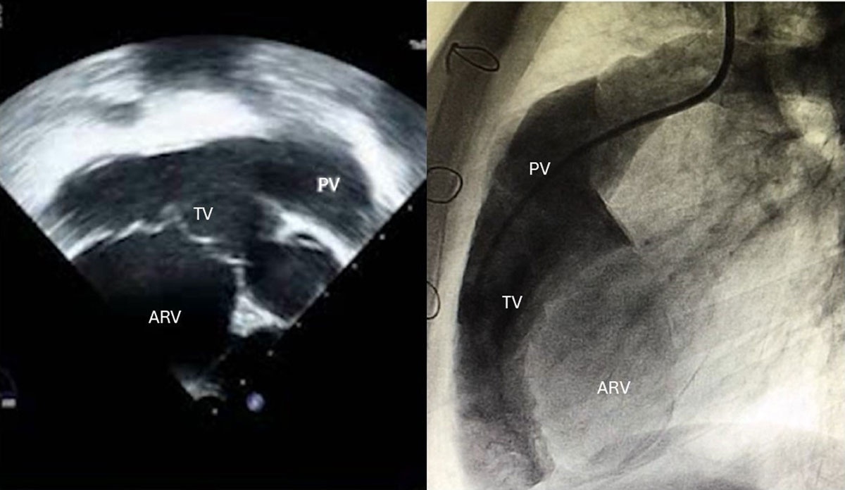
Figure 2
(A) Echocardiogram and (B) heart catheterization images of a cyanotic patient, showing a rotated tricuspid valve into the right ventricular (RV) outflow tract, with a small functional RV. PV: pulmonary valve; TV: tricuspid valve; ARV: atrialized right ventricle
Some diagnostic interventions for patients with discrepant symptoms include exercise hemodynamics and responses to pulmonary vasodilators to decrease pulmonary resistance.
Exercise Testing
In asymptomatic patients with TR and nonenlarged hearts, exercise stress testing can reveal decreased exercise capacity since patients gradually limit their activity, modifying the lifestyle, and may deny symptoms. It can help define the appropriate time for intervention in patients with a gradual, unperceived decline in functional status.
Exercise stress testing has prognostic value; consequently, asymptomatic patients (older children and adults) with EA should undergo measurement of oxygen consumption to unmask occult exercise intolerance in accordance with the AATS expert consensus.4,22,23
Cardiopulmonary exercise testing provides a noninvasive evaluation of functional capacity, oxygen saturation, and heart rate/arrhythmias, allowing longitudinal assessment of patients. It is useful for pre-pregnancy risk assessment and to evaluate changes in functional capacity after surgical intervention.
Patients with EA commonly present lower peak oxygen consumption, shorter exercise duration, and greater breathing effort compared with healthy individuals. These findings may indicate the need for surgical intervention.
Routine Follow-Up
Even asymptomatic patients without dilated chambers or severe TR need regular follow-up with periodic examinations and TTE. The purpose of follow-up in these patients is to prevent irreversible consequences of TR, including RV failure and RA dilation, which can occur in the absence of symptoms. In addition to routine imaging changes, the onset of symptoms or a change in physical examination findings can trigger a surgical indication.
The Natural History of Ebstein Anomaly
Life expectancy in EA patients is significantly lower than in the general population, with a median age of death at 55.8 years (interquartile range [IQR] 27–69 years) according to a recent study.8
Predictors of cardiac-related death include age at diagnosis, degree of echocardiographic severity, presence of congestive heart failure, cyanosis, significant LV dysfunction (LV EF < 40%), severe cardiomegaly (cardiothoracic index > 0.65 at chest radiography), and persistent arrhythmia. Morbidity is mainly related to arrhythmia and refractory late hemodynamic deterioration.8,11,24 Most adult patients will have arrhythmias before 50 years of age, mainly atrial type, probably related to RA dilation secondary to long-term TV regurgitation.
Patients who reach adulthood without intervention frequently have minor valve displacement or minimal regurgitation. Some oligosymptomatic patients have avoided intervention due to fear of needing valve replacement. The surgical risk is higher in early infancy and in adulthood after age 40. Associated comorbidities can increase the surgical mortality in older adults, but the most important factor seems to be the biventricular dysfunction commonly seen in those patients.
Echocardiographic characteristics with prognostic value for mortality were evaluated in a study of 620 adult patients followed from 2003 to 2020 at Mayo Clinic, including 98 who underwent TV repair and 84 who underwent TV replacement.8 RV globular longitudinal strain was the strongest predictor of mortality, but the isolated RV free wall strain was also a prognostic factor for mortality. Patients with prior tricuspid valve intervention were older than those without (42 years [IQR: 31–53 years] vs 34 years [IQR: 22–46 years]; P = .008). This study reinforces the concept of RV myopathy in EA, with progressive RV dysfunction during the lifetime.
Chronic tricuspid regurgitation, RV and RA dilation, and abnormal interventricular interaction can lead to biventricular dysfunction in patients with EA, especially adults. Other factors include intrinsic cardiomyopathy, LV noncompaction, heart rhythm abnormalities causing septal dyskinesia, and RV electromechanical desynchrony.
Also, the myocardium in EA can receive a detrimental blood supply; due to long-term RV dilation and a large tricuspid annulus, the right coronary artery is usually distended in EA adults. The coronary perfusion gradient can be decreased, mainly in those with a severely displaced tricuspid valve without an ASD, who can present with chronic low cardiac output and low aortic pressure associated with elevated RA pressure.
Late LV dysfunction in EA is multifactorial, with chronic cyanosis and elevated filling pressures on the right side of the heart compromising the LV and stretching of the right coronary artery with low coronary perfusion.
Indication for Surgical Intervention in Adults with Primary Tricuspid Regurgitation
Surgical intervention for EA and other tricuspid valve dysplasias is recommended for symptomatic patients. Cyanosis, exertional dyspnea, fatigue not attributable to other causes, and decreased exercise tolerance (lower-than-expected exercise tolerance on exercise testing) are classical indications for TV surgery.
The AATS expert consensus4 is a practical resource for surgical indications in Ebstein anomaly, which we can extrapolate to other forms of primary TV regurgitation. The following are the recommendations for EA with the Class of Recommendation (CoR) and Level of Evidence (LoE):
Surgery is recommended for symptoms that include fatigue (not attributable to other causes), decreasing objective exercise tolerance (ie, lower than expected exercise tolerance on exercise testing), decreased arterial oxygen saturation (cyanosis), and exertional dyspnea (CoR: I; LoE: B-NR).
Cone repair of the tricuspid valve is beneficial when surgery is indicated (CoR: I; LoE: B-NR).
Surgery is recommended when there is severe TR and progressive RV dilation or dysfunction on serial echo or MRI (CoR: I, LoE: B-NR).
Surgery can be beneficial in asymptomatic patients when there is severe TR, moderate RV enlargement, and valve anatomy favorable for repair (CoR: IIa; LoE: C-LD).
Surgical intervention at 3 to 5 years of age is reasonable when there is severe TR, moderate RV enlargement, and a high probability of successful valve repair (CoR: IIa; LoE: C-LD).
Bidirectional cavopulmonary shunt is reasonable when there is severe RV dilation, severe RV systolic dysfunction, RAP:LAP ratio > 1.5, or failure to separate from cardiopulmonary bypass following repair (CoR: IIa; LoE: B-NR).
Concomitant maze procedure at the time of surgery is reasonable when there is paroxysmal or continuous atrial refractory atrial or ventricular arrhythmias.
As previously mentioned, exercise stress testing can unmask reduced exercise capacity, hypoxemia, and cardiac dysfunction in asymptomatic individuals. These data on functional impairment support indications for TV intervention.
The following are recommendations for valve intervention in EA according to the 2025 ACC/AHA/HRS/ISACHDSCAI Guideline for the Management of Adults with Congenital Heart Disease:21
Class I
At least moderate TR and:
Heart failure symptoms
Worsening exercise capacity
Progressive RV systolic dysfunction
Class IIa
Asymptomatic patients with at least moderate TR and:
Progressive RV enlargement
Systemic desaturation from right-to-left atrial shunt
Recurrent arrhythmias
Paradoxical embolism
Severe regurgitation and RA or RV enlargement
In adults with primary TV regurgitation, the Cone repair improves quality of life and carries a low surgical risk. It reestablishes valvar function, promoting increased antegrade pulmonary flow, with consequent increases in LV filling and improved interventricular interaction. This reduces the leftward septal shift in early LV diastole, improving systemic output and biventricular reverse remodeling. Figure 3 demonstrates the surgical steps of the Cone repair.
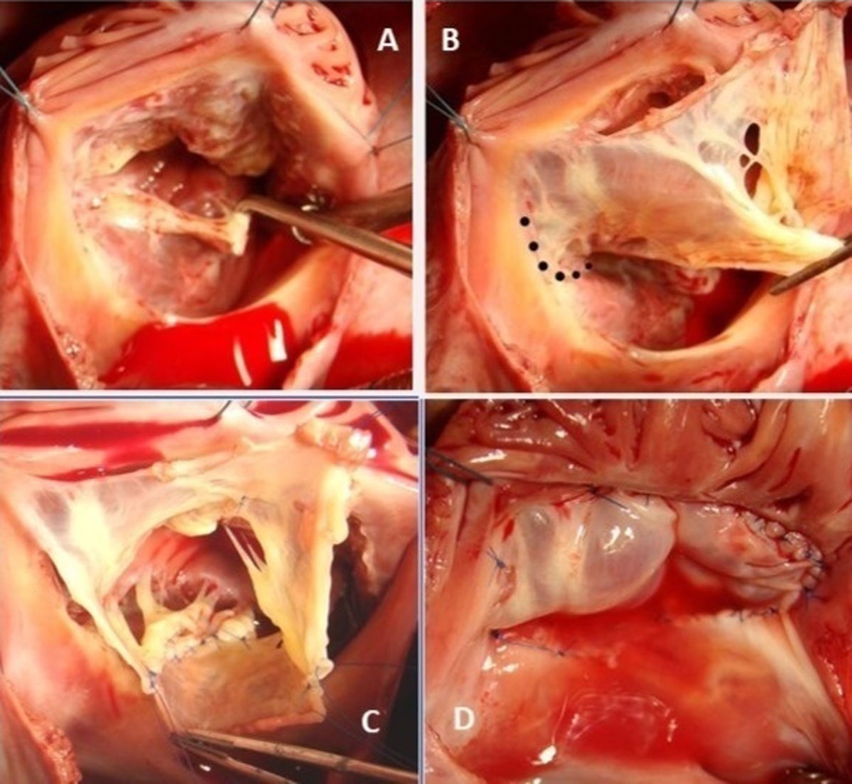
Figure 3
Surgical steps of the Cone repair: (A) Valve inspection shows a small, displaced septal leaflet. (B) The anterior leaflet was detached from the annulus, and the dotted lines show the location to detach the medial portion of the septal leaflet. (C) The Cone was reconstructed by rotating and suturing the valvar leaflets. (D) The base of the Cone was sutured to the truer atrioventricular junction and the saline test shows a competent valve.
Exercise performance deteriorates in non-operated patients with EA, whereas it improves or remains stable in patients after tricuspid valve surgery.25,26
Preoperative functional status, cardiomegaly, age < 1 and > 50 years, and RV dysfunction are associated with increased risk of postoperative complications after EA repair. Severe RV impairment can preclude surgical repair.
Irreversible Biventricular Dysfunction: Considerations for Heart Transplant, ASD Creation, or Cavopulmonary Shunt
The AATS Expert Consensus for management of EA in adults and older children provided additional recommendations:
In patients with severe symptomatic isolated TR, surgical intervention before the onset of severe RV dysfunction or end-organ damage to the liver and kidney may reduce symptoms and recurrent hospitalizations.
TV surgery is not recommended when there is irreversible severe biventricular systolic dysfunction (LV EF < 25%, RV EF < 25%) (CoR: III; LoE: B-NR).
In cases of severe RV dilation and systolic dysfunction (RV EF < 25%), TV surgery may still be feasible if accompanied by interventions such as a bidirectional cavopulmonary shunt or atrial septal fenestration. When severe LV dysfunction (EF < 25%) and significant LV dilation are present, a heart transplant or a ventricular assist device can be recommended.
After TV repair/replacement, RV dysfunction can occur in the early postoperative period, and a small ASD can maintain adequate systemic output by bypassing the RV. In cases of severely dysfunctional RV, a bidirectional cavopulmonary shunt can be considered to decompress the RV, obtaining one-and-a-half ventricle repair.
Given the natural history and the possibility of durable, effective valve repair with the Cone procedure, early surgical intervention is recommended, especially in cyanotic patients, to address the effects of chronic volume overload and RA/RV dilation and prevent deterioration of biventricular function.4
Transcatheter Atrial Septal Defect Closure
The most recent AHA/ACC guidelines consider isolated ASD closure after hemodynamics assessment to improve clinical outcomes in adults with EA with ASD/patent foramen ovale and cyanosis or paradoxical embolism.21 This guideline was probably based on the risk of failed repair requiring implantation of a tricuspid prosthesis at a young age, along with the consequent multiple valvar reinterventions in the long term. These problems in the past had led cardiologists to favor percutaneous ASD closure in institutions not specialized in EA repair to improve O2 saturation. However, with this approach, the tricuspid abnormality persists, and RV dilation continues, ultimately interfering with LV function.
The AATS expert consensus4 states that transcatheter closure of the ASD is not recommended when there is severe TR or a severe form of EA (CoR: III; LoE: C-EO).
In specific subtypes of EA, especially in valves severely rotated towards the RVOT, the echocardiographic TR severity is often underestimated and does not always reflect the true regurgitant volume or its deleterious effect on RV dilation/dysfunction.
In this situation, even with trivial regurgitation, cyanosis and paradoxical embolization can result from the small size of the functional RV cavity, and the isolated ASD closure can lead to low systemic output, especially during exercise, due to low RV stroke volume. Therefore, the Cone repair with surgical ASD closure is the treatment of choice in centers with experience repairing those severely rotated valves, providing biventricular repair.4 Ideally, this repair should be performed at an early age, providing time for reverse remodeling of the ventricles during the growing process.
Patients who undergo ASD device closure may present temporary clinical improvement; however, progressive symptoms of low cardiac output (cold extremities, syncope, reduced exercise performance, etc.) are indications for valve repair.
Cyanosis is a recognized indication for surgical TV repair in EA. Therefore, device closure of an ASD in cyanotic EA patients or patients with a history of paradoxical embolization (right-left shunt) must be avoided. Nowadays, the Cone repair can be performed in specialized centers with low morbidity and mortality and durable results; it should be the preferred treatment.4
Tricuspid Valve Replacement
In adults older than 60 years, TV replacement is reasonable (CoR: IIa; LoE: B-NR).4 Valve repair should always be preferred; however, for adults with severe RV and/or annular enlargement, the complexity of intracardiac repair, RV plication, and ASD closure can result in prolonged cardiopulmonary bypass and cross-clamp times, exacerbating RV dysfunction.
Valve replacement allows for a shorter surgical time and can even be performed on a beating heart if the atrial septum is intact. Valve replacement ensures tricuspid competence, potentially reducing the risk of ventricular dysfunction secondary to prolonged cardioplegia. According to the AATS expert consensus, a porcine bioprosthesis is preferred in the setting of low RA pressure and reduced RV systolic function, as leaflets are more pliable than pericardial leaflets.4
An oversized prosthesis can increase the risk of thrombus formation and accelerated bioprosthetic deterioration due to low mean gradients and reduced leaflet motion. Warfarin anticoagulation therapy is recommended for 3 to 6 months postoperatively, assuming there are no contraindications, followed by lifelong low-dose aspirin.
Maintaining the TV leaflets intact during prosthesis implantation can enable future tricuspid valve repair, as reported in the literature.27
Final Considerations
Despite the advances in TV surgical techniques, many patients with primary tricuspid regurgitation are reaching adulthood with a restrained quality of life, arrhythmias, and enlarged hearts. Performing surgery on younger patients enhances the likelihood of successful biventricular reverse remodeling, as evidenced by MRI evaluations during postoperative follow-up. Consequently, if the surgery can be performed at a center with substantial experience in Cone repair, early referral for surgical evaluation is advised to optimize late outcomes. The ideal age is 3 to 5 years, but in some circumstances (such as cyanosis, low cardiac output, heart failure), the Cone repair can be performed earlier in specialized centers.
The Cone repair is applicable to all types of TV anatomy, offering reliable and lasting valve repair, enhanced biventricular systolic function, and potential long-term reverse remodeling benefits. In the modern era, echocardiographic findings, along with clinical evaluation, exercise stress testing, MRI results, and, in specific cases, heart catheterization and electrophysiological study, should guide the indications for early surgical intervention.
Our efforts should focus on individualized treatment and possible early repair to mitigate or reverse the pathological process leading to RV/LV failure, aiming to improve the long-term outcomes in this rare disease.
Key Points
The Cone repair can be applied to any type of primary tricuspid regurgitation, and the most common diagnoses are Ebstein anomaly and tricuspid valve dysplasia.
Tricuspid regurgitation usually has a silent clinical course, with progressive dilation of the right ventricle and right atrium. The decline of exercise capacity can be unmasked by exercise stress test. Most patients will present arrhythmias at middle adult age and have decreased longevity.
Surgical valve repair with the Cone procedure can be applied to all anatomical presentations and should be performed at an early age in centers with experience to achieve adequate valvar function, improve exercise capacity and quality of life, promote reverse remodeling of the right and left ventricles, and sustain results in the long term.