
Video 1
MRI showing pulmonary valve stenosis; see also at https://vimeo.com/1190188046.
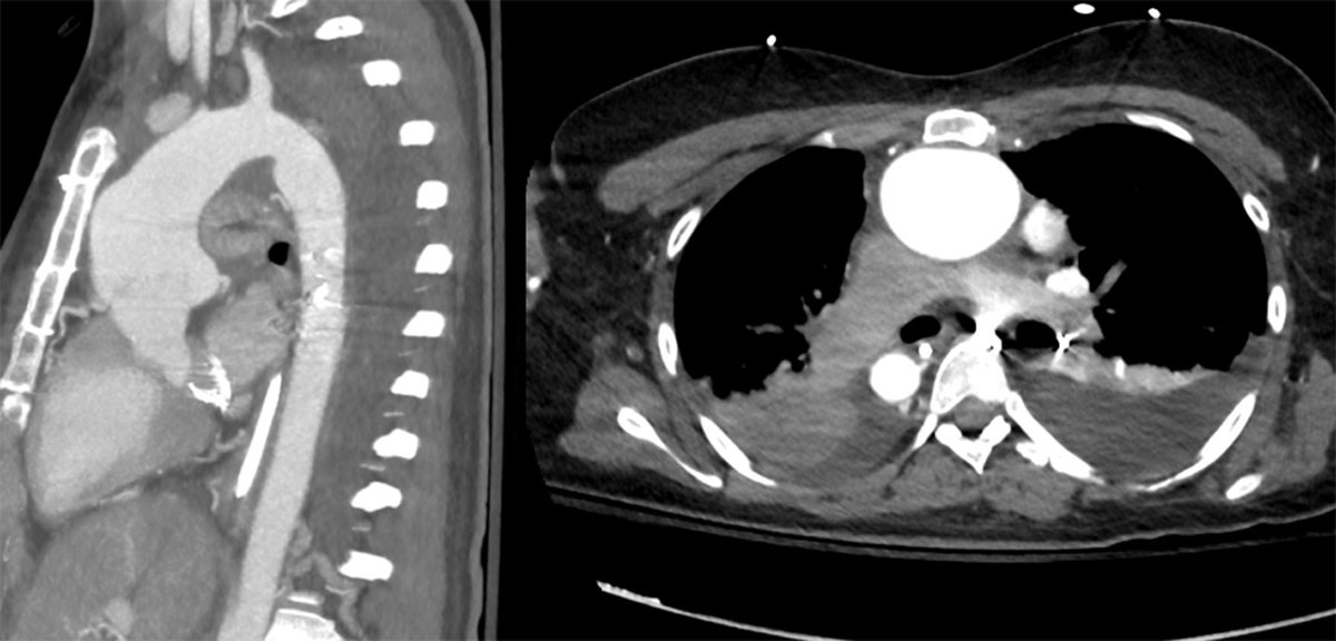
Figure 1
Aortic root and ascending aorta proximity to the sternum.
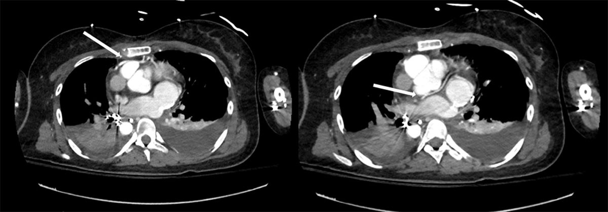
Figure 2
Position of the coronary arteries indicated with arrows.
Table 1
Characteristics of aortic root dilatation in conotruncal anomalies. d-TGA: dextro-transposition of the great arteries; ASO: arterial switch operation; DORV: double outlet right ventricle; RVOT: right ventricular outflow tract
| LESION | ROOT BEHAVIOR | AORTIC REGURGITATION | DISSECTION RISK | SURGICAL IMPLICATION |
|---|---|---|---|---|
| Tetralogy of Fallot | Progressive dilation | Low to moderate | Very low | Combine with RVOT |
| d-TGA (post ASO) | Neo-root dilation | Variable | Low | Valve-sparing feasible |
| Truncus arteriosus | Marked dilation | Common | Very low | Valve pathology drives |
| DORV | Variable | Variable | Low | Depends on physiology |
Video 2
Axillary artery cannulation; see also at https://vimeo.com/1190189108.
Video 3
Cannulation of the innominate vein; see also at https://vimeo.com/1190189731.
Video 4
An additional venous line is connected to the reservoir; see also at https://vimeo.com/1190190613.
Table 2
Surgical strategy selection. AR: aortic regurgitation; RVOT: right ventricular outflow tract
| CLINICAL SCENARIO | PREFERRED STRATEGY |
|---|---|
| Normal valve, mild AR | Valve-sparing (reimplantation) |
| Severe AR, good leaflets | Reimplantation |
| Diseased leaflets | Mechanical Bentall |
| Avoid anticoagulation | Biological Bentall/Wheat |
| Concomitant RVOT surgery | Combined procedure |
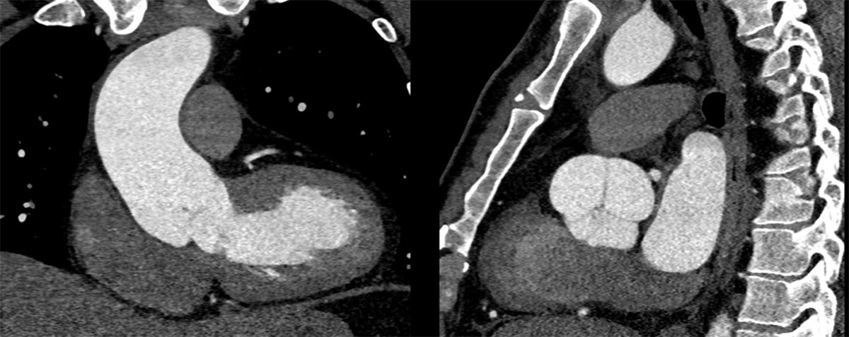
Figure 3
Anomalous aortic valve position in ToF. The LV-aortic valve axis is nearly horizontal. The valve lies posterior to the RVOT, and all three sinuses are visible in the sagittal view. ToF: tetrology of Fallot; LV: left ventricular; RVOT: right ventricular outflow tract
Video 5
David procedure in tetrology of Fallot; see also at https://vimeo.com/1190191390.
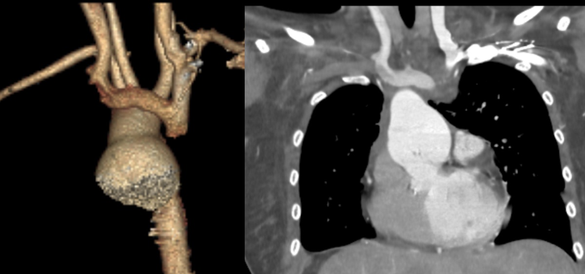
Figure 4
Aortic root dilatation in double outlet right ventricle, Situs Inversus.
Video 6
Double root replacement in double outlet right ventricle; see also at https://vimeo.com/1190194364.
Video 7
David procedure in double outlet right ventricle; see also at https://vimeo.com/1190194946.