
Introduction
Conotruncal anomalies comprise a heterogeneous group of congenital heart defects (CHD) arising from abnormal embryologic development of the cardiac outflow tract and the great arteries. This spectrum includes Tetralogy of Fallot (ToF), dextro-transposition of the great arteries (d-TGA), truncus arteriosus, pulmonary atresia, and double outlet right ventricle.
While the majority of the literature has focused on long-term follow-up and management of right ventricular outflow tract (RVOT) dysfunction, as well as pulmonary valve and pulmonary artery pathology, aortic root dilatation represents a frequent yet comparatively underexplored finding in this population. Despite its potential clinical significance, clear guidelines regarding its surveillance and surgical treatment are limited.
The aim of this study is to evaluate the clinical characteristics, progression, and surgical management of aortic root dilatation in patients with conotruncal anomalies with the goal of contributing to a more comprehensive understanding of this complex condition.
Despite increasing recognition of aortic root dilatation in patients with conotruncal anomalies, there remains no unified framework to guide surveillance thresholds or surgical decision-making. Existing literature is largely descriptive and does not integrate lesion-specific behavior, growth patterns, and operative context into a practical clinical strategy. We propose a structured, clinically applicable approach to guide decision-making in this population.
Pathophysiology
The mechanisms underlying aortic root dilatation are not yet fully clarified but likely reflect the interplay of two closely related processes: chronic hemodynamic stress and intrinsic structural abnormalities of the aortic wall.
A hemodynamic contribution has long been recognized, primarily related to sustained volume overload of the aorta.1 In the presence of a ventricular septal defect (VSD) combined RVOT obstruction (RVOTO), a right-to-left shunt increases blood flow through the aortic valve and ascending aorta. This abnormal flow pattern may explain why aortic root enlargement is already detectable in fetal life. Furthermore, the extent of dilatation correlates significantly with the severity of RVOTO.2 However, hemodynamic factors alone cannot account for the persistence or progression of aortic root enlargement in adults who underwent complete repair during the neonatal period, indicating that additional mechanisms must be involved.
Niwa and colleagues demonstrated that patients with conotruncal anomalies (CTA) consistently exhibit abnormalities of the aortic medial layer, providing evidence for an intrinsic histologic substrate.3,4 These changes include thinning and fragmentation of elastic fibers, increased collagen deposition, accumulation of mucoid extracellular matrix, and disorganization of smooth muscle cells. Such alterations are characteristic of medial degeneration and support the concept of a primary aortopathy. Importantly, medial degeneration appears to be both inherent to the arterial wall and further exacerbated by chronic volume overload, underscoring the dynamic interaction between developmental vulnerability and abnormal flow conditions.
Several risk factors for progressive aortopathy in ToF have been identified, including right aortic arch, prior Blalock-Taussig shunt, delayed repair of the congenital heart defect, male sex, chromosome 22q11 deletion, and severe cyanosis at the time of correction.2
From a morphologic standpoint, dilatation most frequently involves both the sinuses of Valsalva and the sinotubular junction. Ichikawa and colleagues reported an association between CTA and elongation of the geometric and effective heights of the aortic valve leaflets.5 They hypothesized that this elongation may represent a compensatory adaptation to progressive root enlargement, potentially explaining the relatively low incidence of clinically significant aortic regurgitation despite marked dilatation.
Interestingly, there is no clear evidence of aortic root dilatation in unrepaired d-TGA. After Mustard or Senning procedures, the most common indications for reoperation are systemic right ventricular failure or dysfunction of the systemic atrioventricular valve.2 Nevertheless, Yurasek et al. described the presence of aortic root dilatation even after atrial switch operations.6 Following both arterial and atrial switch procedures, progressive enlargement of the aortic root may outpace somatic growth. Histologic studies have identified structural abnormalities of the neo-aortic root that may account for late dilatation after the switch operation.7 In particular, Lalezari S. et al. demonstrated reduced collagen content in the arterial root of patients with d-TGA along with less extensive myocardial embedding of the arterial roots—features that may predispose to progressive enlargement later in life.
In patients with truncus arteriosus, a dilated aortic root is a common finding irrespective of repair status. Carlo et al.8 reported elevated aortic root z-scores in 76 patients, with a mean value of 5.1; only three patients had a z-score of 2. Root dimensions remained stable relative to body surface area and weight during follow-up. In this series, the principal indication for repeat surgery was truncal valve regurgitation, and no cases of aortic dissection were recorded.
Diagnostic Studies
Aortic root dilatation (ARD) may occasionally manifest on chest radiography as mediastinal widening; however, in patients with conotruncal anomalies, it is most often an incidental finding during routine echocardiographic evaluation of the pulmonary valve and the RVOT.
Transthoracic echocardiography remains the first-line imaging modality. It allows accurate measurement of aortic root dimensions, assessment of aortic valve competence, and identification of associated lesions such as residual ventricular or atrial septal defects, ventricular dilatation or dysfunction, and pulmonary valve abnormalities. Given its availability and ability to provide real-time functional assessment, echocardiography represents the cornerstone of longitudinal surveillance.
Once ARD has been identified, further anatomical definition is best achieved with cardiac magnetic resonance imaging (MRI) or computed tomography (CT). Cardiac MRI is frequently preferred because it does not expose the patient to ionizing radiation and provides comprehensive functional assessment of the aortic valve, pulmonary valve, and right ventricle. It enables quantification of ventricular volumes and ejection fraction, evaluation of residual shunts, and precise analysis of blood flow (Video 1). The emerging role of 4-dimensional-flow MRI in assessing the energetics of blood flow across the left ventricular outflow tract represents a promising field of investigation. Nevertheless, MRI is time-consuming and may be contraindicated in patients with pacemakers or implantable cardioverter-defibrillators.
Video 1
MRI showing pulmonary valve stenosis; see also at https://vimeo.com/1190188046.
For surgical planning, particularly in the context of re-sternotomy, CT scanning is often indispensable. Contemporary CT technology offers rapid image acquisition with substantially reduced radiation exposure compared with earlier generations. CT provides detailed information regarding the size and extent of the aneurysm and, crucially, its proximity to the sternum (Figure 1). It also allows identification of anomalous coronary artery anatomy, such as an anterior course of the left anterior descending artery originating from the right coronary artery and crossing the RVOT, a recognized finding in patients with ToF (Figure 2).9 In adults with congenital heart disease, cardiovascular CT additionally serves to exclude concomitant acquired coronary artery disease.
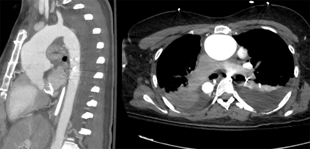
Figure 1
Aortic root and ascending aorta proximity to the sternum.
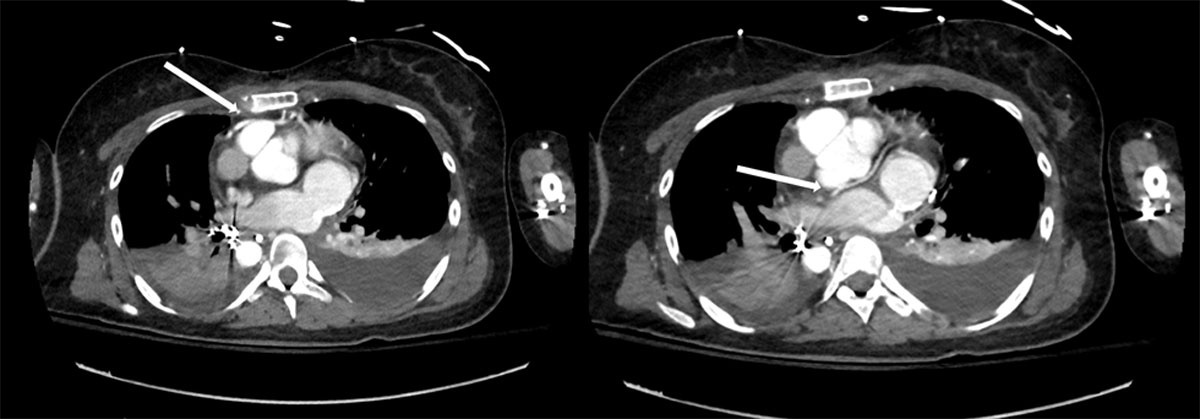
Figure 2
Position of the coronary arteries indicated with arrows.
CT imaging is particularly valuable in assessing the relationship between the sternum and underlying cardiovascular structures before re-entry. In patients who previously underwent a Rastelli repair or truncus arteriosus correction, the right ventricle-to-pulmonary artery conduit is often positioned anteriorly and in the midline, increasing its vulnerability during re-sternotomy. Similarly, following arterial switch repair for transposition of the great arteries, the pulmonary artery may lie immediately behind the sternum and be at risk of injury. CT also delineates the distance between the right ventricle or conduit and the posterior sternal table, information that is critical for operative planning.
Cardiac catheterization retains an important role in selected cases. It remains the gold standard for measuring pulmonary artery pressures, oxygen saturations, and pulmonary vascular resistance, thereby refining surgical risk stratification. Limitations include its invasive nature and the requirement for iodinated contrast, which carries a risk of contrast-induced nephropathy.
In patients with surgical indications, meticulous preoperative assessment of peripheral vascular access is essential. The patency of the femoral vessels and the axillary artery must be confirmed, as peripheral cannulation may be required in the event of hazardous re-entry. Failure to secure adequate peripheral access before re-sternotomy could have catastrophic consequences. The femoral vessels may be compromised by repeated catheterizations or prior surgical procedures, while the axillary artery—although less commonly today—may previously have been used for a classical Blalock–Taussig shunt.
Surgical Indications
The characteristic patterns of aortic root behavior across CTA subtypes are summarized in Table 1. There are no universally established thresholds for surgical intervention solely for ARD in patients with CTA. Isolated ARD is rarely an indication for surgery; in most cases, the primary operative indication remains intervention on the RVOT. Nonetheless, there is broad consensus that ARD should be considered for surgical repair when the aortic diameter exceeds 5.5 cm. This recommendation is supported by several observations: ARD in CTA generally progresses slowly,10 the rate of aneurysm growth declines with age, and minimal enlargement is observed after 40 years. Moreover, the risk of aortic dissection in this population is exceedingly low.
Table 1
Characteristics of aortic root dilatation in conotruncal anomalies. d-TGA: dextro-transposition of the great arteries; ASO: arterial switch operation; DORV: double outlet right ventricle; RVOT: right ventricular outflow tract
| LESION | ROOT BEHAVIOR | AORTIC REGURGITATION | DISSECTION RISK | SURGICAL IMPLICATION |
|---|---|---|---|---|
| Tetralogy of Fallot | Progressive dilation | Low to moderate | Very low | Combine with RVOT |
| d-TGA (post ASO) | Neo-root dilation | Variable | Low | Valve-sparing feasible |
| Truncus arteriosus | Marked dilation | Common | Very low | Valve pathology drives |
| DORV | Variable | Variable | Low | Depends on physiology |
Although aortic dissection has been reported in ToF, large-scale analyses suggest it is extremely uncommon in CTA.11 A study conducted in Texas encompassing 37.9 million hospital admissions between 1999 and 2012 identified 12,016 aortic dissections, of which 214 occurred in patients with congenital heart disease, predominantly associated with bicuspid aortic valve.12 Only six dissections were reported in CTA: three in ToF, two in d-TGA, and one in truncus arteriosus. The study demonstrated a strong association between bicuspid aortic valve and dissection (OR 10.5; CI 8.3-13), but found no correlation between dissection and CTA or aortic coarctation, reflecting the rarity of this complication in congenital heart disease.
In patients with conotruncal anomalies, management of aortic root dilatation should be individualized based on aortic size, growth rate, valve function, and the need for concomitant surgical intervention. In contrast to degenerative aortopathy, isolated aortic root dilatation in CTA rarely mandates intervention due to its slow progression and low risk of dissection. We propose a practical framework: aortic diameter < 4.5 cm = surveillance; 4.5 to 5.0 cm = consider intervention with rapid growth, significant aortic regurgitation, or planned concomitant surgery; 5.0 to 5.5 cm = strong consideration for intervention; and ≥ 5.5 cm = surgical intervention recommended. Importantly, thresholds are often driven by associated pathology rather than root size alone.
The risk of aortic dissection in CTA is markedly lower than in degenerative or connective tissue-related aortopathies and varies by lesion subtype. Available data suggest that dissection is exceedingly rare in ToF and truncus arteriosus and uncommon in d-TGA following arterial switch. As a result, aggressive prophylactic root replacement based solely on size may not be justified, and surgical decision-making should prioritize overall operative context.
Clinically relevant aortic regurgitation is uncommon in CTA. In d-TGA, despite frequent ARD, surgical intervention on the aortic root or neo-aortic valve is rare; freedom from aortic root and neo-aortic valve surgery has been reported at 100% and 95% at 5 and 10 years, respectively.13 Identified risk factors for later intervention in d-TGA include prior pulmonary artery banding, age ≥ 1 year at initial repair, and subaortic left ventricular outflow tract obstruction (LVOTO) surgery.
Finally, surgical consideration may be warranted for aortic diameters below 5 cm in cases of rapid progression, history of aortic dissection, or moderate-to-severe aortic valve regurgitation. In these scenarios, earlier intervention is justified to mitigate the risk of catastrophic complications.
General Surgical Principles
Re-entry represents one of the most critical aspects of surgery in patients with conotruncal anomalies. A detailed preoperative plan is essential to determine whether peripheral cannulation should be established prior to sternotomy or if direct access to the peripheral vessels is safe. There is broad consensus that the femoral vein provides the most reliable venous access for peripheral cardiopulmonary bypass (CPB). When femoral venous access is not feasible, the right internal jugular vein serves as a practical alternative.
Regarding arterial cannulation, we recommend the axillary artery as the first choice when the aorta is closely adherent to the sternum (Video 2). The axillary artery offers versatility, including the ability to provide selective cerebral perfusion in case of inadvertent aortic injury. If damage to the aorta occurs, peripheral CPB combined with systemic cooling becomes crucial. Transesophageal echocardiography is invaluable in detecting aortic regurgitation or left ventricular distension, prompting placement of a left ventricular vent, which can be facilitated via a small additional left thoracotomy if necessary.
Video 2
Axillary artery cannulation; see also at https://vimeo.com/1190189108.
When the right ventricle or an RV–pulmonary conduit lies directly beneath the sternum, femoral arterial access remains a reliable route for emergent CPB. In cases where complex or prolonged surgery is anticipated, central cannulation should be considered, particularly if both arterial and venous peripheral lines would otherwise be established through the same groin. Despite its rarity, leg ischemia has been observed in such scenarios. Once the chest is opened, an additional venous line can be placed in the superior vena cava or the innominate vein to facilitate right-heart exposure (Video 3). This line may be connected directly to the reservoir or via a Y-connector to the femoral venous line (Video 4).
Video 3
Cannulation of the innominate vein; see also at https://vimeo.com/1190189731.
Video 4
An additional venous line is connected to the reservoir; see also at https://vimeo.com/1190190613.
Before sternotomy, it is critical to assess for residual intracardiac shunts such as VSD or atrial septal defect. Peripheral CPB in the presence of residual shunts increases the risk of systemic air embolism in the event of right atrial or right ventricular injury. In such cases, maintaining a positive central venous pressure during CPB until cardiac repair is achieved is essential. The use of CO2 insufflation is strongly recommended, as pockets of air in the left ventricular apex are common and would otherwise require extensive dissection for removal.
Valve and Aortic Root Replacement Considerations
A practical framework for surgical strategy selection is summarized in Table 2. Choosing the optimal surgical technique and prosthetic valve type presents a significant challenge in this patient population, who are often young and have undergone multiple prior operations. Historically, the Bentall procedure with a mechanical valve was widely adopted.14 However, in recent years, valve-sparing techniques have gained prominence and are now commonly applied for aortic root replacement in CTA, with excellent early and mid-term outcomes demonstrating low mortality and durable freedom from aortic regurgitation.15,16
Table 2
Surgical strategy selection. AR: aortic regurgitation; RVOT: right ventricular outflow tract
| CLINICAL SCENARIO | PREFERRED STRATEGY |
|---|---|
| Normal valve, mild AR | Valve-sparing (reimplantation) |
| Severe AR, good leaflets | Reimplantation |
| Diseased leaflets | Mechanical Bentall |
| Avoid anticoagulation | Biological Bentall/Wheat |
| Concomitant RVOT surgery | Combined procedure |
In our practice, when the native aortic valve is well-functioning or exhibits less than moderate regurgitation, valve-sparing root replacement with reimplantation is preferred over the Yacoub remodeling technique, in line with other contemporary centers.16 For severe regurgitation with normal, thin leaflets and a central jet, reimplantation is still favored. Conversely, eccentric jets or diseased leaflets (eg, calcification, retraction, or fenestrations) warrant a mechanical Bentall procedure.
For patients seeking to avoid long-term anticoagulation or with contraindications to warfarin, a biological Bentall or Wheat procedure is considered. Patients should be counseled that valve-in-valve (ViV) interventions in bioprosthetic roots are anatomically constrained and carry a significant risk of needing reoperation. Factors influencing ViV feasibility include the distance between coronary ostia and the prosthetic valve, graft type (straight vs Valsalva), and annular size.17 Reoperation after prior root replacement carries a notable risk, estimated at 3% to 9% depending on the mechanism of failure and type of biological conduit (composite bioconduit versus homograft).18,19 Consequently, we reserve aortic homografts primarily for active endocarditis with root abscess.
If valve-sparing or biological aortic valves are used in conjunction with pulmonary valve replacement, a biological pulmonary valve or homograft is typically preferred. When a mechanical aortic valve is implanted, the choice of pulmonary valve becomes more nuanced. In our practice, mechanical pulmonary valves are avoided due to multiple factors: transcatheter pulmonary valve replacement offers high success and low mortality for degenerated pulmonary valves, anticoagulation targets are higher than for a single aortic valve, and the thromboembolic risk is increased with dual mechanical valves. For patients with both mechanical aortic and pulmonary valves, the international normalized ratio should be titrated according to the more thrombogenic position, analogous to combined aortic-mitral valve replacement.20,21
Nonetheless, other centers have reported favorable outcomes with mechanical pulmonary valves, demonstrating excellent durability and low thrombosis rates, albeit higher than left-sided positions.22,23 Freedom from valve thrombosis has been reported as 91% at 5 years and 86% at 10 years, with thrombolysis success rates up to 88%.22 Larger studies are needed to better define the role of mechanical pulmonary valves, particularly in patients undergoing concomitant mechanical aortic valve replacement.
Specific Considerations in Conotruncal Anomalies
Tetralogy of Fallot
In patients with ToF, the presence of an RVOT conduit can complicate aortic root surgery, often necessitating double root replacement. Careful assessment of coronary artery anatomy is essential. When a valve-sparing procedure is planned, it is important to recognize that, as in other CTA, the plane of the aortic valve is more horizontal and sometimes lies posterior to the RVOT, making optimal visualization more challenging (Figure 3).14 This contrasts with normal anatomy, where the angle between the left ventricle and the aorta is typically 35° to 45°.
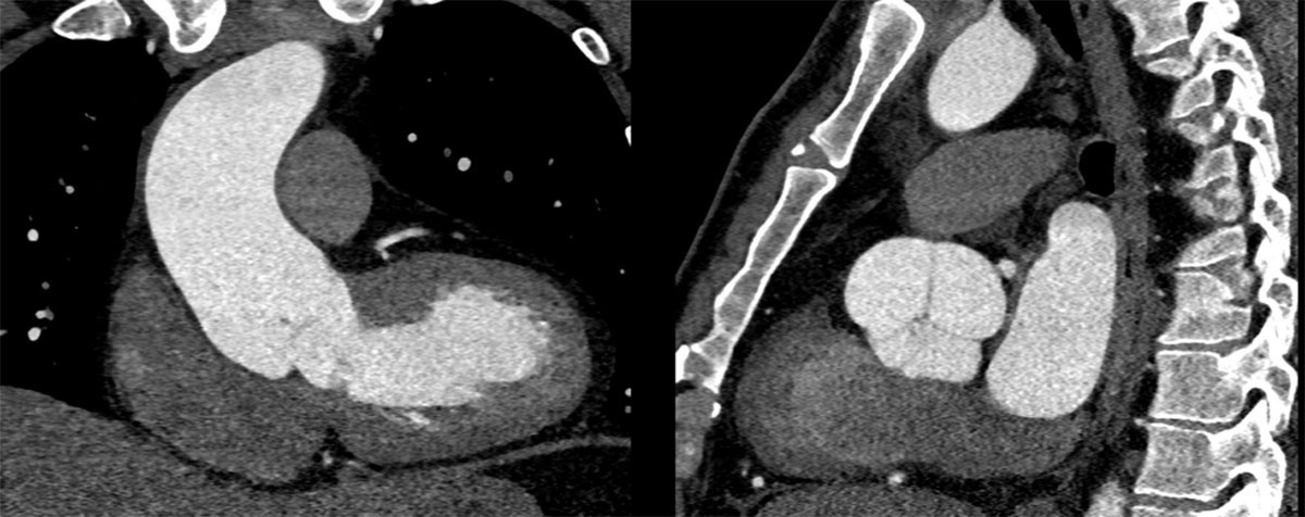
Figure 3
Anomalous aortic valve position in ToF. The LV-aortic valve axis is nearly horizontal. The valve lies posterior to the RVOT, and all three sinuses are visible in the sagittal view. ToF: tetrology of Fallot; LV: left ventricular; RVOT: right ventricular outflow tract
Recent data indicate that valve-sparing procedures in ToF are associated with a low incidence of valve-related complications, including endocarditis and thromboembolic events (Video 5).24,25
Video 5
David procedure in tetrology of Fallot; see also at https://vimeo.com/1190191390.
d-Transposition of the Great Arteries
After arterial switch procedures with the Lecompte maneuver, the aortic root is typically located posterior to the main pulmonary artery. Establishing CPB may require either peripheral cannulation or distal ascending aorta cannulation, the latter often necessitating mobilization of the right pulmonary artery. Surgical exposure of the aortic root usually involves transection of the right pulmonary artery followed by mobilization of the main pulmonary artery. At the conclusion of the procedure, the right pulmonary artery may be reconstructed with a Gore-Tex (W. L. Gore & Associates) conduit. Some centers prefer transection of the main pulmonary artery to reduce the risk of right pulmonary artery complications.
Valve-sparing approaches are particularly appealing in d-TGA because these patients typically do not require subsequent RVOT interventions, unlike other CTA subgroups. However, a David procedure can be challenging due to the anatomical characteristics of the neo-aortic root, which resembles a pulmonic valve. Visualization and assessment of the true neo-aortic valve annulus can be difficult, as the distance from the nadir of the cusp to the ventriculo-arterial junction may be wide and the valve plane unusually horizontal.
Truncus Arteriosus
In truncus arteriosus, aortic root surgery frequently involves the truncal valve, which may exhibit significant regurgitation, quadricuspid morphology, or previous repair. Neo-aortic valve insufficiency is the second most common indication for reintervention in this population.26 Valve-sparing root replacement should be carefully weighed against the risk of valve failure during follow-up. Additionally, the annulus and root are often markedly dilated, making preservation of native valve geometry technically demanding. Despite these challenges, successful valve-sparing root procedures in truncus arteriosus have been reported.27
Double Outlet Right Ventricle
Double outlet right ventricle represents a heterogeneous spectrum of anomalies ranging from ToF-like to TGA-like physiology; therefore, surgical strategies for the aortic root closely mirror those used in these subgroups (Figure 4). In patients with prior double outlet right ventricle repair, careful evaluation of the LVOT is crucial, as residual LVOT obstruction is the most common post-biventricular repair complication, occurring in up to 16% of patients,28 particularly when the VSD is remote. In such cases, aortic root surgery can be combined with LVOTO relief using a modified Konno procedure (Videos 6, 7).
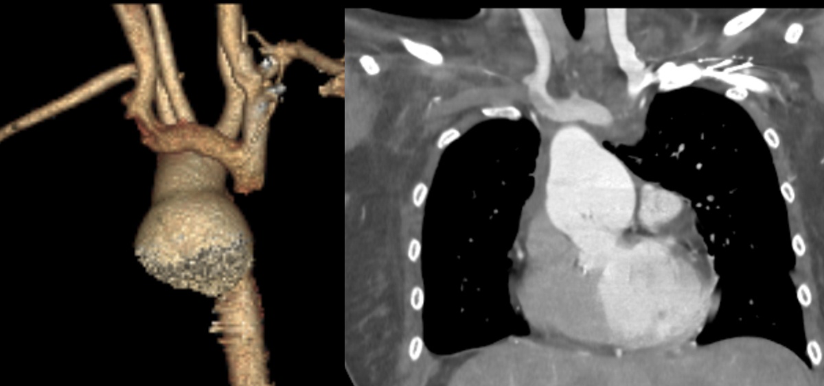
Figure 4
Aortic root dilatation in double outlet right ventricle, Situs Inversus.
Video 6
Double root replacement in double outlet right ventricle; see also at https://vimeo.com/1190194364.
Video 7
David procedure in double outlet right ventricle; see also at https://vimeo.com/1190194946.
Conclusion
Aortic root dilatation is a common but underrecognized feature in patients with conotruncal anomalies, with important long-term implications. Its development reflects a combination of chronic hemodynamic stress and intrinsic abnormalities of the aortic wall. Although ARD may begin early, its persistence after repair highlights an underlying structural vulnerability of the aortic media. The progression of ARD varies by lesion type and is influenced by both anatomical and genetic factors. In most patients, aortic root growth is slow and tends to stabilize in adulthood, with a low overall risk of dissection.
Accurate diagnosis relies on multimodality imaging, with echocardiography as the primary tool and MRI or CT providing additional anatomical and functional detail, particularly in reoperative settings. Surgical management remains highly individualized and is rarely driven by aortic size alone. Instead, decisions should incorporate valve function, growth rate, and the presence of concomitant pathology. Valve-sparing techniques are preferred when feasible, while composite root replacement is reserved for more advanced valve disease. Ultimately, a structured and lesion-specific approach is essential to guide appropriate timing and operative strategy in this complex population.
Key Points
Aortic root dilatation in conotruncal anomalies behaves differently from degenerative aortopathy, with slower progression and low dissection risk.
Surgical intervention is rarely based on root size alone and should incorporate valve function, growth rate, and concomitant pathology.
Thresholds for intervention should be individualized rather than strictly size-based.
Valve-sparing approaches are preferred when anatomy permits; they also offer durable outcomes.
Reoperative strategy must account for prior repairs, adhesions, and associated lesions.
A structured, lesion-specific approach is essential to optimize timing and operative strategy in this complex and heterogeneous population.
Acknowledgements
We extend our heartfelt appreciation to Drs. Valeria Duarte and Cindy Martin for their exceptional contributions and unwavering commitment to the ACHD group.