
Figure 1
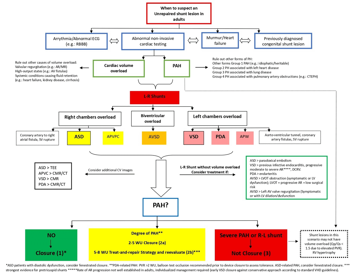
Evaluation and treatment algorithm. Shunt lesions connect the systemic and pulmonary circulations; direction depends on pressure gradients and the pulmonary vascular resistance to systemic vascular resistance ratio. Initially left-to-right with volume overload, they may progress to pulmonary hypertension, then bidirectional flow, and ultimately right-to-left (Eisenmenger syndrome), when closure is no longer feasible.
Considerations in Atrial Septal Defect closure
In moderate-to-large ASDs with Qp:Qs ≥ 1.5 and right ventricular dilation in the absence of pulmonary arterial hypertension (PAH), closure improves functional capacity and reduces long-term morbidity and mortality, even in asymptomatic individuals.
Patients with confirmed precapillary PAH should be managed according to established PAH guidelines. In selected patients with markedly elevated pulmonary vascular resistance (PVR ≥ 5 Wood units, or WU), PAH-directed therapy may allow consideration of ASD closure using treat-to-close or treat-and-repair strategies, although long-term medical therapy and close surveillance are often required. Patients with PAH and PVR of 5 to 8 WU on targeted PAH therapy have also been shown to safely undergo and benefit from ASD closure. However, agreement about the expected reversibility of PAH remains controversial. In cases of progressively increasing pulmonary arterial pressures, fenestrated closure may be considered to preserve a decompressive shunt and mitigate the risk of eliminating the pressure-relief pathway. In adults with unrepaired ASD and Eisenmenger physiology, ASD closure should not be performed to avoid increasing morbidity and mortality.
Considerations in VSD closure in these clinical scenarios
(1) Qp:Qs ≥ 1.5 with PVR ≤ 2 Wood units (WU) and significant (at least moderate) or progressive left ventricular (LV) dilation (Class 1).
(2) Qp:Qs ≥ 1.5 with PVR > 2 but < 5 WU and LV dilation (Class 2a), where closure may preserve LV function and reduce the risk of progressive PAH, balanced against perioperative risks and the risk for long-term progressive RV dysfunction and heart failure in patients with PAH after ventricular septal defect (VSD) closure.
(3) Qp:Qs ≥ 1.5 with PVR 5 to 8 WU, in the absence of hypoxemia, and reduction of PVR to < 5 WU after at least 3 months of PAH therapy (Class 2b), following a “treat-and-repair” strategy, for which evidence is strongest in atrial septal defects.
(4) Qp/Qs ≤ 1.5 with progressive moderate-to-severe aortic regurgitation (AR) (Class 2a): In children, progressive AR may justify VSD closure to prevent valve intervention. In adults, the rate of progression is not well established, therefore management should be individualized, weighing the risks and benefits of early surgery against conservative follow-up according to standard aortic valve replacement guidelines.
(5) Qp:Qs ≤ 1.5 with a history of infective endocarditis involving the VSD (Class 2b).
(6) NO VSD closure: Patients with pressure- and volume-restrictive defects without evidence of volume overload should not undergo closure, nor should those with large defects complicated by severe PAH or Eisenmenger physiology (Class 3).
Considerations in Patent Ductus Arteriosus closure
(1) A small patent ductus arteriosus (PDA) without a hemodynamically significant shunt derives no benefit from closure (Class III).
(2) PDA with a hemodynamically significant shunt should be evaluated for PAH. If pulmonary vascular resistance (PVR) is < 2 Wood units (WU), closure is recommended (Class I). If PVR is > 2 WU, balloon test occlusion is recommended prior to device closure to assess tolerance, including the development of symptoms, an increase in pulmonary artery pressure, or a decrease in aortic pressure. If balloon test occlusion is favorable and PVR is < 5 WU, closure is recommended (Class IIa). For patients with PVR between 5 and 8 WU, PAH-specific therapy may be initiated, and if PVR decreases to < 5 WU, closure may be considered, although the available evidence in this scenario is limited (Class IIb).
(3) PDA with severe PAH (> 10 WU) or Eisenmenger physiology should not undergo closure (Class III).
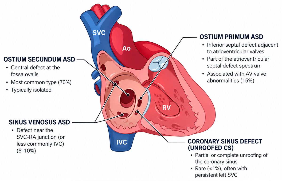
Figure 2
Anatomical classification of atrial septal defect. ASD: atrial septal defect; AV: atrioventricular; SVC: superior vena cava; SVC-RA: superior cavoatrial junction; IVC: inferior vena cava; CS: coronary sinus; Ao: aorta; RV: right ventrical
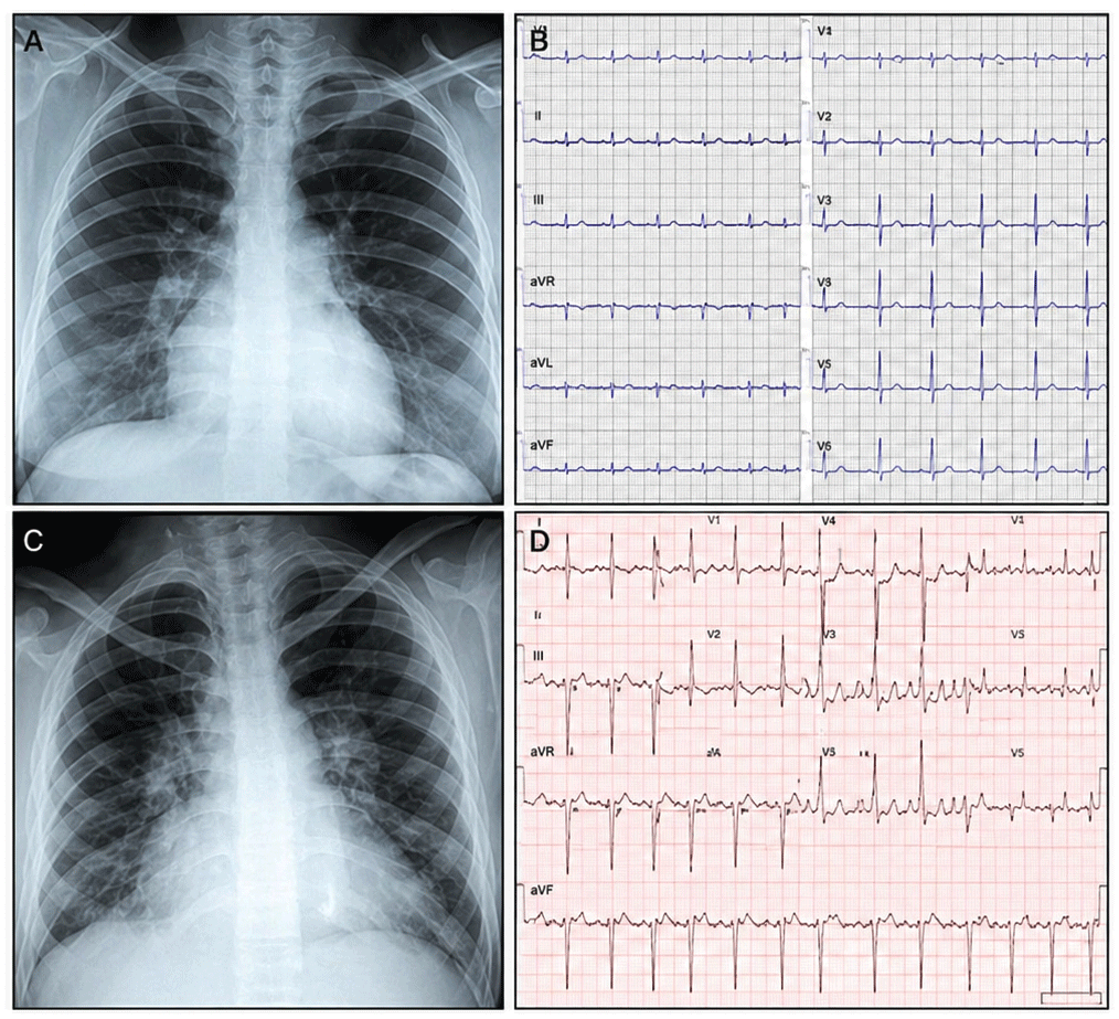
Figure 3
(A, B) A 35-year-old woman with a secundum-type atrial septal defect. (A) Posteroanterior chest radiograph shows moderate cardiomegaly predominantly involving the right-sided cardiac chambers, a prominent pulmonary artery segment, and increased pulmonary vascularity consistent with pulmonary overcirculation. (B) Twelve-lead electrocardiogram demonstrates sinus rhythm, incomplete right bundle branch block, and T-wave inversion extending to lead V4. (C, D) A 60-year-old patient with systemic arterial hypertension, pulmonary hypertension, and left ventricular diastolic dysfunction. (C) Posteroanterior chest radiograph shows severe cardiomegaly with signs of marked pulmonary overcirculation and a prominent pulmonary artery segment. (D) Twelve-lead electrocardiogram demonstrates atrial flutter with variable ventricular response.
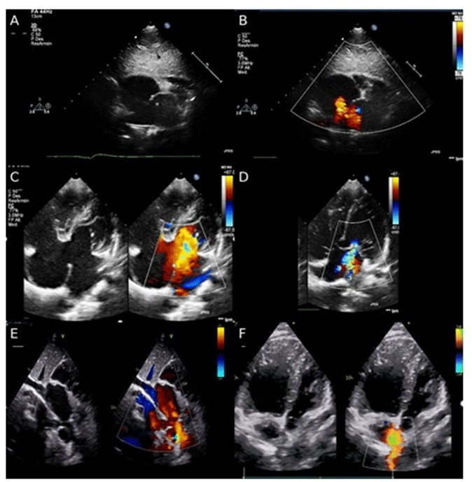
Figure 4
Two-dimensional (2D) and color Doppler echocardiography illustrates different types of atrial septal defects. (A) Subcostal view shows a 2D image of an ostium secundum atrial septal defect. (B) In the same view, color Doppler demonstrates a central left-to-right shunt. (C) Apical four-chamber view shows comparative 2D and color Doppler images of an ostium primum atrial septal defect with inferoposterior left-to-right shunting. (D) Apical four-chamber view demonstrates mitral regurgitation secondary to a cleft of the anterior mitral leaflet. (E) Subcostal view along the caval axis shows a superior sinus venosus atrial septal defect with drainage of the right upper pulmonary vein into the junction of the superior vena cava and the right atrium. (F) Apical four-chamber view of a sinus venosus atrial septal defect with color Doppler demonstrates left-to-right shunting at the superior portion of the interatrial septum.
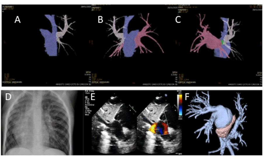
Figure 5
(A-C) Computed tomography (CT) angiography with three-dimensional (3D) reconstruction in a patient with a diagnosis of sinus venosus atrial septal defect associated with partial anomalous right pulmonary venous return. (A) White: right upper pulmonary vein draining into the superior vena cava. (B) Pink: fusion image of the remaining three pulmonary veins draining normally into the left atrium. (C) Green: image of a small sinus venosus–type atrial septal defect. (D-F) Patient with a diagnosis of Scimitar syndrome. (D) Chest X-ray in Scimitar Syndrome: cardiac dextroposition, right lung hypoplasia, and a curvilinear linear opacity along the right cardiac border extending from the hilum toward the cardiophrenic angle, consistent with the characteristic “scimitar sign.” (E) Subcostal color Doppler echocardiographic view demonstrates right pulmonary venous drainage into inferior vena cava. (F) CT scan demonstrates all right pulmonary venous drainage into inferior vena cava.
Video 1
Comparative parasternal long-axis color Doppler imaging of two clinical spectra in adult patients with ventricular septal defects. Left: restrictive ventricular septal defect with a left-to-right shunt. Right: large ventricular septal defect with a bidirectional shunt in a patient with Eisenmenger syndrome; also see at https://vimeo.com/1182784596?share=copy&fl=sv&fe=ci.
Video 2
Apical four-chamber two-dimensional echocardiographic image: 22-year-old female patient with complete atrioventricular canal who progressed to Eisenmenger syndrome. Note the ostium primum atrial septal defect, posterior ventricular septal defect, common atrioventricular valve, and right ventricular hypertrophy; also see at https://vimeo.com/1182785759?share=copy&fl=sv&fe=ci.
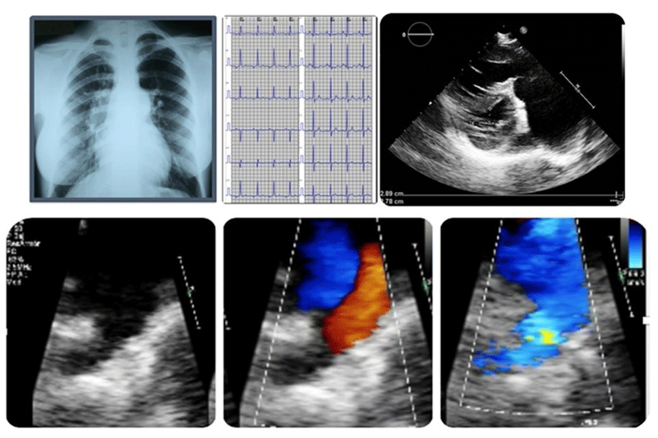
Figure 6
A 21-year-old patient with a diagnosis of Eisenmenger syndrome. (A) Chest radiograph (frontal view): prominent main pulmonary artery segment with asymmetric pulmonary blood flow shows increased central hilar vascularity and reduced peripheral perfusion, consistent with a typical “pruned tree” appearance. (B) Electrocardiogram shows right axis deviation and right ventricular hypertrophy. (C) Parasternal short-axis echocardiographic view demonstrates right ventricular hypertrophy, interventricular septal flattening, and severe dilation of the main pulmonary artery and its branches. (D) Two-dimensional echocardiographic image with zoom in the high parasternal short-axis view demonstrates a large patent ductus arteriosus. (E, F) Color Doppler echocardiography shows a bidirectional shunt through patent ductus arteriosus, with flow reversal during systole and diastole.