
Introduction
The presentation of shunt lesions in adulthood may be related to their high prevalence in childhood and the heterogeneity of their clinical presentation, with some lesions persisting or remaining undiagnosed despite the existence of well-defined diagnostic and treatment algorithms for congenital heart disease (CHD). They usually present in an isolated form, although they may be part of complex CHD. This review focuses on the isolated forms.
Shunt lesions allow communication between the systemic and pulmonary circulations. Given the pathophysiology of moderate and large defects, there is progression to pulmonary arterial hypertension (PAH), particularly in post-tricuspid shunts, where this evolution is often unavoidable in the absence of timely treatment, secondary to pulmonary vascular disease in advanced stages (Eisenmenger syndrome, or ES). Therefore, it is important to understand and analyze the different scenarios in which shunt lesions remain unrepaired.
The direction and magnitude of shunting are determined primarily by differences in chamber compliance and capacitance and by the relationship between pulmonary vascular resistance (PVR) and systemic vascular resistance (SVR). In childhood, these conditions are present with left-right shunt, since PVR is lower than SVR. Moderate and large defects initially have hemodynamic overload and pulmonary overcirculation, resulting in increased pulmonary venous return and volume overload of the cardiac chambers. Defects proximal to the atrioventricular (AV) valves (communications at the venous or atrial level) overload the right chambers, whereas defects distal to the AV valves (at the ventricular level or in the great vessels) predominantly overload the left chambers. The magnitude of the shunt reflects the relative volume of flow crossing the communication and is quantified by the ratio between pulmonary and systemic blood flow (Qp/Qs). We refer to nonrestrictive defects as those that are sufficiently large enough to allow pressures on both sides of the defect to be equal or nearly equal without significant restriction to flow. In shunts proximal to the AV valves (such as atrial septal defect or partial anomalous pulmonary venous return), elevated Qp/Qs values may be observed in the presence of normal pulmonary pressures. In these lesions, shunting occurs during both systole and diastole and is influenced by differences in atrial and ventricular compliance and capacitance, particularly the greater distensibility of the right ventricle. In these cases, pulmonary artery pressure does not usually increase significantly until adulthood. In contrast, nonrestrictive shunts distal to the AV valves (such as ventricular septal defect or patent ductus arteriosus) expose the pulmonary circulation to higher pressures and are more likely to induce early pulmonary vascular disease. A left-to-right shunt is considered hemodynamically significant when the pulmonary-to-systemic blood flow ratio (Qp:Qs) is ≥ 1.5 and is associated with cardiac chamber enlargement.1 In the absence of an increased Qp:Qs, findings such as chamber enlargement or pulmonary arterial hypertension should prompt careful evaluation to exclude alternative causes (Figure 1).
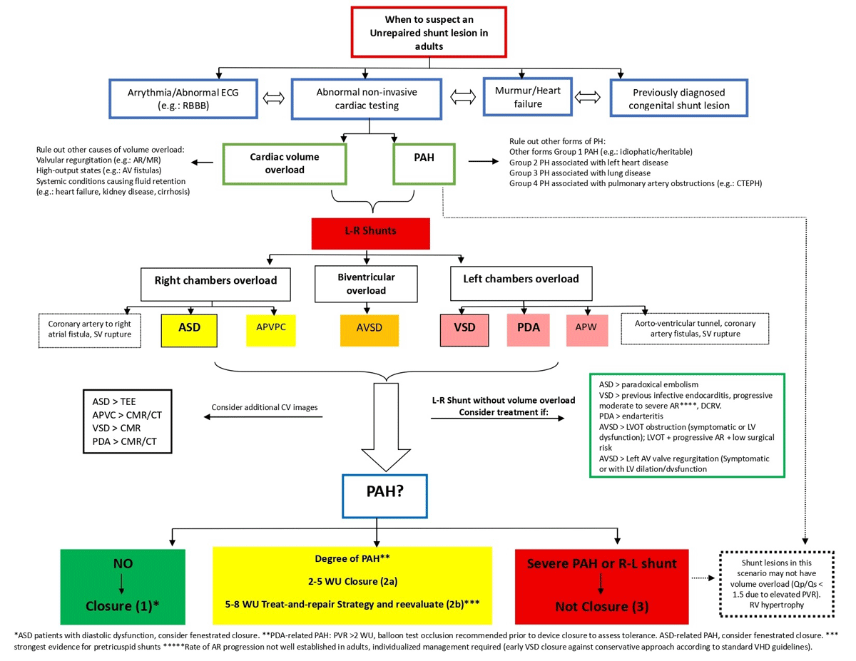
Figure 1
Evaluation and treatment algorithm. Shunt lesions connect the systemic and pulmonary circulations; direction depends on pressure gradients and the pulmonary vascular resistance to systemic vascular resistance ratio. Initially left-to-right with volume overload, they may progress to pulmonary hypertension, then bidirectional flow, and ultimately right-to-left (Eisenmenger syndrome), when closure is no longer feasible.
Considerations in Atrial Septal Defect closure
In moderate-to-large ASDs with Qp:Qs ≥ 1.5 and right ventricular dilation in the absence of pulmonary arterial hypertension (PAH), closure improves functional capacity and reduces long-term morbidity and mortality, even in asymptomatic individuals.
Patients with confirmed precapillary PAH should be managed according to established PAH guidelines. In selected patients with markedly elevated pulmonary vascular resistance (PVR ≥ 5 Wood units, or WU), PAH-directed therapy may allow consideration of ASD closure using treat-to-close or treat-and-repair strategies, although long-term medical therapy and close surveillance are often required. Patients with PAH and PVR of 5 to 8 WU on targeted PAH therapy have also been shown to safely undergo and benefit from ASD closure. However, agreement about the expected reversibility of PAH remains controversial. In cases of progressively increasing pulmonary arterial pressures, fenestrated closure may be considered to preserve a decompressive shunt and mitigate the risk of eliminating the pressure-relief pathway. In adults with unrepaired ASD and Eisenmenger physiology, ASD closure should not be performed to avoid increasing morbidity and mortality.
Considerations in VSD closure in these clinical scenarios
(1) Qp:Qs ≥ 1.5 with PVR ≤ 2 Wood units (WU) and significant (at least moderate) or progressive left ventricular (LV) dilation (Class 1).
(2) Qp:Qs ≥ 1.5 with PVR > 2 but < 5 WU and LV dilation (Class 2a), where closure may preserve LV function and reduce the risk of progressive PAH, balanced against perioperative risks and the risk for long-term progressive RV dysfunction and heart failure in patients with PAH after ventricular septal defect (VSD) closure.
(3) Qp:Qs ≥ 1.5 with PVR 5 to 8 WU, in the absence of hypoxemia, and reduction of PVR to < 5 WU after at least 3 months of PAH therapy (Class 2b), following a “treat-and-repair” strategy, for which evidence is strongest in atrial septal defects.
(4) Qp/Qs ≤ 1.5 with progressive moderate-to-severe aortic regurgitation (AR) (Class 2a): In children, progressive AR may justify VSD closure to prevent valve intervention. In adults, the rate of progression is not well established, therefore management should be individualized, weighing the risks and benefits of early surgery against conservative follow-up according to standard aortic valve replacement guidelines.
(5) Qp:Qs ≤ 1.5 with a history of infective endocarditis involving the VSD (Class 2b).
(6) NO VSD closure: Patients with pressure- and volume-restrictive defects without evidence of volume overload should not undergo closure, nor should those with large defects complicated by severe PAH or Eisenmenger physiology (Class 3).
Considerations in Patent Ductus Arteriosus closure
(1) A small patent ductus arteriosus (PDA) without a hemodynamically significant shunt derives no benefit from closure (Class III).
(2) PDA with a hemodynamically significant shunt should be evaluated for PAH. If pulmonary vascular resistance (PVR) is < 2 Wood units (WU), closure is recommended (Class I). If PVR is > 2 WU, balloon test occlusion is recommended prior to device closure to assess tolerance, including the development of symptoms, an increase in pulmonary artery pressure, or a decrease in aortic pressure. If balloon test occlusion is favorable and PVR is < 5 WU, closure is recommended (Class IIa). For patients with PVR between 5 and 8 WU, PAH-specific therapy may be initiated, and if PVR decreases to < 5 WU, closure may be considered, although the available evidence in this scenario is limited (Class IIb).
(3) PDA with severe PAH (> 10 WU) or Eisenmenger physiology should not undergo closure (Class III).
The natural history of unrepaired moderate and large shunt lesions is the development of PAH due to pulmonary vascular disease. As PVR increases, shunt magnitude may decrease as a sign of progression toward advanced pulmonary vascular disease. With increasing PVR, the shunt may become bidirectional. In clinical practice, exercise-induced hypoxemia in the presence of an isolated shunt suggests bidirectional flow.1 To confirm this, the evaluation should be completed with additional complementary tests; furthermore, assessing the response to supplemental oxygen administration can be useful to help differentiate intracardiac shunting from other causes, such as lung parenchymal disease. In advanced stages, when PVR equals or exceeds SVR, the shunt reverses and becomes predominantly right-to-left, leading to systemic hypoxemia and cyanosis, characteristic findings of ES, marking the impossibility of defect closure. It should be noted that, in certain cases, progression to PAH may be accelerated by comorbidities such as associated genetic syndromes (eg, Down syndrome).2
In adulthood, unrepaired shunt lesions typically present with one or more of the following scenarios: heart murmur, abnormal electrocardiogram (ECG), arrhythmias, chamber overload, PAH, heart failure (HF), preconception counseling screening, or in the setting of pregnancy-related complications. Shunt lesions may infrequently present as transient ischemic attack.3
In summary, unrepaired shunt lesions in adulthood represent a spectrum of clinical presentations. This spectrum ranges from small, with mild or even no hemodynamic impact lesions, to larger defects with variable PAH, and up to fixed pulmonary vascular resistance causing ES with shunt reversal.
Determining the optimal approach—including the indication and feasibility of defect closure and PAH management—remains challenging. While each lesion is addressed individually, Eisenmenger syndrome (ES, the feared final stage in unrepaired patients) serves as a key determinant for guiding management.
Atrial Septal Defect
Atrial septal defect (ASD) is one of the most common CHDs, accounting for approximately 7% to 10% of all CHD in childhood, with female predominance. Because it is often asymptomatic during childhood and early adulthood, diagnosis is frequently delayed, making ASD one of the most commonly diagnosed structural CHDs in adults within the ACHD population, accounting for approximately 25% to 30% of cases in specialized series.4,5,6
While historically regarded as a benign or “simple” lesion, growing evidence demonstrates that unrepaired ASD in adulthood is associated with substantial morbidity and reduced long-term survival.7,8 Beyond its traditional characterization as a right-sided volume overload lesion, ASD exerts complex effects on biventricular interaction, pulmonary vasculature, and atrial electrophysiology. Consequently, ASD in adulthood has emerged as a paradigm for CHD, illustrating how delayed recognition and suboptimal timing of intervention may lead to complex clinical scenarios.1,9,10,11,12,13
The most common anatomical subtype is ostium secundum ASD (> 75%), followed by ostium primum 15%, sinus venosus 5% to 10%, and the rare coronary sinus defect (< 1%) (Figure 2).
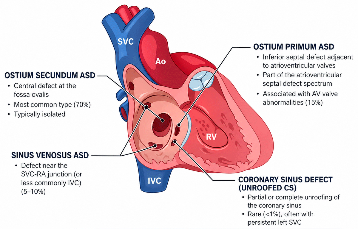
Figure 2
Anatomical classification of atrial septal defect. ASD: atrial septal defect; AV: atrioventricular; SVC: superior vena cava; SVC-RA: superior cavoatrial junction; IVC: inferior vena cava; CS: coronary sinus; Ao: aorta; RV: right ventrical
The magnitude of left-to-right shunting is determined by defect size, ventricular compliance, and the balance between PVR and SVR. Chronic shunting results in progressive right atrial and right ventricular (RV) volume overload, myocardial remodeling, and atrial enlargement. Over time, these changes predispose patients to atrial tachyarrhythmias, reduced exercise capacity, HF, and thromboembolic events. In a subset of patients, prolonged pulmonary overcirculation leads to pulmonary vascular remodeling and PAH, with the possibility of progression to ES (see below).14,15,16
Diagnosis
Clinical presentation in adulthood is variable. Some patients develop symptoms such as exertional dyspnea, reduced exercise tolerance, palpitations, or atrial arrhythmias related to chronic right-sided volume overload. However, many adults with ASD remain asymptomatic for years, and diagnosis is often made incidentally after detection of a cardiac murmur or abnormal ECG or chest X-ray findings. As patients age, exertional dyspnea and palpitations become increasingly common.
Physical examination typically reveals an RV heave, fixed splitting of the second heart sound, and a systolic flow murmur over the pulmonary area. Electrocardiography frequently demonstrates incomplete right bundle branch block and signs of right atrial enlargement, with a superior axis in ostium primum defects, while chest radiography shows right heart enlargement and pulmonary plethora (Figure 3). Atrial arrhythmias are the most common complication of unrepaired ASD; atrial fibrillation is the most frequent in adults, increases with age, and often persists after closure, especially when repair occurs after age 40. This likely reflects the long-standing effects of atrial volume overload and atrial remodeling associated with chronic left-to-right shunting, which predispose to electrical instability.1,9,10,11,16,17,18
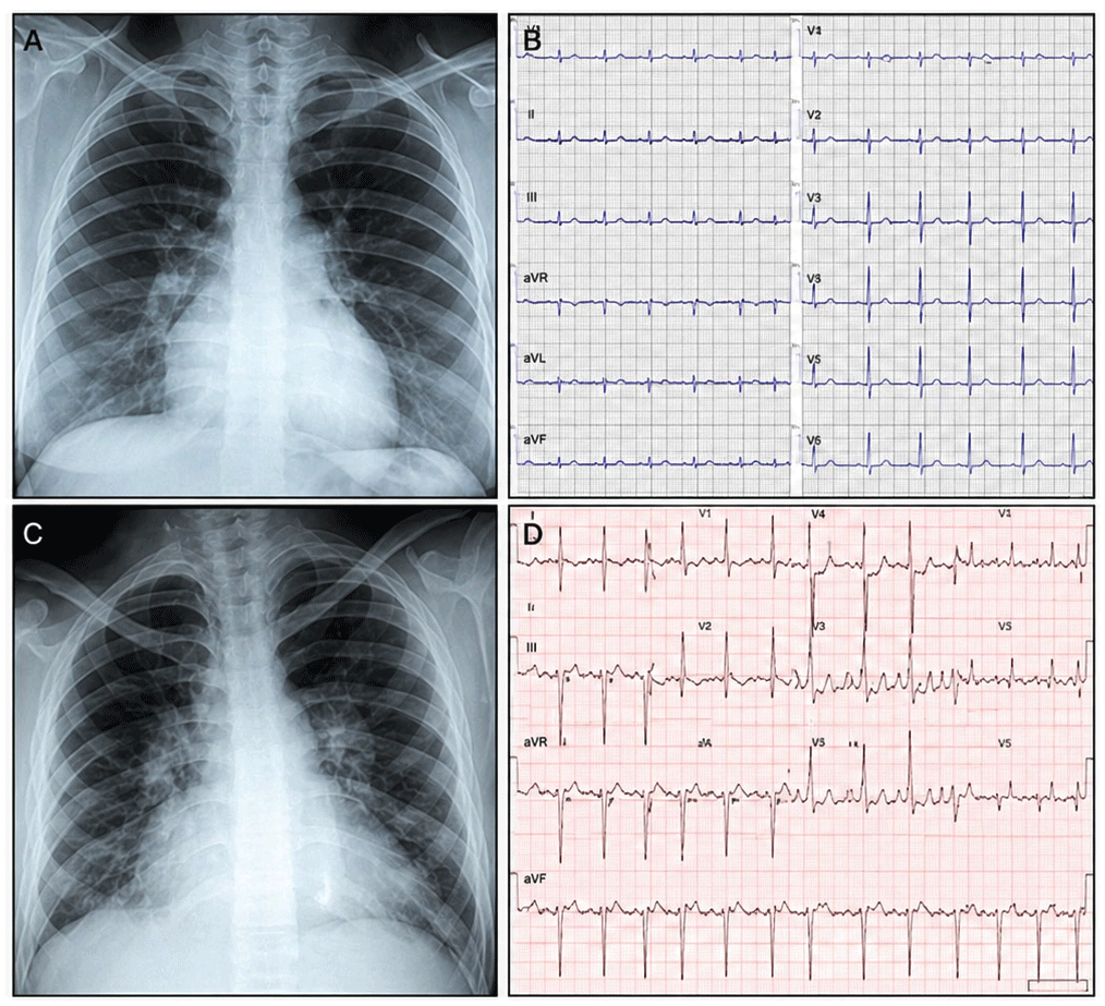
Figure 3
(A, B) A 35-year-old woman with a secundum-type atrial septal defect. (A) Posteroanterior chest radiograph shows moderate cardiomegaly predominantly involving the right-sided cardiac chambers, a prominent pulmonary artery segment, and increased pulmonary vascularity consistent with pulmonary overcirculation. (B) Twelve-lead electrocardiogram demonstrates sinus rhythm, incomplete right bundle branch block, and T-wave inversion extending to lead V4. (C, D) A 60-year-old patient with systemic arterial hypertension, pulmonary hypertension, and left ventricular diastolic dysfunction. (C) Posteroanterior chest radiograph shows severe cardiomegaly with signs of marked pulmonary overcirculation and a prominent pulmonary artery segment. (D) Twelve-lead electrocardiogram demonstrates atrial flutter with variable ventricular response.
Specific conduction abnormalities correlate with anatomical subtypes: sinus node dysfunction is more common in superior sinus venosus defects, while AV block predominates in ostium primum ASDs, regardless of surgery, due to inferior displacement of the AV conduction system (Figure 4).
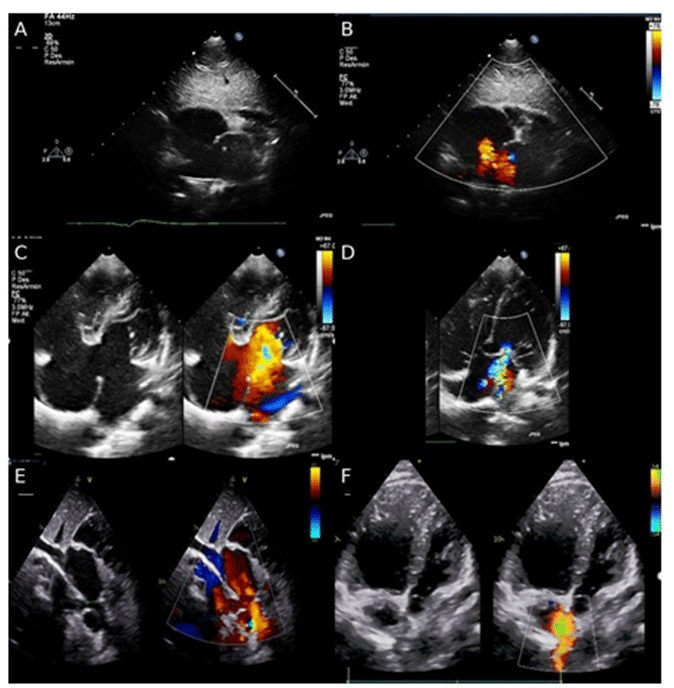
Figure 4
Two-dimensional (2D) and color Doppler echocardiography illustrates different types of atrial septal defects. (A) Subcostal view shows a 2D image of an ostium secundum atrial septal defect. (B) In the same view, color Doppler demonstrates a central left-to-right shunt. (C) Apical four-chamber view shows comparative 2D and color Doppler images of an ostium primum atrial septal defect with inferoposterior left-to-right shunting. (D) Apical four-chamber view demonstrates mitral regurgitation secondary to a cleft of the anterior mitral leaflet. (E) Subcostal view along the caval axis shows a superior sinus venosus atrial septal defect with drainage of the right upper pulmonary vein into the junction of the superior vena cava and the right atrium. (F) Apical four-chamber view of a sinus venosus atrial septal defect with color Doppler demonstrates left-to-right shunting at the superior portion of the interatrial septum.
Accurate diagnosis requires a multimodality imaging approach. Transthoracic echocardiography (TTE) remains the first-line modality but may be limited in defining atrial septal anatomy and pulmonary venous connections in adults. Transesophageal echocardiography improves visualization of septal rims and is particularly valuable for identifying sinus venosus defects and associated anomalous pulmonary venous return. Cardiac magnetic resonance (CMR) imaging provides radiation-free quantification of shunt magnitude (Qp:Qs) and ventricular volumes, whereas cardiac computed tomography (CCT) offers high-resolution anatomical detail for interventional or surgical planning (Figure 5).19,20 All adults with unrepaired ASD should be systematically screened for PAH since the presence of PAH guides management. Disease may be clinically silent at rest. Evaluation includes clinical assessment, ECG, echocardiography, biomarkers, exercise testing, and invasive hemodynamic testing when PAH is suspected.21,22,23
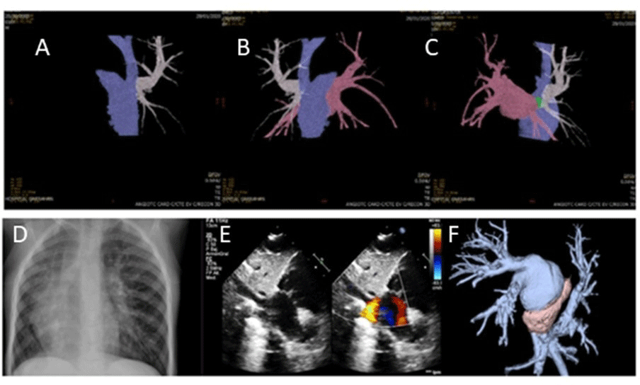
Figure 5
(A-C) Computed tomography (CT) angiography with three-dimensional (3D) reconstruction in a patient with a diagnosis of sinus venosus atrial septal defect associated with partial anomalous right pulmonary venous return. (A) White: right upper pulmonary vein draining into the superior vena cava. (B) Pink: fusion image of the remaining three pulmonary veins draining normally into the left atrium. (C) Green: image of a small sinus venosus–type atrial septal defect. (D-F) Patient with a diagnosis of Scimitar syndrome. (D) Chest X-ray in Scimitar Syndrome: cardiac dextroposition, right lung hypoplasia, and a curvilinear linear opacity along the right cardiac border extending from the hilum toward the cardiophrenic angle, consistent with the characteristic “scimitar sign.” (E) Subcostal color Doppler echocardiographic view demonstrates right pulmonary venous drainage into inferior vena cava. (F) CT scan demonstrates all right pulmonary venous drainage into inferior vena cava.
Management of ASD in adulthood is highly individualized and depends on shunt magnitude, ventricular remodeling, PVR, and comorbid conditions. The management of adults with ASD is summarized in Figure 1.
In patients who are candidates for closure (Figure 1), transcatheter closure may be preferred in suitable anatomy because of its less invasive nature and favorable procedural outcomes, including lower morbidity and shorter hospital stay compared with surgery in observational series. Surgical closure remains an appropriate option when anatomical features are not suitable for device closure or when additional cardiac surgical procedures are required.
ASDs amenable to percutaneous closure are ostium secundum ASDs with adequate rims and a defect size less than 35 mm.24,25 Selected superior sinus venosus defects can be treated with percutaneous closure using covered stents in expert CHD centers. Surgical repair remains appropriate for ostium primum, sinus venosus, and coronary sinus defects, or when anatomy precludes device closure.26,27
In adults, associated conditions such as significant left ventricular (LV) diastolic dysfunction may require modification of the algorithm, as ASD closure can be contraindicated and a fenestrated patch may be safer; they also may require modification of the management algorithm since abrupt closure may increase left-sided filling pressures. In such cases, careful hemodynamic assessment should guide individualized decision-making. In selected cases, fenestrated closure strategies may be considered, although supporting data remain limited.1
Partial Anomalous Pulmonary Venous Return
In partial anomalous pulmonary venous return (PAPVR) one or more pulmonary veins drain into the systemic venous circulation or the right atrium. The most common drainage sites are the superior vena cava (SVC), right atrium, and inferior vena cava (IVC). Right-sided anomalies are more frequent than left-sided ones.
Several anatomical variants have been described. The most prevalent is drainage of the right superior pulmonary vein, often together with the middle lobe vein, into the SVC, which is strongly associated (> 90%) with sinus venosus-type ASD.28 Other variants include drainage of right pulmonary veins into the IVC (Scimitar syndrome), left pulmonary veins draining into the innominate or left vertical vein, and left pulmonary veins draining into the coronary sinus.
PAPVR is frequently an incidental finding in asymptomatic patients. Unexplained dilation of right cardiac chambers in the absence of an ASD should prompt evaluation for this condition. The hemodynamic significance depends on the number of anomalous veins, the presence of an associated ASD, and the PVR. The physical exam and ECG are similar to those of the ASD. Atrial arrhythmias are more common in patients over 40 years of age.
TTE assesses right chamber dilation and PAH. CMR imaging allows detailed visualization of anomalous pulmonary venous connections and quantification of shunt magnitude, while CCT is the imaging modality of choice for precise anatomical delineation (Figure 5).29 Percutaneous approaches are limited to selected cases and are only available in a few experienced centers.
Scimitar Syndrome
Scimitar syndrome is a rare variant of PAPVR characterized by anomalous drainage of most or all right pulmonary veins into the IVC, although partial anomalous drainage may also occur. It is commonly associated with hypoplasia of the right PA and right lung, often with systemic collateral blood supply.
Clinical presentation ranges from exertional dyspnea and palpitations to recurrent pulmonary infections and hemoptysis, depending on shunt magnitude and associated pulmonary sequestration. Chest radiography typically shows the characteristic curvilinear “scimitar” sign.30 Cardiac MRI and CCT provide comprehensive assessment of venous anatomy, collateral circulation, PA size, and lung parenchyma.31 Right heart catheterization is indicated in patients with PAH (Figure 5).
Surgical correction, consisting of redirection of the anomalous vein to the left atrium, is most beneficial in patients with right heart dilation and no significant pulmonary sequestration.32,33 Long-term prognoses is generally favorable, with atrial arrhythmias being the most common late complication.
Ventricular Septal Defect
Ventricular septal defect (VSD) is the most common CHD in childhood and the second most frequent in adults, largely due to spontaneous closure in early life. It results from incomplete development of the interventricular septum during embryogenesis. VSDs are classified by anatomic location into four types: perimembranous, outlet, inlet, and muscular.34 Outlet VSDs are associated with aortic cusp prolapse and regurgitation, inlet defects are associated with atrioventricular canal abnormalities, and muscular VSDs, which occur in the trabecular septum, are more likely to close spontaneously.
The hemodynamic significance of a VSD is determined by defect size and location as well as pulmonary vascular resistance, which together influence both the magnitude and direction of intracardiac shunting. In congenital VSD, this physiology typically results in volume overload of the left-sided chambers due to increased pulmonary venous return. By contrast, in acquired ventricular septal rupture following myocardial infarction, right ventricular compliance and capacitance may also influence the magnitude of shunting, and right ventricular dilatation may occur as the ventricle accommodates the acute pressure and volume overload.
Long-standing large shunts may induce irreversible pulmonary vascular remodeling, consequently resulting in persistent PAH.
In adults, the clinical spectrum of isolated unrepaired VSD includes:1
Small VSDs (pressure- and volume-restrictive): no left-sided chamber enlargement or PAH.
Moderate VSDs (pressure-restrictive but not volume-restrictive): may be associated with LV dilation, PAH, or both.
Large nonrestrictive VSDs: often associated with ES in adulthood.
Complications that may occur in the natural history of isolated VSDs include endocarditis, aortic regurgitation, RV outflow tract obstruction (RVOTO), and the development of HF due to increased shunting from comorbidities.
Diagnosis
Murmur intensity correlates with flow velocity across the defect. Smaller VSDs are often associated with louder murmurs (holosystolic or protomesosystolic) and may produce a palpable thrill. In contrast, larger defects usually generate softer murmurs, particularly in the presence of elevated pulmonary pressures. Large defects without significant shunting or those with Eisenmenger physiology may lack an audible systolic murmur.
ECG is frequently normal in patients with small VSDs. As shunt magnitude increases, findings may include evidence of left ventricular volume overload and hypertrophy. As pulmonary pressure increases, signs of right ventricular hypertrophy may appear.
TTE is the cornerstone for VSD evaluation, enabling assessment of defect morphology, hemodynamic significance (chamber enlargement and PAH), associated complications (aortic regurgitation, double-chambered right ventricle), and concomitant congenital anomalies (Video 1).
Video 1
Comparative parasternal long-axis color Doppler imaging of two clinical spectra in adult patients with ventricular septal defects. Left: restrictive ventricular septal defect with a left-to-right shunt. Right: large ventricular septal defect with a bidirectional shunt in a patient with Eisenmenger syndrome; also see at https://vimeo.com/1182784596?share=copy&fl=sv&fe=ci.
When indicated, VSD repair may be achieved surgically or percutaneously (Figure 1). Transcatheter closure is feasible in most muscular VSDs and selected perimembranous defects.
In patients with more than mild aortic regurgitation, follow-up is recommended to monitor for progression of aortic valve dysfunction and to assess the potential need for future aortic valve intervention.
In unrepaired VSDs without PAH, RVOTO may develop, resulting in a double-chambered right ventricle. It can be caused either by the presence of anomalous muscle bands, by hypertrophy of endogenous trabecular tissue, or occasionally by an aberrant moderator band. Pathologic turbulent flow may act as the initial stimulus for hypertrophic changes to the trabeculae, even in the presence of small VSD.
When this causes significant pressure overload of the subpulmonary chamber, surgical relief is indicated to preserve RV function.
Long-term follow-up is guided by the patient’s physiological stage. Asymptomatic patients without volume overload, arrhythmias, or PAH may be followed every 36–60 months with clinical evaluation, ECG, and TTE. Patients with a history of HF, arrhythmias, PAH, or infective endocarditis require closer surveillance, including annual ECG and TTE, with outpatient follow-up intervals ranging from 3 to 12 months depending on clinical status.
Although VSD was considered a “simple” congenital heart disease for many years, its diagnosis is associated with significant long-term consequences. Patients with isolated VSD—whether unrepaired or surgically closed— carry a substantial burden of cardiovascular morbidity throughout life including arrhythmias, infective endocarditis, HF, aortic regurgitation, RVOTO and PAH.35 Morbidity in unrepaired patients remains low until around the fourth decade of life, after which it rises markedly. Patients with unrepaired small VSDs diagnosed in adulthood have a two-fold higher hazard ratio for mortality compared with matched peers.36 These findings reinforce current guidelines recommending regular follow-up in specialized adult congenital heart disease clinics.
Patent Ductus Arteriosus (PDA)
PDA is one of the most common CHD, accounting for approximately 5% to 10% of all CHD, excluding premature infants. The diagnosis of PDA in adults is uncommon, as most cases are identified and treated during childhood; it accounts for approximately 1.3% of adults with CHD receiving care at a single tertiary center in the United States.37
PDA represents persistent patency of a normal fetal vascular structure connecting the PA and the descending aorta. The pathophysiology involves systemic-to-pulmonary blood shunting. The magnitude of the shunt depends on the size of the PDA (length and diameter) as well as the relative PVR and SVR. In large PDA, increased venous return to the left atrium and LV results in volume overload and chamber enlargement. Increased pulmonary blood flow may lead to PAH, which, if left untreated, can progress to ES. Although rare, PDA aneurysm may develop in adulthood, with potential compression of adjacent structures and risk of rupture. PDA calcification is another complication observed in adults.
The clinical spectrum of isolated PDA in adults includes:
Small PDA with no volume overload and normal PA pressure, typically asymptomatic.
Moderate PDA with predominant LV dilation and preserved or reduced systolic function, or predominant PAH with RV pressure overload.
Large PDA with Eisenmenger physiology, often associated with differential cyanosis due to ductal right-to-left shunting and lower-extremity hypoxemia.38
In moderate PDA with low PVR, continuous murmur is heard along the upper left sternal border. In smaller shunts, only a systolic component may be present. Clinical findings in the presence of pulmonary arterial hypertension are similar to those described in ES.
ECG may be normal, or demonstrate left-sided chamber enlargement, with evidence of biventricular hypertrophy in cases complicated by PAH.
TTE remains the first-line imaging modality for the assessment of PDA, allowing diagnosis and evaluation of hemodynamic significance through assessment of chamber enlargement and PAH. However, in adults, a poor acoustic window may compromise diagnostic accuracy. The identification of PDA with right-to-left shunting in the setting of ES may be challenging. The use of agitated saline contrast, with visualization from the suprasternal view in the PA and descending aorta, may unmask the diagnosis;7 however, a high index of suspicion is required. CMR allows noninvasive flow assessment, Qp:Qs calculation, quantification of LV volumes and function, and improved delineation of ductal anatomy. When CMR is not feasible, CCT may be considered, particularly when detailed delineation of PDA anatomy or the aortic arch is required prior to consideration of transcatheter device closure. For management, see Figure 1.
When indicated, percutaneous closure can be performed with minimal risk and is preferred over surgical closure. A calcified PDA carries an increased risk with surgical closure due to a higher risk of life-threatening rupture and bleeding, making device closure the method of choice, even when cardiac surgery is indicated for other concomitant CHDs.
Special Considerations
In PDA-associated endarteritis, closure may be considered to reduce risk for recurrent infection.
Asymptomatic and hemodynamically insignificant PDAs carry a small but lifetime risk of infectious endarteritis, most often affecting the pulmonary end of the duct. Whether to close these defects to prevent this rare but potentially devastating complication remains controversial, and it could be considered taking into consideration each center experience.
In adults, most PDAs are hemodynamically insignificant, small, and detected incidentally; however, larger PDAs can also be identified. Left-sided chamber volume overload or PAH should raise suspicion of PDA. More evidence is needed to define optimal timing of intervention to prevent persistent LV systolic dysfunction and to guide treatment and prognosis in patients with significant PAH.
Aortopulmonary Window
Aortopulmonary window is a rare CHD characterized by an abnormal communication between the ascending aorta and the PA in the presence of two separate semilunar valves arising from distinct subarterial ventricular outflow tracts. It represents a high-pressure left-to-right shunt that rapidly leads to HF in most untreated patients during infancy. If left undiagnosed and unrepaired, progressive pulmonary vascular disease develops, resulting in severe PAH; consequently, most untreated adults present as ES.
Atrioventricular Septal Defect
Atrioventricular septal defects (AVSD) are characterized by the combination of an AV septal defect and AV valve abnormalities. They include partial and complete forms, with complete AVSD (CAVSD) being the most frequent and strongly associated with Down syndrome. CAVSD consists of an ostium primum ASD, a large inlet ventricular septal defect (VSD), and a common AV valve with a single AV orifice.1,2,3,4,5,6,7,8,9,10 Partial AVSD includes an ostium primum ASD and a valvar cleft, generally of the left anterior AV valve, often associated with significant regurgitation.39 Left ventricular outflow tract obstruction may occur. The natural history of the ostium primum ASD was addressed in the ASD section. On ECG, a left anterior hemiblock is typically observed in all AVSDs. AVSDs are usually surgically corrected in childhood, and adults with unrepaired CAVSD present as ES (Video 2).40,41
Video 2
Apical four-chamber two-dimensional echocardiographic image: 22-year-old female patient with complete atrioventricular canal who progressed to Eisenmenger syndrome. Note the ostium primum atrial septal defect, posterior ventricular septal defect, common atrioventricular valve, and right ventricular hypertrophy; also see at https://vimeo.com/1182785759?share=copy&fl=sv&fe=ci.
Therapeutic Strategies and Clinical Management
Shunt lesions share a common management approach guided by the hemodynamic impact of the left-to-right shunt and the presence and magnitude of PAH. Based on these parameters, the defect may be completely closed, closed with a fenestrated patch, require medical treatment before closure, or be deemed not suitable for closure (Figure 1). Patients with significant shunts who have already developed PAH should be evaluated and treated at a center with ACHD and PAH expertise.42
Eisenmenger Syndrome
ES represents the most advanced and irreversible form of pulmonary vascular disease associated with CHD characterized by longstanding left-to-right shunting. It is defined by the development of severe PAH with PVR at SVR levels, leading to bidirectional or right-to-left shunt reversal and resultant systemic hypoxemia (Figure 6). The diagnosis of ES precludes shunt closure. ES is a multisystem disorder with clinical manifestations that extend beyond the cardiovascular system, and it requires a lifelong specialized care. Although its incidence has declined, ES remains a relevant clinical entity worldwide, particularly among adults with unrepaired CHD.21,22,43
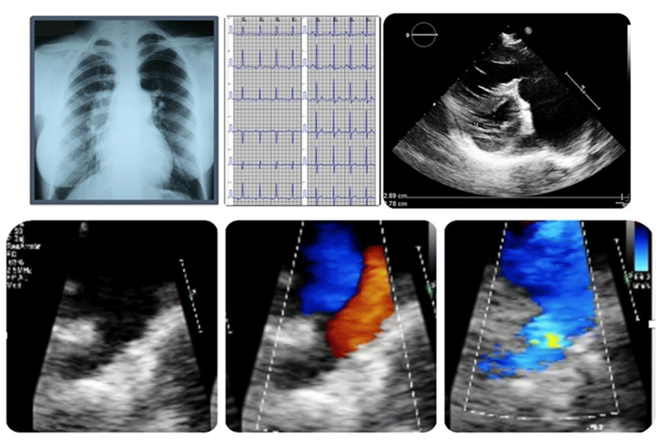
Figure 6
A 21-year-old patient with a diagnosis of Eisenmenger syndrome. (A) Chest radiograph (frontal view): prominent main pulmonary artery segment with asymmetric pulmonary blood flow shows increased central hilar vascularity and reduced peripheral perfusion, consistent with a typical “pruned tree” appearance. (B) Electrocardiogram shows right axis deviation and right ventricular hypertrophy. (C) Parasternal short-axis echocardiographic view demonstrates right ventricular hypertrophy, interventricular septal flattening, and severe dilation of the main pulmonary artery and its branches. (D) Two-dimensional echocardiographic image with zoom in the high parasternal short-axis view demonstrates a large patent ductus arteriosus. (E, F) Color Doppler echocardiography shows a bidirectional shunt through patent ductus arteriosus, with flow reversal during systole and diastole.
Clinical Manifestations and Natural History of ES
Central cyanosis, exertional dyspnea, fatigue, and reduced exercise tolerance are common presenting features. With disease progression, patients may develop syncope, arrhythmias, signs of RV dysfunction, and overt right HF.44
Chronic hypoxemia leads to secondary erythrocytosis, a compensatory mechanism aimed at maintaining adequate tissue oxygen delivery. Iron deficiency frequently coexists and may blunt this adaptive response, resulting in worsened functional capacity and adverse outcomes. Hemostatic abnormalities are common, with complex alterations in platelet function and coagulation pathways that predispose patients to both thrombotic and hemorrhagic events. Hemoptysis, paradoxical embolism, cerebrovascular accidents, and PA thrombosis contribute substantially to morbidity and mortality.45,46,47,48,49,50,51
Extra-cardiac organ involvement includes renal dysfunction, hyperuricemia, osteoarticular manifestations, cholelithiasis, and an increased susceptibility to infections. High-risk situations such as pregnancy, major noncardiac surgery, and abrupt reductions in SVR are associated with significant morbidity and mortality and should be avoided whenever possible.46,52,53,54,55
ES remains associated with reduced life expectancy compared with the general population. Contemporary cohorts show improved survival due to advances in supportive care and targeted PAH therapies, but right HF remains the leading cause of death.56,57,58
Management of ES requires a comprehensive, multidisciplinary approach focused on symptom control, prevention of complications, and optimization of quality of life. General measures include avoidance of dehydration, correction of iron deficiency, cautious management of erythrocytosis, prevention of air embolism, and prompt treatment of infections. Routine anticoagulation is not recommended in the absence of specific indications due to the fragile balance between thrombotic and hemorrhagic risk.1,52,53,59 Examples of indications for anticoagulation include the presence of heart failure, sustained arrhythmias, thrombosis, or a history of embolic events.
Heart-lung transplantation or bilateral lung transplantation with concomitant repair of the cardiac defect represents the only definitive therapy for end-stage disease.
Conclusions
In summary, unrepaired shunt lesions in adulthood illustrate how simple CHD may evolve into complex clinical conditions requiring individualized management in specialized ACHD centers. The main challenge related to shunt lesions is to optimize timely diagnosis and treatment to prevent long-term complications of the natural course.
Key Points
Shunt lesions are the most common congenital heart disease and may progress to pulmonary arterial hypertension (PAH) and Eisenmenger syndrome if unrepaired, particularly in moderate and large defects.
Shunt direction and magnitude are determined by pressure gradients and the balance between pulmonary and systemic vascular resistance, with left-to-right shunting in childhood and potential reversal as pulmonary vascular resistance increases.
Unrepaired shunt lesions in adulthood present a wide clinical spectrum, ranging from asymptomatic disease to PAH, heart failure, arrhythmias, thromboembolic events, and Eisenmenger syndrome.
Defect location influences chamber overload and disease progression: proximal shunts primarily overload right chambers and often delay PAH, whereas distal nonrestrictive shunts expose the pulmonary circulation to higher pressures and earlier vascular disease.
Management is individualized and guided by shunt significance and PAH severity, with Eisenmenger syndrome representing an irreversible stage that precludes defect closure and requires lifelong specialized care.