
Introduction
Adults with congenital heart disease (ACHD) represent a growing population of patients in the United States due to advances in pediatric interventions and congenital cardiac surgery. Patients with repaired congenital heart lesions require lifelong surveillance by expert ACHD cardiologists to mitigate late complications and ensure optimal outcomes.1 However, many unrepaired obstructive lesions, such as pulmonic stenosis, branch pulmonary stenosis, coarctation of the aorta, and subaortic stenosis, may frequently manifest in adulthood with common symptoms including chest pain, shortness of breath, palpitations, and murmurs discovered incidentally or accentuated during pregnancy. These common presentations highlight the critical need for the general cardiologist to identify congenital heart lesions in common clinical scenarios. This review highlights these common presentations and synthesizes their pathophysiology, diagnostic approaches, and management through representative adult cases.
Pulmonic Stenosis
A 53-year-old cisgender man presented to the emergency department (ED) with new onset of chest pain and palpitations that began the prior day while performing his normal exercise routine. He noted his heart rate did not decrease once he stopped exercising. His symptoms persisted, interfering with his sleep. He then went to the ED for evaluation. Upon arrival, his heart rate was 150 beats per minute with otherwise normal vital signs. His exam was notable for tachycardia with regular rhythm, normal jugular venous pressure without hepatojugular reflux, IV/VI systolic murmur with widely split S2 and soft P2 at the left second intercostal space without radiation, right ventricular (RV) heave, nondisplaced pulse of maximal impulse, palpable liver at the costal margin, no ascites, and no bilateral lower extremity edema. His distal pulses were 2+ and his extremities were warm. An electrocardiogram (ECG) showed atrial flutter with 2:1 block, right axis deviation, and right ventricular hypertrophy (RVH) with strain pattern. He underwent elective direct current cardioversion (DCCV) with restoration of normal sinus rhythm and relief of his symptoms. An echocardiogram was obtained after DCCV and demonstrated normal left ventricular (LV) size and systolic function, a mildly dilated RV with hypertrophy and normal systolic function, normal tricuspid leaflets with moderate regurgitation, thickened pulmonic leaflets with doming, peak velocity 4 meters per second with associated gradient of 64 mm Hg at the level of the pulmonary valve (PV), trace pulmonary regurgitation, dilated main pulmonary artery (MPA), and dilated right atrium (Figure 1). He started on long-acting metoprolol and direct oral anticoagulation and was referred to outpatient cardiology clinic for further management.
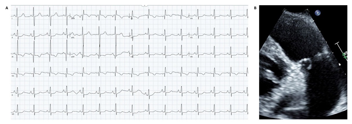
Figure 1
(A) Electrocardiogram after cardioversion demonstrating normal sinus rhythm with right axis deviation and right ventricular hypertrophy. (B) Echocardiogram frame (enlarged) during mid-systole showing thickened pulmonary valve leaflets with doming.
Congenital pulmonary stenosis (PS) is relatively common, affecting 1 in 2,000 live births globally.2 It can occur as an isolated lesion or be associated with other congenital anomalies, including tetralogy of Fallot, D-loop and congenitally corrected transposition of the great arteries, tricuspid atresia, and double outlet right ventricle. It may also be linked to congenital maternal rubella or genetic syndromes such as Noonan, Williams-Beuren, or Alagille syndromes.3 Valvar PS is more common than subvalvular or supravalvular stenosis and is often characterized by doming or conical leaflets (noted during systole) due to fusion of the usually thin leaflets at the commissures.4 The pathophysiology of PS relates to pressure overload within the RV, increasing wall stress and contractility to maintain normal cardiac output; the ventricle itself then hypertrophies to compensate for decreased compliance, leading to increased volumes and elevated end diastolic pressure and eventual dysfunction.5
The presentation of PS depends on the severity of the stenosis. Patients with mild or even moderate PS may be asymptomatic. Those with moderate or severe stenosis may develop symptoms when physiologic reserve—characterized by an increase in contractility with compensatory RVH, augmented end-diastolic volumes, and elevated end diastolic pressure—maximizes relative to the degree of obstruction. In this scenario, the RV is unable to augment cardiac output, leading to syncope, chest pain, dyspnea on exertion, or even sudden cardiac death.4,6 Post-stenotic MPA is common and may cause angina if it compresses an adjacent coronary artery (often the left main and/or left anterior descending artery).3 Elevated right heart filling pressures may also lead to peripheral edema and abdominal fullness.7
The physical exam often involves elevated a-wave noted in the jugular venous pulsations (due to atrial contraction against a restrictive RV), ascites, peripheral edema, and liver congestion.7 Left parasternal heave may be palpated due to RVH, and while mild stenosis is characterized by a normal first heart sound (S1), an ejection click, and a normal to increased second heart sound (S2), the murmur as the stenosis increases to severe is a harsh, medium-to-high, crescendo-decrescendo sound during systole with a soft or inaudible P2 component and widely split S2.3,8 As opposed to the murmur of aortic stenosis, the pulmonary stenosis murmur will increase with inspiration, does not radiate, and does not affect carotid pulsation. A right-sided S4 gallop may also be heard.4
Evaluating the patient may require several different testing methods to quantify severity. Electrocardiograms in patients with mild stenosis may be normal while patients with severe disease may have features typical of right atrial enlargement, right axis deviation, and RV hypertrophy with large amplitude R waves in V1 and aVR, deep S waves in V6, and an R:S ratio of < 1.9 Chest x-ray may demonstrate conspicuous right heart border or pulmonary arteries although the findings are not specific enough to be diagnostic.3 Echocardiography remains the preferred test as it often is adequate to visualize the specific valve morphology, surrounding structures, and origin and degree of stenosis while being readily available and safely and quickly performed. Elevated estimated RV systolic pressure must be interpreted thoughtfully in the presence of pulmonary stenosis as this will reflect the degree of pulmonary stenosis rather than estimated pulmonary artery systolic pressure. Transesophageal echocardiogram, which could have been performed on the case patient, can also evaluate the pulmonary valve, although ultrasound beam angle can often be insufficient to generate a valid peak Doppler gradient due to the anterior position of the valve.5 However, cardiac computed tomography (CT) and/or magnetic resonance imaging (MRI) may be used if echocardiographic windows are difficult or inadequate to quantify stenosis or if the patient has complicated anatomy. PS severity on echocardiogram is characterized as mild with peak gradient < 36 mm Hg or velocity < 3 meters per second (m/s), moderate with gradient 36 to 64 mm Hg or velocity 3 to 4 m/s, and severe with peak gradient > 64 mm Hg, mean gradient > 35 to 40 mm Hg, or velocity > 4 m/s. 10,11 CT and MRI are useful for anatomic and functional assessment of the PV, RV outflow tract, branch pulmonary arteries (BPAs), and RV.12 Cardiac catheterization is no longer preferred for diagnosing stenosis.
Treatment in patients with moderate or severe stenosis is recommended for those with symptoms and for those who are asymptomatic but demonstrate at least moderate tricuspid regurgitation or any degree of RV dysfunction to improve ventricular function and reduce pressure overload.10 In those with asymptomatic severe stenosis, it is reasonable to intervene if there is impaired ventricular function or evidence of pressure overload.10 When performed by a congenital interventionalist, balloon valvuloplasty (BV) is favored in patients with domed rather than dysplastic valves, although recurrent stenosis may warrant repeat interventions.3 As part of interventional planning, pulmonary annulus size should be measured during mid-systole from the inner edge to inner edge of the valve insertion hinge points, with normal size ≤ 2.5 cm; of note, mean diastolic MPA parameters by CMR are 22.9 ± 2.4 mm for males and 21.2 ± 2.1 mm for females (based on binary sex categories).13,14 Young age, severe PS, low weight, increased RV:systemic pressure ratio (a marker of stenosis severity), smaller pulmonary annulus diameter, and higher initial systolic gradient across the PV are identified risk factors for development of moderate pulmonary regurgitation after BV.15 Surgical intervention is indicated for those who remain symptomatic after BV or have mixed stenosis and regurgitation, hypoplastic pulmonary annulus, subvalvar stenosis, supravalvar stenosis, dysplastic valves, concomitant moderate or greater tricuspid regurgitation, or require additional surgical intervention.3,10 Transcatheter pulmonary valves are now used in native RV outflow tracts and may be more suitable for patients who are not surgical candidates or prefer nonsurgical intervention.4
In this case, the patient did not have adequate cardiac reserve to compensate for the tachycardia associated with atrial flutter, which could develop in the setting of RV remodeling associated with his severe PS. Proceeding with BV would be reasonable assuming adequate annular size since he has only trace associated pulmonary regurgitation. Surgical and/or transcatheter-based intervention could be pursued if he remained symptomatic, had restenosis of his valve, developed worsening degree of TR, or required surgical ablation of his atrial flutter.
Branch Pulmonary Artery Stenosis
A 26-year-old cisgender woman, who is gravida 1 para 0 at 20 weeks gestational age presented to the cardiology clinic upon referral from her obstetric clinic after she was noted to have a murmur on auscultation during the first trimester of her pregnancy. She reported a lifelong history of a murmur and was seen during childhood by a pediatric cardiologist; she was lost to follow-up after age 16 and did not remember her childhood medical history. She noted fatigue and mild dyspnea on exertion and attributes her symptoms to pregnancy; she felt unlimited with regard to activity prior to pregnancy. On exam, her vital signs were normal. She was well appearing, with normal jugular venous pulsations and no hepatojugular reflux. Her lungs were clear to auscultation bilaterally without wheeze or rhonchi, but an II/VI holosystolic murmur was noted in the peripheral lung fields. Her S1 and S2 were normal with II/VI holosystolic murmur heard in the second intercostal space bilaterally; she had an RV heave without apical displacement. There was no hepatosplenomegaly, ascites, or peripheral edema. Distal pulses were intact throughout. ECG showed normal sinus rhythm with incomplete right bundle branch block and RVH. Echocardiogram showed normal LV size and systolic function; however, she had systolic septal flattening, normal RV size, systolic function, and RVH, estimated RV systolic pressure of 50 mm Hg, normal tricuspid leaflets with mild regurgitation, normal pulmonary valve, normal MPA size, and narrowing of the distal BPAs bilaterally with a peak gradient of 46 mm Hg in the right pulmonary artery and 50 mm Hg in the left (Figure 2). She inquired about management of pregnancy and plan for delivery.
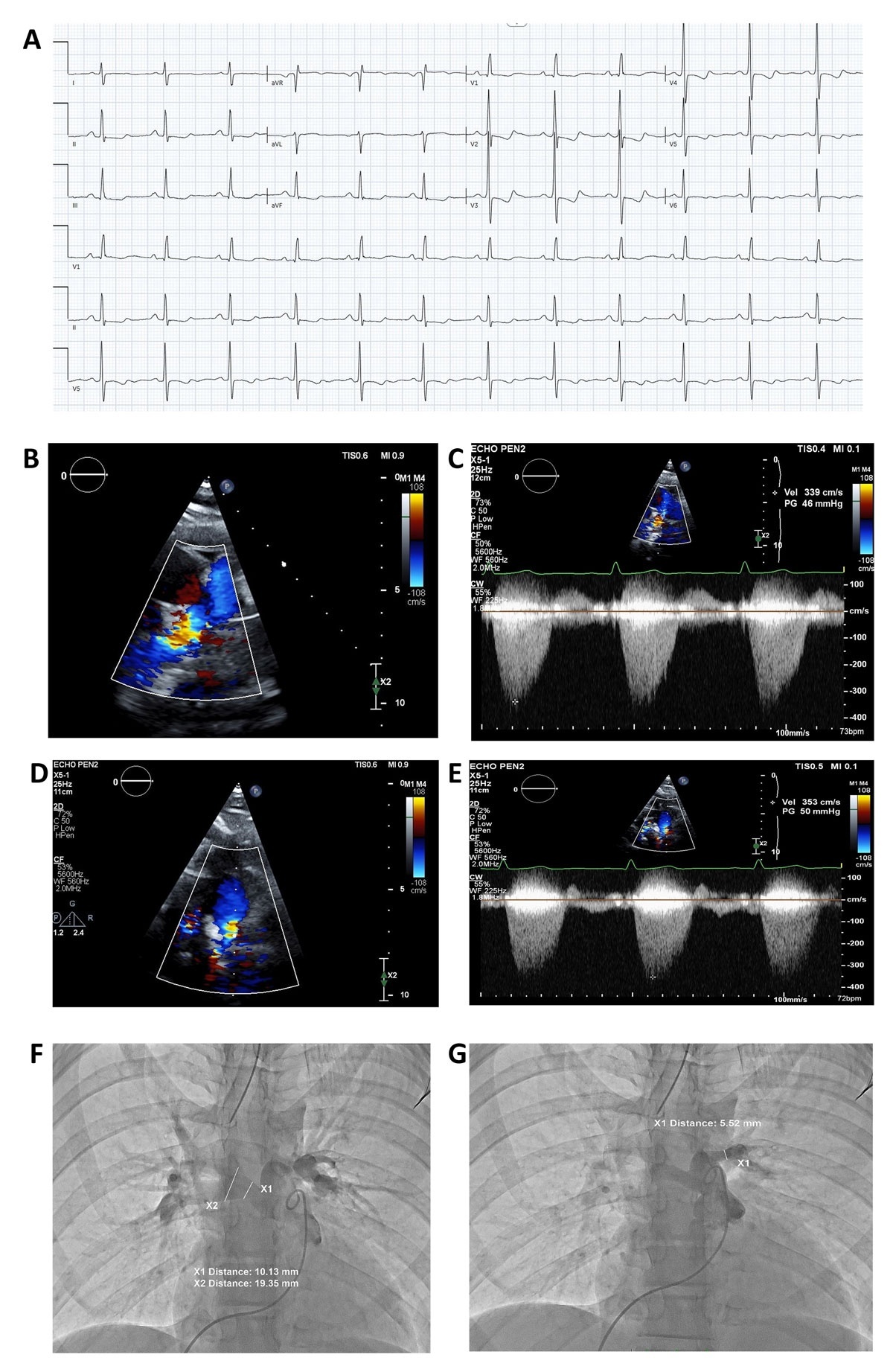
Figure 2
(A) Electrocardiogram demonstrates normal sinus rhythm with incomplete right bundle branch block and right ventricular hypertrophy. (B) Echocardiographic frame shows flow acceleration with color Doppler of the long axis of the right pulmonary artery. (C) Echocardiographic frame shows continuous wave Doppler of the long axis of the right pulmonary artery with peak velocity of 3.39 meters per second and peak gradient of 46 mm Hg. (D) Echocardiographic frame shows flow acceleration with color Doppler of the long axis of the left pulmonary artery. (E) Echocardiographic frame shows continuous wave Doppler of the long axis of the left pulmonary artery with peak velocity of 3.53 meters per second and peak gradient of 50 mm Hg. (F) Angiographic frame (anterior-posterior projection) shows injection of pigtail catheter into the main pulmonary artery with measurement of the proximal right pulmonary artery of 10.13 mm and distal right pulmonary artery of 19.35 mm. (G) Angiographic frame (anterior-posterior projection) shows injection of pigtail catheter into the main pulmonary artery with measurement of the proximal left pulmonary artery of 5.52 mm.
Congenital branch pulmonary stenosis (CBPS) is found in 2% to 3% of congenital heart disease patients and is associated with four types: stenosis within the MPA, stenosis at the bifurcation of the BPAs, multiple stenoses within the BPAs, and stenoses of the MPA and BPAs.16 BPS can be seen in syndromes such as Williams-Beuren, Alagille, Keutel, cutis laxa, congenital rubella, Marfan, scimitar, Turner, and Ehlers Danlos but also can be associated with vasculitis or compression due to adjacent structures, such as mediastinal tumors.16,17,18 Presentation varies relative to the degree of stenosis and physiologic reserve of the RV to adapt to increased workload. Diagnostic imaging, including echocardiogram, cardiac MRI, and cardiac CT, is crucial in differentiating the etiology of CBPS from other diseases, including chronic thromboembolic pulmonary hypertension. Interventions for patients with CBPS include catheter-based balloon angioplasty and PA stenting as well as surgical augmentation of artery stenosis.19 Serial interventions may be needed for those with recurrent stenosis.
Management of congenital heart disease during pregnancy can be challenging and requires knowledge of the physiologic changes of pregnancy as well as careful consideration of the cardiovascular adaptations relative to the congenital lesion. Maternal cardiovascular changes in pregnancy include systemic vasodilation with a concomitant decrease in systemic vascular resistance (SVR), decrease in pulmonary vascular resistance (PVR), augmentation in stroke volume and heart rate related to increased cardiac output, and rise in blood volume.20,21 Due to its strong negative predictive value, serial measurements of natriuretic peptides (brain natriuretic peptide [BNP] and N-terminal proBNP) can be useful in distinguishing the symptoms of normal pregnancy from those of cardiac pathology because rising or elevated levels may foretell worsening cardiac function.22,23 Medications, such as diuretics, antiarrhythmics, and antihypertensives, may be used safely to alleviate symptoms and lower maternal and fetal risk.24 Delivery should be planned in consultation with a cardio-obstetric team consisting of a cardiologist (preferably specializing in ACHD), maternal fetal medicine specialist, perinatologist, obstetric anesthesiologist, and specialized nurses.23
In this case, the patient had developed symptoms relative to her degree of stenosis and current gestational age. Comprehensive echocardiographic evaluation is crucial to assess proximal BPA dimensions, peak Doppler gradients, RVSP, and RV systolic function.19 If her anatomy could not be well assessed via echocardiogram, CMR would be an excellent alternative to improve estimation of chamber size and function as well as pulmonary vascular anatomy.25 BNP or pro-BNP measurement would be prudent, and diuretics could be used to alleviate symptoms or volume overload. If she were to decompensate hemodynamically, transcatheter balloon angioplasty could be considered with appropriate planning for limiting maternal and fetal fluoroscopic exposure. Surgical intervention should be avoided unless critical for maternal health because cardiopulmonary bypass increases maternal and fetal risk.24
Coarctation of the Aorta
A 40-year-old cisgender woman presented to the ED with acute onset chest pain that began earlier that morning and progressed over the day. She described the pain as sharp, radiating to her back and left arm, and associated with a mild headache. She denied shortness of breath, nausea, and diaphoresis. She reported a history of hypertension since age 20, managed intermittently with lifestyle modifications, and a childhood heart murmur, but she was lost to follow-up after pediatric cardiology evaluation. On arrival, her blood pressure was 180/100 mm Hg in the right arm, 176/96 mm Hg in the left arm, 140/80 mm Hg in the left lower extremity, and 145/90 mm Hg in her right lower extremity. Her heart rate was 90 beats per minute with otherwise normal vital signs. Physical exam revealed bounding carotid and brachial pulses with radio-femoral delay, palpable thrill over the left upper back, II/VI systolic murmur heard best at the left interscapular area radiating to the suprasternal notch, normal S1 and S2 without clicks or gallops, no jugular venous distension, clear lungs, soft nontender abdomen, and symmetric and diminished lower extremity pulses without bruits or edema. ECG showed normal sinus rhythm with left ventricular hypertrophy (LVH) and strain pattern. CT angiography confirmed severe coarctation of the aorta distal to the left subclavian artery, post-stenotic dilatation, and prominent collaterals. A transthoracic echocardiogram showed normal LV size and systolic function with concentric LVH, normal aortic valve leaflets with trace regurgitation, and a peak gradient of 46 mm Hg by continuous wave Doppler in the proximal descending thoracic aorta (Figure 3). She received beta-blockers and pain control and was referred for evaluation and intervention.
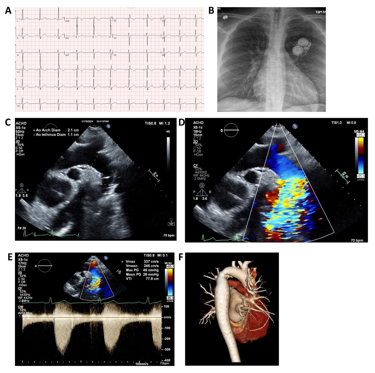
Figure 3
(A) Electrocardiogram demonstrating normal sinus rhythm with left atrial enlargement and left ventricular hypertrophy. (B) Chest x-ray (anterior-posterior projection) with “figure 3” sign showing dilated left subclavian artery and aortic arch (upper curve), site of coarctation of the aorta (middle curve), and post-stenotic dilation of the descending thoracic aorta (lower curve). (C) Echocardiogram frame of the long axis of the aortic arch (diameter 2.1 cm) and area of coarctation (diameter 1.1 cm). (D) Echocardiogram frame of the long axis of the aortic arch showing flow acceleration in the distal arch/area of coarctation by color Doppler. (E) Echocardiogram frame showing continuous wave Doppler flow in the area of coarctation with peak velocity 3.37 meters per second and peak gradient 46 mm Hg. (F) Three-dimensional reconstruction of cardiac magnetic resonance angiography (posterolateral projection) demonstrating area of coarctation.
Coarctation of the aorta (CoA) is a discrete narrowing typically at the aortic isthmus near the ductus arteriosus insertion site, occurring in 1 in 2,500 live births and accounting for 5% to 8% of congenital heart defects.26 It can occur as an isolated defect or be associated with other lesions such as bicuspid aortic valve, ventricular septal defects, mitral valve abnormalities, intracranial aneurysms, or syndromes such as Turner, Noonan, or Williams-Beuren.10 Adults with unrepaired CoA can present with hypertension, headache, chest pain, claudication. or heart failure. These nonspecific presentations warrant exclusion of other common differentials, including acute coronary syndrome and aortic dissection prior to focusing on congenital lesion.
The pathophysiology of CoA involves upper body hypertension proximal to the lesion, lower body hypoperfusion distal to the lesion, and progressive collateral vessel development often via the internal mammary and/or intercostal arteries. In patients with extensive collaterals, a Doppler or catheter gradient may underestimate anatomic severity, so the absence of a very high gradient does not exclude clinically significant obstruction when the anatomy and collateral burden are severe. Classic exam findings include upper-lower extremity blood pressure discrepancy (> 20 mm Hg), delayed/weak femoral pulses (radio-femoral delay), brachial-femoral murmur, or thrill posteriorly.
In this patient, chest pain workup should include serial troponins, ECG to evaluate for ischemia, and imaging to exclude acute aortic syndrome and coronary artery disease because patients with CoA are also at increased risk for premature coronary artery disease. ECG may show signs of left atrial or LV pressure overload, LVH with strain, or ischemia patterns. Chest x-ray may show rib notching from collaterals and “figure 3 sign” beneath the aortic knob. On echocardiogram, the aortic isthmus may not be well visualized in adults; however, color Doppler imaging can identify turbulent flow pattern in the descending aorta while spectral Doppler can identify increased flow rates distal to the coarctation and diastolic runoff pattern. A mean gradient greater than 20 mm Hg in the descending aorta suggests significant CoA. Cross-sectional imaging with cardiac MRI or CT allows for visualization of the entire aorta, determination of the severity of CoA (by calculating the ratio of the aortic isthmus diameter to descending aorta at the level of the diaphragm), aortic arch anatomy, and burden of collaterals. Three-dimensional reconstruction of CT scans can be helpful for interventional planning and post-procedural follow-up.27 In this case, a CTA is especially useful as it can define coarctation anatomy, evaluate the entire thoracic aorta, assess for dissection, and describe atherosclerotic disease in the coronary arteries.
Intervention is indicated for patients with hemodynamically significant CoA, classified by upper to lower extremity systolic BP gradient greater than 20 mm Hg, mean gradient greater than 20 mm Hg across the lesion by echocardiogram or cardiac catheterization, or aortic isthmus to aorta ratio < 0.5 to 0.7 with collateral arteries on cross-sectional imaging.10 Intervention can also be considered at lower gradients if there is LV systolic dysfunction, significant collaterals, or associated regurgitation.10
CoA repair should be performed by cardiovascular surgeons or interventional cardiologists with expertise in ACHD. In adults, catheter-based stenting is generally preferred when anatomy is suitable while surgery is reserved for complex arch anatomy, associated aneurysm, or other situations where endovascular therapy is less favorable.28 Long-term complications after intervention can include re-coarctation, aneurysms proximal or distal to the repair, fracture of the stent, and persistent hypertension.29,30,31
The life expectancy of a patient with repaired CoA is reduced compared to the general population despite excellent early and intermediate survival. Long-term cohort studies show survival rates of approximately 93% at 10 years, 86% at 20 years, and 74% at 30 years post-repair.32 Complications post repair can include re-coarctation, aortic dissection, aneurysm formation at the repair site, and premature coronary artery disease.29,33
In this case, the patient’s chest pain stems from acute hypertension exacerbating LV pressure overload from unrepaired CoA. The patient’s headache is clinically important because it may reflect severe proximal hypertension, but in this setting, it also raises concern for cerebrovascular complications, including intracranial aneurysm. Screening for intracranial aneurysm is therefore relevant in selected patients with coarctation, particularly those with neurologic symptoms or additional risk factors. In this case, the combination of marked upper-extremity hypertension, diminished lower-extremity pulses, collateral vessels, and confirmatory imaging supported the diagnosis of severe native coarctation of the aorta with secondary LV hypertrophy and symptomatic systemic hypertension. Catheter-based stenting is indicated for her age, anatomy, and symptoms. Long term, she will require lifelong surveillance with cross-sectional imaging and management for hypertension to goal systolic BP < 130 mm Hg.
Subaortic Stenosis
A 57-year-old cisgender female presented to the cardiology clinic with progressive shortness of breath on exertion over the prior 6 months, now limiting climbing two flights of stairs. She denies exertional chest pain, palpitations, syncope, orthopnea, or leg edema. She has mild hypertension controlled with lisinopril. Vital signs show blood pressure 140/85 mm Hg, heart rate 75 beats per minute, and normal oxygen saturation. Exam revealed normal jugular venous pressure without hepatojugular reflux, clear lungs, III/VI crescendo-decrescendo systolic murmur at the right upper sternal border radiating to carotids with late-peaking quality, normal S1 and paradoxically split S2, sustained apical impulse with bifid quality, and no peripheral edema. Distal pulses were equal and brisk. ECG demonstrated normal sinus rhythm with LVH, left atrial enlargement, and T-wave inversions in the lateral leads. Echocardiogram was notable for concentric LVH with normal systolic function, a discrete subaortic fibrous membrane one cm below the aortic annulus associated with peak gradient 64 mm Hg across the LV outflow tract (LVOT). The aortic valve was trileaflet with mild aortic regurgitation and normal aortic root, sinotubular junction, and ascending aorta size (Figure 4). She was referred for further evaluation.
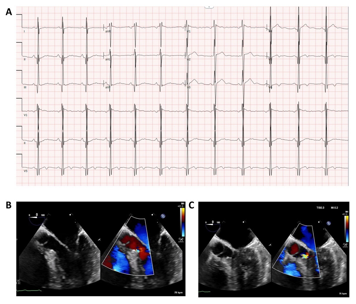
Figure 4
(A) Electrocardiogram demonstrating normal sinus rhythm, left axis deviation, and left ventricular hypertrophy with strain pattern. (B) Transesophageal echocardiogram frame from the mid-esophageal view showing flow acceleration by color Doppler within the left ventricular outflow tract in end-systole. (C) Transesophageal echocardiogram frame from the mid-esophageal view showing flow acceleration by color Doppler within the left ventricular outflow tract in early systole.
Subaortic stenosis (SAS) is a rare defect characterized by a discrete fibrous membrane or ridge beneath the aortic valve, leading to a fixed LVOT obstruction due to discrete membrane narrowing the flow within the outflow tract (70-90% cases) or a fibromuscular tunnel-like diffuse narrowing within the outflow tract (10-30%).34 The high velocity and turbulent jet can then damage the aortic leaflets, leading to aortic regurgitation and valvular dysfunction in 50% to 80% of patients. While commonly diagnosed in childhood with a male predominance, there is an increasing prevalence of adults with SAS and slow progression of LVOT obstruction, either as an isolated lesion or in association with other congenital lesions such as patent ductus arteriosus (34%), bicuspid aortic valve (23%), ventricular septal defect, and arch abnormalities such as interrupted aortic arch or coarctation of aorta (23%).35
SAS can be found incidentally on imaging or when patients present with symptoms of exertional dyspnea, angina, or syncope from LV pressure overload. The physical exam often reveals a harsh systolic ejection murmur at the right upper sternal border, soft A2, or a S4 gallop from LVH. A diastolic rumble of an AR murmur may be present. ECG can have an LVH pattern in moderate to severe cases. Echocardiography is diagnostic and often shows a membrane location under the aortic valve, associated with flow acceleration and LVOT outflow peak gradient of greater than 50 mm Hg. It can help identify the degree of AR if present, LV size, and LV hypertrophy. If there is suspicion for LVOT obstruction and subaortic stenosis without well-visualized membrane, a transesophageal echocardiogram can often clarify the anatomy and site of obstruction (Figure 3).36
Surgical intervention is indicated for patients with symptoms attributable to subaortic stenosis, asymptomatic patients with LV dysfunction or hypertrophy to mitigate further LV worsening, and those with at least mild AR to prevent progression. Surgical intervention typically involves membrane resection with or without myectomy or aortic valve intervention as indicated.10 The Canadian guidelines also add that peak instantaneous gradient > 50 mm Hg is indication for intervention.37 There is a 15% to 25% risk of recurrent LVOT obstruction, especially in females and in patients with increased peak LVOT gradients ≥ 50 mm Hg, as well as a 4% to 5% risk of complete heart block postoperatively.38,39
In this case, her progressive dyspnea reflected reduced cardiac reserve from her fixed LVOT obstruction due to unrepaired SAS. Surgical resection was indicated given her symptoms, peak gradients greater than 50 mm Hg, and mild AR to halt progression and preserve LV function. If she were asymptomatic and this was an incidental diagnosis, an exercise stress echocardiogram may have been helpful to identify symptoms attributable to the congenital lesion, as patients often compensate due to slow membrane growth, concomitant morbidities, lifetime experience of living with the lesion, and sedentary lifestyle.
Conclusion
Common congenital heart disease lesions such as pulmonic stenosis, branch pulmonary artery stenosis, coarctation of the aorta, and subaortic stenosis are usually diagnosed in childhood but can present as an unrepaired lesion in adulthood with common symptoms such as chest pain, shortness of breath, palpitations, and murmur during pregnancy. It is imperative for the general cardiologist to keep these lesions as a differential, particularly in pregnant patients or those with unexplained symptoms, and to promptly refer to an ACHD specialist for long-term surveillance and management.
Key Points
Unrepaired obstructive congenital lesions still present in adulthood with common cardiology complaints (chest pain, dyspnea, palpitations, and murmurs), so general cardiologists must keep congenital heart disease on the differential and trigger timely specialist referral.
Pulmonic stenosis in adults is driven by right ventricle pressure overload and remodeling, which can precipitate arrhythmias and right-sided failure physiology; echocardiogram is the cornerstone for diagnosis and grading (velocity/gradient thresholds), and balloon valvuloplasty is the first line for appropriate valve morphology with surgery/transcatheter options for mixed disease or complex anatomy.
Branch pulmonary artery stenosis requires multimodality imaging to confirm anatomy and exclude mimics; management commonly involves catheter-based angioplasty/stenting, and pregnancy care hinges on cardio-obstetric planning, symptom surveillance, and fetal/maternal risk mitigation.
Coarctation of the aorta is a high-yield adult diagnosis in patients with early-onset or resistant hypertension, classic pulse/blood pressure gradients, and collateral signs; computed tomography/magnetic resonance imaging define severity and anatomy, and intervention is often transcatheter stenting followed by lifelong surveillance for re-coarctation/aneurysm and aggressive blood pressure control.
Subaortic stenosis causes fixed left ventricular outflow tract obstruction and can progressively injure the aortic valve, leading to aortic regurgitation; echo establishes diagnosis and gradients, and surgical membrane resection is indicated with symptoms, significant gradients/left ventricular response, or aortic regurgitation balanced against recurrence and conduction disease risk counseling.