
Table 1
Distribution of causative microorganisms in infective endocarditis among patients with congenital heart disease across population studies.
| STUDY (ref) | STREPTOCOCCI (%) | STAPHYLOCOCCUS AUREUS (%) | OTHER STAPHYLOCOCCI (CONS) (%) | ENTEROCOCCI (%) | OTHER PATHOGENS (%) |
|---|---|---|---|---|---|
| Niwa et al.15 | 43.3 | 29.8 | — | 5.5 | 10.2 |
| Snygg-Martin et al.8 | 19.3 | — | 22.4 | 7.8 | 9.8 |
| Verheugt et al.14 | 46.0 | — | 28.0 | — | 16.9 |
| Havers-Borgersen et al.7 | 16.4 (viridans) | 17.5 | — | 4.4 | 5.8† |
| Van Melle et al.40 | 29.6 | 22.7 | 8.7 | 7.0 | 10.1 |
[i] †Includes Coxiella burnetii, Streptococcus bovis, and other rare pathogens
Table 2
Rare microbiological agents causing infective endocarditis. spp: species
| Brucella spp | C. burnetiid | Bartonella spp | T. whipplei |
| Mycoplasma spp | Legionella spp | Fungi | Mycobacteria |
Table 3
2023 European Society of Cardiology revised criteria for diagnosis of infective endocarditis (IE).
| MAJOR CRITERIA | ||
|---|---|---|
| Blood culture positive for IE | ||
| Typical IE-related organisms isolated from two independent blood cultures: Oral streptococci, Streptococcus gallolyticus (formerly S. bovis), HACEK group, S. aureus, E. faecalis | Persistent positivity of blood cultures for microorganism compatible with IE, defined as:
| A single positive blood culture for C. burnetii or a phase I IgG antibody titer greater than 1:800 |
| Imaging positive for IE | ||
| Structural or metabolic abnormalities suggestive of IE identified on cardiac valves, perivalvular or periprosthetic regions, or intracardiac foreign material. These findings may be detected using: echocardiography (either transthoracic or transesophageal), cardiac computed tomography, 18F-fluorodeoxyglucose positron emission tomography, white blood cell single photon emission tomography/computed tomography | ||
| MINOR CRITERIA | ||
| Risk factors (ie, predisposing heart condition at high or intermediate risk of IE or people who inject drugs) | ||
| Fever (body temperature above 38°C) | ||
| Embolic vascular dissemination (including clinically apparent or incidentally detected lesions) | ||
| Systemic or pulmonary emboli or infarcts, abscesses | Hematogenous osteoarticular septic complications | Mycotic aneurysms |
| Intracranial ischemic or hemorrhagic lesions | Conjunctival hemorrhages | Janeway’s lesions |
| Immunological phenomena | ||
| Glomerulonephritis | Osler nodes, Roth spots | Rheumatoid factor |
| Microbiological evidence | ||
| Positive blood culture that do not meet the definition of a major criterion | Serological evidence of active infection caused by a microorganism compatible with IE | |
| Infective endocarditis classification | ||
| Definite infective endocarditis | ||
| 2 major criteria | 1 major criterion and at least 3 minor criteria | 5 minor criteria |
| Possible | ||
| 1 major criterion and 1 or 2 minor criteria | 3-4 minor criteria | |
| Rejected | ||
| Does not meet criteria for definite or possible at admission with or without a firm alternative diagnosis | ||
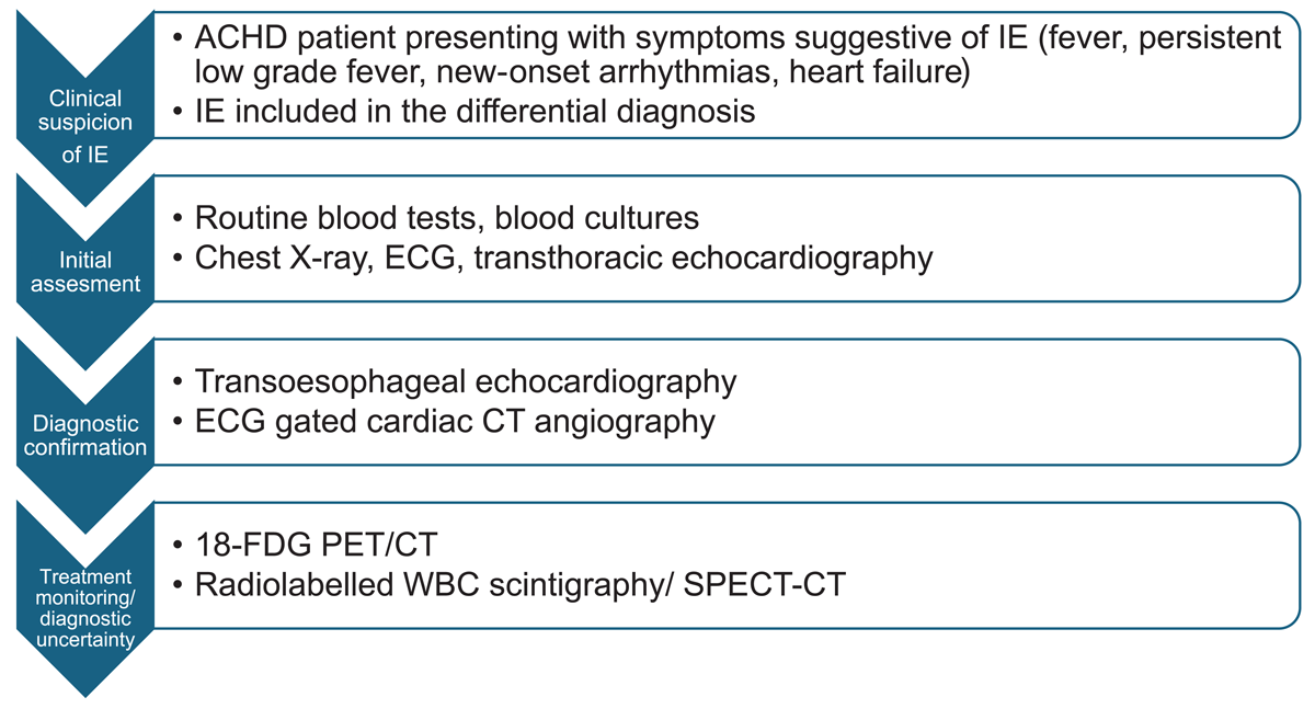
Figure 1
Diagnostic pathway for suspected infective endocarditis (IE) in adults with congenital heart disease (ACHD). ECG: electrocardiogram; CT: computed tomography; 18-FDG PET: 18F-fluorodeoxyglucose positron emission tomography; WBC: white blood cell; SPECT: single-photon emission computed tomography
Table 4
Surgical indication for infective endocarditis based on the 2023 European Society of Cardiology guidelines. NVE: native valve endocarditis; PVE: prosthetic valve endocarditis; HF: heart failure; AVB: atrioventricular block; HACEK: Haemophilus, Aggregatibacter, Cardiobacterium, Eikenella, and Kingella
| Emergency surgery is indicated in patients with aortic or mitral NVE or PVE when acute severe regurgitation, valve obstruction, or fistula formation leads to refractory pulmonary edema or cardiogenic shock (I – B) | Urgent surgery is indicated in aortic or mitral NVE or PVE when severe acute regurgitation or obstruction cause HF symptoms or echocardiographic evidence of poor hemodynamic tolerance (I – B) |
| Urgent surgery is indicated when there is evidence of uncontrolled local infection (abscess, false aneurysm, fistula, enlarging vegetation, prosthetic dehiscence, new AVB) (I – B) | In IE caused by fungal or multidrug-resistant pathogens, surgery is recommended urgently or electively depending on the hemodynamic status of the patient (I – C) |
| Urgent surgery should be considered in patients with IE and persistent positive blood cultures for more than one week or ongoing sepsis despite appropriate antimicrobial therapy and adequate management of metastatic infectious foci (IIa – B) | Urgent surgery should be considered in PVE caused by S. aureus or non-HACEK gram-negative bacteria (IIa – C) |
| Urgent surgery is indicated in aortic or mitral NVE or PVE when vegetations remain ≥ 10 mm after one or more embolic events despite appropriate antimicrobial treatment (I – B) | Urgent surgery is recommended in IE with vegetation ≥ 10 mm when another indication for surgery is present (I – C) |
| In low-risk surgical candidates with aortic or mitral IE, urgent surgery may be considered for vegetations ≥ 10 mm even in the absence of severe valve dysfunction or clinical embolic events (IIb – B) |
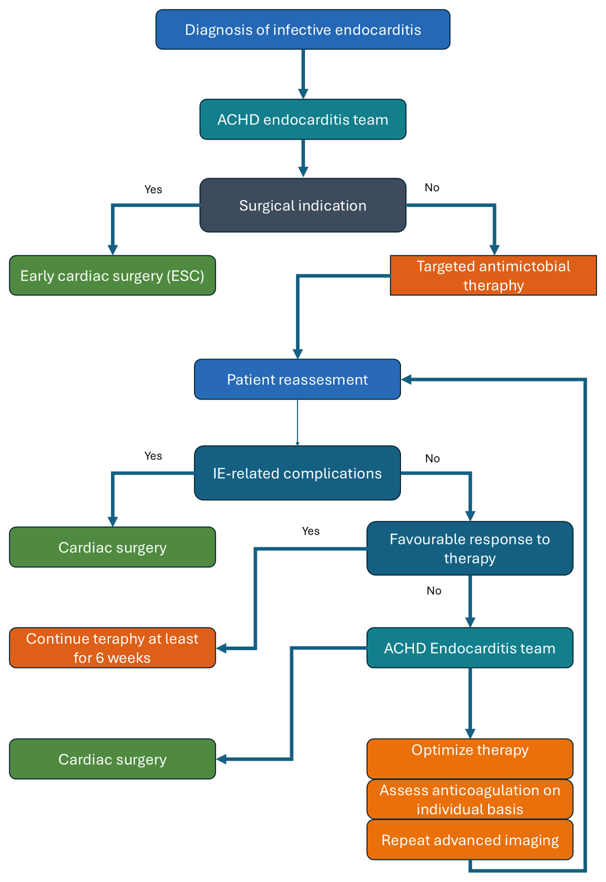
Figure 2
Proposed management pathway for infective endocarditis (IE) in adults with congenital heart disease (ACHD).