
Introduction
Infective endocarditis (IE) is an infection of the heart’s endocardial surface that involves native or prosthetic cardiac valves, patches or conduits, the mural endocardium, septal defects, or an indwelling intracardiac device.1 Current international guidelines describe IE as a major public health challenge because of its increasing incidence, an expanding at-risk population, and rising antimicrobial resistance.2
Congenital heart disease (CHD) has an estimated prevalence of approximately 9 per 1,000 live births. Advances in diagnostic techniques, surgical repair, and long-term medical management have led to a dramatic improvement in survival, with more than 90% of these patients surviving into adulthood. As a consequence, the global population of adults with congenital heart disease (ACHD) is steadily increasing.3
Over the past two decades, the incidence of IE has increased substantially. In the general population, the estimated incidence in 2019 was 13.8 cases per 100,000 person-years compared with 3 to 10 cases per 100,000 person-years reported in the 2009 European Society of Cardiology (ESC) guidelines on IE.4 Men are more frequently affected than women, although mortality rates are higher among female patients. The incidence of IE increases with advancing age, reflecting the growing burden of comorbidities and prosthetic devices in the older population.1,2
Studies focusing on ACHD populations have reported a 50- to 100-fold increased risk of developing IE compared with the general population.5,6,7 This risk is higher in adulthood than in childhood,6 and pediatric population studies suggest that IE occurs predominantly in patients with complex lesions, particularly those with cyanotic CHD, which represents the highest risk substrate for developing IE.8
Data from international registries, including the EURO-ENDO and ICE-PCS registries, identify Staphylococcus aureus as the most frequent causative microorganism of IE in the general population, followed by oral streptococci and coagulase-negative staphylococci.2
In patients with CHD, the distribution of causative microorganism shows modest but clinically relevant differences across studies, as summarized in Table 1. Although the streptococcal species and staphylococcus aureus remain the most commonly identified pathogens, some variability is observed across studies. Overall, streptococci and staphylococci consistently account for the majority of IE cases in patients with CHD, whereas enterococci and other microorganisms represent a smaller but clinically relevant proportion. These epidemiological patterns should be carefully considered when initiating empirical antimicrobial therapy.
Table 1
Distribution of causative microorganisms in infective endocarditis among patients with congenital heart disease across population studies.
| STUDY (ref) | STREPTOCOCCI (%) | STAPHYLOCOCCUS AUREUS (%) | OTHER STAPHYLOCOCCI (CONS) (%) | ENTEROCOCCI (%) | OTHER PATHOGENS (%) |
|---|---|---|---|---|---|
| Niwa et al.15 | 43.3 | 29.8 | — | 5.5 | 10.2 |
| Snygg-Martin et al.8 | 19.3 | — | 22.4 | 7.8 | 9.8 |
| Verheugt et al.14 | 46.0 | — | 28.0 | — | 16.9 |
| Havers-Borgersen et al.7 | 16.4 (viridans) | 17.5 | — | 4.4 | 5.8† |
| Van Melle et al.40 | 29.6 | 22.7 | 8.7 | 7.0 | 10.1 |
[i] †Includes Coxiella burnetii, Streptococcus bovis, and other rare pathogens
The reported rate of blood culture-negative IE ranges from 20% to 50% of cases, depending on the study. Rare causes of IE are summarized in Table 2. Although uncommon, these etiologies should be considered in selected clinical scenarios, such as patients with high clinical suspicion and relevant risk factors—including immunocompromised status, recent dental procedures, or repeated antibiotic exposure—but negative blood cultures.
Infective Endocarditis Pathophysiology
The initial event in the development of IE is the adhesion of bacteria or fungi to the endocardial surface. Under physiological conditions, the intact endothelial layer is resistant to microbial adhesion. However, the presence of prosthetic material, intracardiac devices, turbulent flow—as commonly observed in CHD—or repeated episodes of bacteremia, such as those occurring in people who inject drugs, may facilitate pathogen adhesion. This process triggers a localized inflammatory response and subsequent vegetation formation.1 Endocardial damage is a prerequisite for microbial adhesion. Disruption of the endothelial layer exposes subendothelial matrix proteins and promotes platelet and fibrin deposition, leading to the formation of a nonbacterial thrombus that can subsequently be colonized by circulating microorganism.
Prosthetic material is particularly prone to fibrinogen and platelet adhesion, thereby increasing susceptibility to microbial colonization. This favorable environment is further amplified by the host response to foreign material and the propensity for biofilm formation. These mechanisms help explain why medical therapy alone is often insufficient to achieve complete eradication of infection in patients with prosthetic material.9
Earlier clinical observations identified lesions characterized by high-velocity blood flow from a high-pressure chamber into a low-pressure chamber as particularly susceptible to IE. In these settings, the sudden drop in lateral pressure after the jet enters the low-pressure chamber results in localized endothelial malperfusion, creating a vulnerable substrate for bacterial colonization.10
Additional risk factors for IE in patients with CHD include the high number of surgical and transcatheter procedures and an increased likelihood of colonization with multidrug-resistant organisms.11 Patients with CHD also exhibit a higher prevalence of genetic conditions associated with intrinsic immunological defects, such as DiGeorge syndrome.12 Another potential contributor to immune disfunction is thymectomy, which is frequently performed during congenital heart surgery in neonates and infants to facilitate surgical exposure through median sternotomy. The long-term immunological consequences of early thymectomy remain incompletely understood. A recent systematic review by Cavalcanti and colleagues including 23 predominantly small cohort studies demonstrated persistent immunological alteration but did not identify a clear increase in autoimmune disorders or consistently elevated risk of infectious complications.13
Diagnosis of Infective Endocarditis
The clinical presentation of IE is highly variable and may be acute, subacute, or chronic. In some cases, the diagnosis is established only during the investigation of complications related to the primary infectious process, such as septic embolization leading to organ ischemia. Given the elevated risk in patients with CHD, a high index of suspicion must always be maintained. Fever, often low-grade and prolonged, may be the only presenting symptom. Positive blood cultures are frequently observed, and a history of recent high-risk procedures may precede symptoms onset.
The diagnosis of IE has historically relied on the modified Duke criteria. However, these criteria have important limitations, particularly in patients with prosthetic materials, intracardiac devices, or complex congenital anatomy.
To address these limitations, the 2023 ESC guidelines introduced revised diagnostic criteria that incorporate advanced imaging modalities and emphasize the perioperative context. According to these criteria, diagnosis of IE can be classified as definite, possible, or rejected, based on a combination of major and minor criteria (Table 3).
Table 3
2023 European Society of Cardiology revised criteria for diagnosis of infective endocarditis (IE).
| MAJOR CRITERIA | ||
|---|---|---|
| Blood culture positive for IE | ||
| Typical IE-related organisms isolated from two independent blood cultures: Oral streptococci, Streptococcus gallolyticus (formerly S. bovis), HACEK group, S. aureus, E. faecalis | Persistent positivity of blood cultures for microorganism compatible with IE, defined as:
| A single positive blood culture for C. burnetii or a phase I IgG antibody titer greater than 1:800 |
| Imaging positive for IE | ||
| Structural or metabolic abnormalities suggestive of IE identified on cardiac valves, perivalvular or periprosthetic regions, or intracardiac foreign material. These findings may be detected using: echocardiography (either transthoracic or transesophageal), cardiac computed tomography, 18F-fluorodeoxyglucose positron emission tomography, white blood cell single photon emission tomography/computed tomography | ||
| MINOR CRITERIA | ||
| Risk factors (ie, predisposing heart condition at high or intermediate risk of IE or people who inject drugs) | ||
| Fever (body temperature above 38°C) | ||
| Embolic vascular dissemination (including clinically apparent or incidentally detected lesions) | ||
| Systemic or pulmonary emboli or infarcts, abscesses | Hematogenous osteoarticular septic complications | Mycotic aneurysms |
| Intracranial ischemic or hemorrhagic lesions | Conjunctival hemorrhages | Janeway’s lesions |
| Immunological phenomena | ||
| Glomerulonephritis | Osler nodes, Roth spots | Rheumatoid factor |
| Microbiological evidence | ||
| Positive blood culture that do not meet the definition of a major criterion | Serological evidence of active infection caused by a microorganism compatible with IE | |
| Infective endocarditis classification | ||
| Definite infective endocarditis | ||
| 2 major criteria | 1 major criterion and at least 3 minor criteria | 5 minor criteria |
| Possible | ||
| 1 major criterion and 1 or 2 minor criteria | 3-4 minor criteria | |
| Rejected | ||
| Does not meet criteria for definite or possible at admission with or without a firm alternative diagnosis | ||
Risk stratification plays an important role in the long-term management of patients with CHD, allowing estimation of the lifetime risk of developing IE. A risk score derived from the national CONCOR (Congenital Corvitia) registry has been developed to predict the probability of IE over time.14 This model incorporates a range of clinical and anamnestic variables, including gender, the presence, number and type of congenital heart defects, and complications occurring in childhood. Based on these parameters, the score estimates the cumulative risk of developing IE up to 40 and 60 years of age.
Establishing a diagnosis of IE in patients with ACHD may be particularly challenging since they often have suboptimal acoustic windows, resulting in low-quality echocardiographic imaging. This is a frequently observed problem and may also be related to thoracic deformity, multiple previous sternotomy at a young age, syndromic features, repeated surgery, and postoperative fibrous adhesion. Even when adequate acoustic windows are available, important diagnostic challenges remain.
Many patients have undergone surgery 20, 30, or even 40 years earlier, where surgical reports are often unavailable and previous imaging data may be missing—making it difficult to compare with prior morphology. In some cases, a fold in a patch, a residual chordae tendineae, or a surgical knot may be difficult to distinguish from an endocarditic vegetation.
Patients with ACHD frequently have calcified prosthetic material or a mechanical valve, which may generate acoustic shadowing and further complicate echocardiographic assessment. IE involving a prosthetic pulmonary valve may be particularly difficult to assess because of its anterior position and prosthetic valve artefacts, especially in the setting of stented valves or calcified conduits. Notably, patients with ACHD may develop vegetation in anatomical regions that are difficult to visualize, such as right ventricle-pulmonary artery extracardiac conduits, pulmonary artery branches, or stents implanted after procedure such as the LeCompte maneuver.
For these reasons, virtually all patients with ACHD and suspected IE require second-level imaging. In this setting, the diagnostic pathway often includes electrocardiogram-gated cardiac computed tomography (CT) angiography. Cardiac CT allows assessment of cardiac and great vessel morphology, particularly in patients with complex or poorly documented anatomy. It can also help identify septic embolization, extracardiac infectious foci, local complication, and intracardiac thrombus.
The role of cardiac magnetic resonance imaging (CMR) is less well established. Although tissue characterization may theoretically be useful, endocarditic vegetations are often highly mobile, making reliable tissue characterization difficult.
In case of persistent diagnostic uncertainty, fludeoxyglucose F18 positron emission tomography/CT or radiolabeled white blood cell scintigraphy may be considered. However, the use of nuclear imaging in ACHD-related IE has several limitations. A recent surgery or transcatheter procedure may lead to a false-positive finding, while calcified patches and prosthetic material may reduce diagnostic sensitivity. Overall, a reasonable diagnostic pathway is represented in Figure 1.
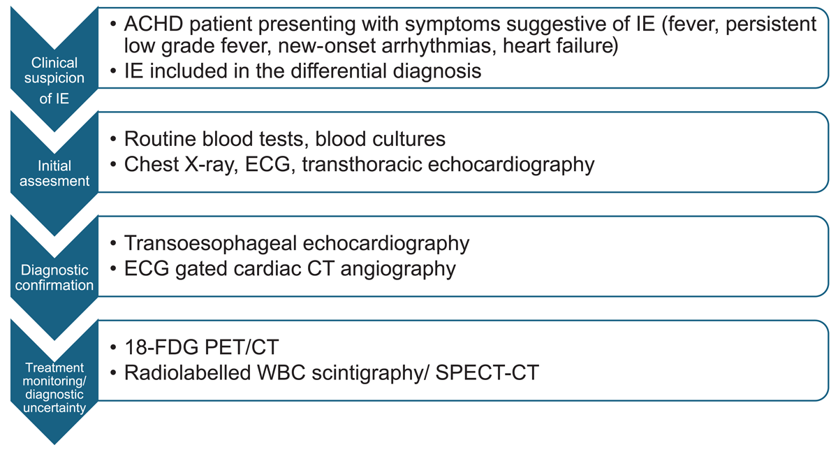
Figure 1
Diagnostic pathway for suspected infective endocarditis (IE) in adults with congenital heart disease (ACHD). ECG: electrocardiogram; CT: computed tomography; 18-FDG PET: 18F-fluorodeoxyglucose positron emission tomography; WBC: white blood cell; SPECT: single-photon emission computed tomography
Infective Endocarditis in Different Clinical Contexts
In the following sections, IE is discussed across different clinical settings. It should be acknowledged that heterogeneity in CHD classification across studies limits the generalization of the data. Some classification systems categorize congenital lesions according to anatomical location, such as left-sided versus right-sided lesions, whereas others are based on surgical status or the type of prior intervention.
Here we adopt a clinically oriented approach and discuss IE in patients with CHD according to disease status: unrepaired CHD, repaired CHD, and palliated CHD. A brief discussion of prosthetic valve endocarditis and cardiac implantable electronical devices is also provided.
Infective Endocarditis in Unrepaired CHD
Patients with unrepaired CHD represent a high-risk group for the development of IE, particularly those with unrepaired cyanotic lesions.7 The reported incidence of IE in patients with unrepaired CHD varies according to the underlying lesion. Estimated cases per 1,000 patient-years include 2.4 of ventricular septal defect (VSD), 2 of aortic valve stenosis, and 1.7 of atrioventricular septal defect.9
A large Japanese cohort study including both adult and pediatric patients with CHD identified a relatively simple lesion, such as isolated VSD, combined VSD and atrial septal defect (ASD), and patent ductus arteriosus (PDA), as the lesions most frequently associated with IE in unrepaired CHD. Valvular lesions, particularly involving the mitral valve more often than aortic valve, were also commonly represented.15 Notably, these defects are among the most prevalent congenital heart lesions, with an estimated incidence per 1,000 live births of 2.62 for VSD, 1.64 for ASD, and 0.87 for PDA.16
With regard to VSD, a Swedish national study reported a high incidence of IE in adults, accounting for 2% of the cohort, including patients with small and hemodynamically restrictive shunts.17
These findings raise an important clinical dilemma regarding whether all shunt lesions, including small restrictive VSD or small PDA, should be electively closed to reduce the risk of endocarditis. For cases in which the procedure is technically straightforward, shunt closure may be considered. However, current American Heart Association guidelines for ACHD do not recommend closure of restrictive VSDs solely for the purpose of IE prophylaxis and suggest that interventions be reserved for secondary prevention.18
A special subgroup within unrepaired CHD is represented by patients with bicuspid aortic valve. This relatively common condition is associated with an increased risk of IE and a more aggressive disease course, including a higher incidence of perivalvular complications such as abscess formations.19 Despite these observations, current ESC guidelines classify bicuspid aortic valve as an intermediate risk lesion.
In clinical practice, the most commonly encountered patients with unrepaired CHD and IE are often those with relatively simple restrictive shunt lesion, such as VSD, ASD, or PDA.
In some cases, IE may represent the first clinical event leading to the diagnosis of CHD. This scenario may occur when imaging performed during the diagnostic work-up of IE incidentally reveals an underlying CHD.
This presentation is less common in Western settings and is particularly uncommon in patients with complex CHD, who typically present earlier in life with other manifestations, such as heart failure, cyanosis, arrhythmias, or neurological events. Nevertheless, in rare cases, IE may be the first manifestation leading to diagnosis of complex CHD.
In patients with unrepaired CHD complicated by IE, repair of the defects should be considered after resolution of the infectious process. However, specific data on the risk of IE after defect closure in this setting are lacking.
Infective Endocarditis in Repaired CHD
Correction of congenital heart defects is generally expected to reduce the risk of IE. In the setting of transcatheter closure procedures such as PDA or ASD closure, the long-term risk of IE appears to be very low once complete device endothelialization has occurred, typically after approximately 6 months.
The risk profile differs substantially in patients whose CHD repair involves prosthetic material, including patches, conduits, or prosthetic valves. Foreign material provides a substrate for bacterial adhesion and vegetation formation. The quality and completeness of surgical or transcatheter repair are therefore of paramount importance, as complete repair may reduce the risk of subsequent future endocarditis. The cumulative risk of IE is further influenced by the number of prior surgical or transcatheter interventions and by the presence of other indwelling devices, such as pacemaker leads.
Historically, homografts have been considered less susceptible to infection compared with synthetic conduits or other prosthetic materials. However, a recent meta-analysis evaluating aortic homograft implantation for aortic valve endocarditis in adults without CHD found no significant differences in long-term major clinical outcomes.20 In contrast, studies focusing on CHD populations have reported a lower incidence of endocarditis when pulmonary or aortic homografts are used in the pulmonary position rather than other prosthetic conduit.19,21 The clinical relevance of those mixed findings remains debated. In many tertiary centers, homografts are preferred when available because of their favorable durability and hemodynamic performance, both of which may contribute to a lower risk of endocarditis. Conversely, dysfunction of a homograft valve may itself represent a risk factor for IE. In addition, the absence of synthetic material may theoretically reduce susceptibility to bacterial adhesion.
Interpretation of IE rates in repaired CHD is subject to several important limitations. Residual defects or shunts represent major risk factors for IE, yet their presence and hemodynamic significance are often difficult to quantify accurately in population-based studies. In addition, many studies do not adequately account for the anatomical complexity of CHD or for the cumulative burden of multiple previous surgical and transcatheter procedures over a patient’s lifetime.
Available data regarding IE in repaired CHD are therefore heterogeneous. One study identified corrected tetralogy of Fallot, followed by VSD, as the most common lesion associated with IE.15 Similar findings were reported in a Swedish nationwide ACHD study that observed the highest incidence of IE in surgically corrected conotruncal defects, including common arterial trunk, transposition of the great arteries, tetralogy of Fallot, and aortopulmonary septal defects, and the lowest incidence in patients with repaired ASD.8
Data from a Canadian ACHD population-based database identified left-sided lesions, including aortic coarctation, aortic stenosis or insufficiency, and mitral valve disease, as the lesions most frequently involved in IE, with an incidence of 1.61 case per 1,000 person-years. In contrast, right-sided CHD—including Ebstein anomaly, pulmonary valve or artery anomalies, and congenital tricuspid valve disease—as well as atrial septal defects and patent ductus arteriosus were associated with substantially lower incidence rates of 0.35, 0.28, and 0.24 cases per 1.000 person-years, respectively.
A Danish population study reported the highest incidence of IE in patients with repaired tetralogy of Fallot and complex CHD, including truncus arteriosus, transposition of the great arteries, and univentricular heart, with incidence rates of 23.6 and 18.1 cases per 1,000 person-years, respectively.7
It should be noted that surgical techniques for several defects, such as the Da Silva procedure for Ebstein anomaly, have evolved over time; therefore, historical data should be interpreted with caution. The implantation of non-valved devices, such as closure device, has been associated with an increased risk of IE during the first 6 months after implantation.22 This observation is consistent with the time required for complete device endothelialization. Beyond this early period, the risk appears to decrease and to approach that of the general population.
Overall, available data do not support the definition of a single “typical” ACHD patient profile for IE. All patients with ACHD carry an increased risk of endocarditis, but the risk is higher in those with more complex CHD, multiple prior interventions, residual defects, or prosthetic material. Simple lesions, such as repaired ASD or PDA, are associated with lower risk, whereas complex lesions and conotruncal defects appear to carry a substantially higher risk. The surgical era of correction should also be considered, although it has rarely been specifically addressed in available studies. Modern surgical techniques and materials may reduce the risk of residual defects and may involve materials less prone to bacterial contamination or colonization.
Finally, the growing number of aging ACHD patients represents an emerging challenge. As patients with CHD survive into older age, comorbidities such as chronic kidney disease, diabetes, obesity, and malignancy may further increase susceptibility to IE.
Infective Endocarditis in Palliated CHD
Palliation of CHD encompasses a wide spectrum of physiological conditions, ranging from definite palliation, such as the Fontan circulation, to temporary palliative procedures, including modified Blalock-Taussig shunts in neonates.
Data on the incidence of IE in palliated ACHD are limited, and available registry studies often lack a specific focus on this subgroup. Paradoxically, several studies have reported a very low incidence of IE in adult patients with Fontan circulation.23 These finding have led some authors to question the routine indication for antibiotic prophylaxis in this population.24
However, although IE in patients with Fontan circulation appears to be rare, cases have been reported in multiple studies. Havers-Borgersen et al. reported 11 cases of IE among patients with univentricular heart.7 Niwa et al. identified four cases of IE in patients with Fontan circulation,15 Verheugt et al. reported four additional cases in a large population-based study.14 Although these data suggest that IE is an uncommon complication of Fontan circulation, robust evidence remains lacking. Therefore, caution is warranted before reconsidering prophylaxis strategies in this population.
The apparent rarity of IE in patients with Fontan circulation is intriguing, and no definitive explanation has been established. It may be speculated that the direct flow of systemic venous blood to the pulmonary circulation, an immunological reservoir, bypassing the heart, could contribute to the low incidence of IE. The mechanism might be particularly relevant to establish because this patient may have several risk factors for IE, including cyanosis, prosthetic material, low flow circulation, vascular malformation.
Prosthetic Valve Endocarditis
The 2023 ESC guidelines confirm the traditional distinction between early and late prosthetic valve endocarditis (PVE) while emphasizing that “the time to IE onset is prognostically less important than the connection of IE to the perioperative period or to specific pathogens.”
Early PVE is typically related to perioperative contamination and often involves sutures, patches or prosthetic rings. In the context of valve surgery, infection may extend to the valvular annulus, leading to severe local complications such as abscess formation or prosthetic dehiscence. Late PVE shares a pathogenetic mechanism similar to native valve endocarditis, with bacteremia representing the main initiating event. Vegetations typically develop in areas of flow turbulence, such as paravalvular leaks or commissural regions. Infective endocarditis involving a prosthetic valve more frequently presents with valve regurgitation than stenosis.
The presence of a prosthetic heart valve in a CHD patient represents a significant risk factor for the development of IE.25 In a recent retrospective study by the Mayo Clinic, the incidence of PVE in adults with CHD was 5.2 cases per 1,000 person-years, with a 10-year cumulative incidence of 5.2%. The incidence was similar between biological and mechanical prostheses, whereas left-side prosthetic valves were associated with a higher risk. This study also confirmed an increased risk of IE associated with the Melody™ (Medtronic) valve, as discussed below. In addition, the incidence of PVE was similar between surgical implanted bioprostheses and transcatheter Sapien (Edwards Lifesciences) valves.26
Transcatheter Pulmonary Valve Replacement
The reported incidence of IE following transcatheter pulmonary valve replacement (TPVR) ranges from 1.6% to 4%.2 Real-world data, however, suggest higher cumulative incidence rates, reaching 9.5% at 5 years and 16.9% at 8 years. These findings are largely derived from cohorts with a high proportion of procedures performed using valves derived from bovine jugular veins, which have been associated with increased susceptibility to IE.27,28,29
Multiple studies and meta-analyses have consistently identified the Melody valve as being associated with a higher risk of IE compared with other TPVR platforms.30 The underlying mechanisms are not fully understood but may relate to valve material, flow dynamics, and host-prosthesis interactions. Follow-up data on relatively newer valve systems suggest potentially lower rates of IE, although experience remains limited.
A follow-up study of the Venus P-valve (BVM Medical), a self-expanding transcatheter pulmonary valve, reported four IE cases (7.3%) at 1 year and one additional case during extended follow-up, resulting in a cumulative incidence of 9% at 5 years.31 Another study of 81 patients reported a 3-year IE incidence of 1.2%, with the only IE case occurring during the first year after implantation.32 Short-term follow-up data for the Harmony (Medtronic) valve showed an IE incidence of 2.6% at 1 year.33
Early results from studies evaluating the Alterra Adaptive Prestent system (Edwards Lifesciences) have not reported cases of IE; however, longer follow-up and larger patient cohorts are required to determine whether this system modifies the long-term risk of infection.34 Nevertheless, a case of IE has been reported in a patient who underwent pulmonary valve implantation using this system and developed endocarditis 2 months later. The infection was localized on the valve, and the role of the present system in the infectious process remains unclear. The key message of this case report focused on the technical difficulty of surgical explantation, particularly because of aortic compression. Therefore, long-term data are needed, and caution remains warranted.35
An important determinant of prosthetic valve durability and IE risk following TPVR is the presence of a residual transvalvular gradient after implantation. Higher residual gradients are associated with prosthetic dysfunction and may contribute to increased susceptibility to infection.36
Cardiac Implantable Electronic Devices
In patients with CHD and particularly adults with CHD, implantation of a cardiac implantable electronic device (CIED) is frequently required at some point during the disease course. CIED-related infections represent one of the most serious complications of device therapy and are associated with significant mortality, morbidity, and financial healthcare burden.37
CIED infection may present as localized infections, such as superficial or isolated pocket infection, or as systemic infections, with or without concomitant pocket and lead involvement. Local infections are typically related to perioperative contamination, and the most commonly involved pathogens are skin microorganism.
Several risk factors for CIED infection are particularly prevalent in the ACHD population. These include the use of epicardial leads, abdominal device pocket, multiple device generator replacements, and younger age at first implantation. The diagnostic approach to CIED infection has been standardized in a manner analogous to that for IE. According to the 2019 International CIED Infection Criteria, cases may be classified as definite, possible, or rejected.37
Current consensus documents and international guidelines place strong emphasis on infection prevention, highlighting the importance of careful assessment of patient-related, procedure-related, and device-lead related factors to minimize the risk of CIED infection.37,38 Device pocket hematoma represent a major contributor to the risk of CIED infection. This issue is particularly relevant in ACHD patients, who frequently require antiplatelet therapy or long-term anticoagulation.
The use of an antibacterial mesh envelope has been shown to reduce the risk of CIED infection in selected populations. However, patients with ACHD were not specifically addressed in the trials evaluating this strategy; therefore, its potential role in this population remains to be defined in future studies.39
Therapy
Medical Therapy
Medical management of IE is based on targeted antimicrobial therapy. The choice of antimicrobial agent should be guided by blood culture results, microbiological susceptibility profiles, prior antibiotic exposure, and individual patient risk factors. In patients with CHD, antimicrobial therapy is typically prolonged, most commonly for 4 to 6 weeks or longer depending on the causative organism, the presence of prosthetic material, and clinical response. Given the anatomical complexity of CHD and the frequent presence of intracardiac prosthetic material and additional patient risk factors, therapeutic decisions should be discussed within a dedicated endocarditis team with specific expertise in CHD.
The concept of an expert endocarditis team is strongly emphasized in the latest ESC guidelines. In the setting of ACHD, this team should include at least a clinical cardiologist, an imaging expert, either a cardiologist or radiologist, an infectious disease specialist, an interventional cardiologist, and a cardiac surgeon, all with extensive experience in managing CHD.
When patients are referred from another center for the treatment of confirmed or suspected IE, the referring center should be involved in the discussion whenever possible to better reconstruct the clinical history, previous interventions, and imaging and laboratory findings.
Medical therapy is usually the first-line approach in patients without heart failure and those with endocarditis without vegetations who are judged to be at high risk of embolization because of their size, morphology, or mobility. Empirical antimicrobial therapy is usually started while awaiting microbiological identification and susceptibility testing and should subsequently be tailored according to results and antimicrobial stewardship principles.
Whether anticoagulation should be started in patients without another indication remains controversial. In some centers, anticoagulation is considered in patients with IE with the aim of limiting further fibrin deposition on existing vegetations and reducing their potential growth. However, other centers avoid routine anticoagulation because of concerns regarding a possible increase in embolic or hemorrhagic complications. This issue is not specifically addressed by current guidelines and should therefore be evaluated on an individual basis. Relevant factors include the location of the vegetation, left- or right-sided, native or prosthetic valve, patch or conduit involvement, its morphology, its mobility, and the estimated embolic risk.
Discussion of the medical therapy should also consider the local microbiological epidemiology, the presence or absence of prosthetic material, recent potential sources of bacteremia such as dental procedures, possible intestinal translocation in patients with chronic low output states, and colonization by multidrug-resistant microorganisms.
Surgical Therapy
Surgical management of IE patients with CHD poses substantial challenges. These patients have often undergone multiple prior surgical and transcatheter interventions, resulting in a higher cumulative procedural risk. In addition, many ACHD patients underwent surgery 20 to 40 years earlier, and surgical reports and postoperative diagnostic imaging may be difficult to retrieve, making preoperative anatomical reconstruction more challenging.
The presence of extensive adhesions, prosthetic materials, calcifications, epicardial pacemaker leads and prosthetic valves significantly increases technical complexity. In addition, patients with CHD frequently present with relevant comorbidities, including chronic kidney disease and hepatic dysfunction, further increasing perioperative risk. As a result, each case requires meticulous preoperative planning and individualized decision-making. Surgical indications generally follow the principles outlined in the ESC guidelines (Table 4), with the primary objectives being management of heart failure, control of the infectious source, and prevention of embolic events. These criteria are well suited to hearts with normal anatomy, but their application in the context of CHD may be challenging. While indications for surgery in left-side endocarditis are relatively well established, recommendations for right-sided IE and extracardiac prosthetic material are less clearly defined.
Table 4
Surgical indication for infective endocarditis based on the 2023 European Society of Cardiology guidelines. NVE: native valve endocarditis; PVE: prosthetic valve endocarditis; HF: heart failure; AVB: atrioventricular block; HACEK: Haemophilus, Aggregatibacter, Cardiobacterium, Eikenella, and Kingella
| Emergency surgery is indicated in patients with aortic or mitral NVE or PVE when acute severe regurgitation, valve obstruction, or fistula formation leads to refractory pulmonary edema or cardiogenic shock (I – B) | Urgent surgery is indicated in aortic or mitral NVE or PVE when severe acute regurgitation or obstruction cause HF symptoms or echocardiographic evidence of poor hemodynamic tolerance (I – B) |
| Urgent surgery is indicated when there is evidence of uncontrolled local infection (abscess, false aneurysm, fistula, enlarging vegetation, prosthetic dehiscence, new AVB) (I – B) | In IE caused by fungal or multidrug-resistant pathogens, surgery is recommended urgently or electively depending on the hemodynamic status of the patient (I – C) |
| Urgent surgery should be considered in patients with IE and persistent positive blood cultures for more than one week or ongoing sepsis despite appropriate antimicrobial therapy and adequate management of metastatic infectious foci (IIa – B) | Urgent surgery should be considered in PVE caused by S. aureus or non-HACEK gram-negative bacteria (IIa – C) |
| Urgent surgery is indicated in aortic or mitral NVE or PVE when vegetations remain ≥ 10 mm after one or more embolic events despite appropriate antimicrobial treatment (I – B) | Urgent surgery is recommended in IE with vegetation ≥ 10 mm when another indication for surgery is present (I – C) |
| In low-risk surgical candidates with aortic or mitral IE, urgent surgery may be considered for vegetations ≥ 10 mm even in the absence of severe valve dysfunction or clinical embolic events (IIb – B) |
A common site of IE in CHD is the right ventricle to pulmonary artery conduit, either homograft or prosthetic. Vegetation localized on a suture, patch, or conduit may not fit easily within standard ESC criteria. Therefore, the decision to proceed with surgery cannot be based solely on general guideline criteria and should be individualized.
When left-side IE occurs with CHD, standard ESC surgical criteria are generally applicable.
Conversely, IE localized on a conduit, a tricuspid prosthesis, or patch represent a more complex clinical scenario. All such cases should be discussed within a multidisciplinary ACHD endocarditis team.
In the presence of complications, including local extension, heart failure, or arrhythmias, the surgical indication is usually straightforward, even in right-sided IE. The most challenging cases are those involving right-sided IE without overt complications, with vegetation of intermediate size (5-10 mm) that are relatively mobile but not associated with documented embolization. In this “intermediate-high risk” scenario, advanced imaging should be performed to assess for subclinical embolization, which may influence the indication for surgery. Valve function, whether involving a homograft, conduit valve or prosthetic valve, should be assessed with serial echocardiography.
In the absence of complications, our institutional approach is to start targeted antibiotic therapy with appropriate antimicrobial stewardship. If clinical, laboratory, and imaging responses are favorable, antibiotic therapy should be continued for at least 6 weeks from the first negative blood cultures. In the event of complications, worsening or persistence of vegetation despite tailored antimicrobial therapy, or valve disfunction, surgical should be reconsidered. Nevertheless, each case has its own anatomical and clinical complexity, therefore a multidisciplinary team discussion remains of paramount importance.
The available literature indicates that the rate of surgical intervention for IE in patients with CHD is higher than that observed in patients without CHD.40 This difference is partly attributable to the type of prosthetic material commonly used in CHD repair, which is particularly susceptible to microbial contamination and may hinder complete eradication of the infectious source.9 In addition, patients with CHD appear to have a higher incidence of complications at the time of the initial diagnosis.41
Reported rates of surgical intervention vary widely across studies. A recent meta-analysis reported a pooled surgical intervention rate of 58.1%.25 In contrast, registry-based studies have documented substantially higher rates, ranging from 74.9% to 81.1%, whereas other cohorts have reported lower rates, such as 35.7%.40,41,42
These marked differences likely reflect heterogeneity in geographical practice patterns, patient selection, lesion complexity, referral bias, and institutional surgical thresholds. Notably, high rates of surgical intervention have also been observed in control populations without CHD in some studies, suggesting that broader differences in clinical practice may contribute to this variability. In addition, tertiary referral centers may show higher intervention rates because they often receive patients with complications or cases unresponsive to medical therapy.
In our experience, at least half of the ACHD patients presenting with IE undergo surgical intervention because of heart failure, relevant embolization risk such as homograft vegetation becoming thinner and more mobile during medical therapy, or failure to achieve resolution of vegetation despite prolonged antimicrobial therapy. A pragmatic management flow chart is proposed in Figure 2.
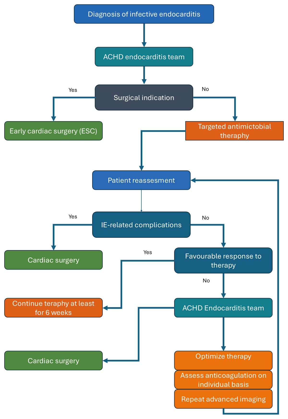
Figure 2
Proposed management pathway for infective endocarditis (IE) in adults with congenital heart disease (ACHD).
Outcomes
Infective endocarditis remains a major determinant of adverse outcomes in patients with CHD. The reported in-hospital mortality rate is estimated at 9.0%,40 with early post-discharge mortality estimated at around 5%. The most common cause of death is heart failure, accounting for up to 75% of fatal events, while cerebral complications occur in approximately 6% of cases.
Another contemporary cohort study reported an in-hospital mortality rate of 8.8% and a 1-year mortality rate of 2.4%. Hospitalization was frequently complicated by heart failure (40.3%), arrhythmias (26.1%), and embolic events (9.5%).41 Across multiple studies, 1-year mortality rates range from 5.7% to 9.9%.6,14,15,42
Recurrent IE is not uncommon, with multiple episodes reported in approximately 15% of patients in CONCOR registry and in 13% of patients in a Danish nationwide cohort.42
A first episode of IE represents a high-risk clinical event associated not only with in-hospital complications but also adverse outcomes after discharge. These patients should therefore undergo individualized and structured follow-up, similarly to what is recommended after hospitalization for heart failure.
Prevention
Patients with CHD are classified as a high-risk population for IE, making preventive strategies a cornerstone of management. All patients should adhere to general preventive measures, including meticulous skin and dental hygiene and appropriate wound care.
Antibiotic prophylaxis is recommended for high-risk dental procedures, particularly those involving manipulation of the gingiva, dental extractions, and oral surgery. Prophylaxis typically consist of a single dose of antibiotic administered 30 to 60 minutes before the procedure, targeting oral streptococci. For other potentially high-risk procedures, recommendations are less well defined, and clinical judgement is required.2
Notably, dental procedures performed in the month preceding the diagnosis of IE are frequently reported, underscoring the importance of proper dental care and patient education.40
The potential risk of endocarditis associated with tattooing and body piercing should be discussed with the patient. When patients choose to proceed with such practices, strict adherence to appropriate hygienic standards is essential. The role of antibiotic prophylaxis in this setting has not been systematically studied, and current evidence is limited to case reports and small series. Overall, the risk of IE following tattooing appears to be low but not negligible.43
Body piercings may theoretically carry a higher risk due to the relatively high incidence of local infection, which could facilitate bacteremia. However, the available literature remains limited to anecdotal reports.43
Conclusion
Infective endocarditis in ACHD is an increasingly relevant problem because the number of patients at risk continues to grow. Although the pathophysiological mechanisms of IE are broadly similar in ACHD and non-ACHD populations, important differences exist. Clinical presentation may be subtle, with prolonged and nonspecific symptoms in patients who may already have impaired baseline clinical status. Vegetations may be difficult to identify, particularly when involving calcified right ventricle-pulmonary artery conduits or extra-anatomic conduits that may occur after complex congenital heart disease repair or redo surgery. Blood cultures, although central to diagnosis, may be negative in a substantial proportion of patients. In addition, anatomy and surgical history may be difficult to reconstruct, particularly when patients have undergone surgery or transcatheter interventions at different centers. Overall, IE in ACHD is a complex and challenging disease that may be difficult to suspect, even more difficult to prove, and often difficult to treat.
As in patients without CHD, treatment of IE in ACHD is based on eradication of septic focus. This may be particularly challenging because of the high prevalence of prosthetic material, prone to biofilm formation. Surgery in this setting is particularly complex. First, patients have an active systemic infection, which increases the risk of organ dysfunction during extracorporeal circulation. Second, many patients have already undergone two or more sternotomies, resulting in higher baseline risk. Therefore, the anatomy should be extensively evaluated before surgery using appropriate imaging modalities to establish a clear surgical plan and minimize procedural risk and invasiveness.
Prevention remains the cornerstone of management. Patients and caregivers must be carefully counseled regarding the importance of personal hygiene, dental care, and adherence to prophylactic recommendations. A comprehensive, patient-centered approach integrating prevention, early diagnosis, and individualized therapy is essential to improve outcomes in this complex and high-risk population.
Key Points
The incidence of infective endocarditis is significantly higher in patients with congenital heart disease than in the general population.
A high index of suspicion must be maintained in this patient population.
In patients with congenital heart disease, infective endocarditis frequently requires surgical reintervention as conservative management may often be insufficient.
The management of these high-risk and clinically complex patients requires experienced multidisciplinary teams.
Prevention, including meticulous dental and skin hygiene and appropriate use of antibiotic prophylaxis, is of paramount importance.
Patient education and clear communication regarding preventive measures are essential.