
Clinical Epidemiology of RVOT Dysfunction in Adults with Congenital Heart Disease
Right ventricular outflow tract (RVOT) dysfunction represents one of the most prevalent and clinically consequential late sequelae among adults with congenital heart disease (ACHD), often occurring after repair of conotruncal anomalies, Ross intervention, or isolated pulmonary valve disease. Owing to major advances in pediatric cardiac surgery and perioperative care over the past decades, survival into adulthood now exceeds 90% for many complex congenital substrates. Consequently, the epidemiology of ACHD has shifted from primary defect-related mortality toward chronic postoperative morbidities, among which surgical RVOT and conduit dysfunction are particularly prominent.1,2,3,4,5
Repaired tetralogy of Fallot (rToF) constitutes the largest subgroup of patients affected by RVOT dysfunction. ToF is the most common cyanotic congenital heart defect, with prevalence estimates of 3 to 6 per 10,000 live births. The etiology of ToF is complex, involving both genetic and nongenetic factors. Of note, approximately 20% to 25% of individuals with ToF exhibit identifiable chromosomal abnormalities, with the most prevalent being trisomy 21 and DiGeorge syndrome (ie, 22q11 deletion syndrome).6 Thanks to advances in treatment for ToF, the outcome has shifted from a high morbidity and mortality congenital malformation in infancy to a remarkable survival rate > 94% at 25 years after repair, even if genetic abnormalities seem to be associated with a 3- to 4-times higher risk of mortality during both early and late follow-up periods.4,6 The historical use of transannular patches and valveless reconstructions results in chronic pulmonary regurgitation (PR) that often becomes hemodynamically significant during adolescence or early adulthood. The prevalence of hemodynamically significant RVOT dysfunction is as high as 50% to 70% in long-term follow-up of rToF, often decades after initial surgery.7,8 Risk factors contributing to progressive RVOT dysfunction include the type of initial surgical intervention (transannular patch or valve-sparing technique), the presence of residual stenosis following surgery, and the progressive degeneration of an RV to pulmonary artery (RV-PA) conduit. In patients treated with RVOT transannular patch, postoperative severe PR is nearly always present and usually well tolerated, although it invariably leads to a complex pathophysiological cascade determined by gradual RV dilation, fibrosis, contractile dysfunction, and subsequent arrhythmias.4 It should be noted that the modern approach to ToF surgical repair includes surgical techniques aiming to respect the integrity of the pulmonary valve as much as possible in selected cases using transatrial/transpulmonary repair with limited muscle resection and anatomy preservation.4 It is anticipated that, in the future, we will be able to progressively reduce the hemodynamic burden of longstanding RV volume loading in rToF. Current long-term outcome studies (enrolling historical cohorts), mainly focused on rToF, reveal that the progression of RV dysfunction develops gradually over several years, with the majority of patients remaining asymptomatic until they reach adolescence or adulthood.8,9 This topic is of pivotal importance since in many ACHD centers RVOT dysfunction is the leading indication for reoperation or catheter-based intervention, with 20% to 40% of rToF patients requiring pulmonary valve replacement (PVR) by early adulthood. In this scenario, early identification of clinical and imaging biomarkers through echocardiography, cardiac magnetic resonance (CMR), or exercise testing is crucial to prevent irreversible RV remodeling.10,11,12,13
Additional populations at risk include patients with ToF with pulmonary atresia, truncus arteriosus, pulmonary valve stenosis, Ross procedure recipients, and those with RV–PA conduits or bioprosthetic valves. Across these anatomies, progressive stenosis, regurgitation, or mixed lesions inevitably occur because of somatic growth, prosthetic degeneration, calcification, or infection.
The burden of RVOT dysfunction is lifelong. Many patients require multiple reinterventions across decades, creating a cumulative exposure to surgery, catheterization procedures, and prosthetic materials. This trajectory highlights that RVOT disease should not be conceptualized as a single procedural problem but rather as a chronic condition requiring anticipatory management within specialized ACHD programs.1,3,4
Pathophysiology
RVOT dysfunction may manifest as isolated PR, pulmonary stenosis (PS), or mixed disease. Each hemodynamic lesion exerts distinct but overlapping effects on RV structure and function.
Chronic PR produces persistent volume overload. The RV initially compensates through dilation and increased compliance, preserving stroke volume at the expense of progressive chamber enlargement. Over time, however, excessive dilation leads to impaired contractile efficiency, increased wall stress, and reduced systolic and diastolic performance. Ventricular interdependence further compromises left ventricular filling and systemic output.
Conversely, PS or conduit obstruction imposes pressure overload, promoting hypertrophy, increased myocardial oxygen demand, and reduced coronary reserve. Longstanding pressure loading may result in fibrosis and eventual systolic dysfunction. Mixed lesions combine adverse features of both states, accelerating maladaptive remodeling.13,14
Beyond global indices and pulmonary valvar function, regional abnormalities of the RVOT, such as aneurysmal dilation or akinetic segments, play a critical role in determining RV performance. These noncontractile regions contribute to ineffective systolic ejection, increased wall stress, and adverse ventricular remodeling, often leading to disproportionate RV dilation relative to PR severity. Importantly, RVOT dysfunction also exacerbates interventricular interaction; septal shift and altered ventricular geometry impair left ventricular (LV) filling and output, thereby amplifying the hemodynamic burden. CMR-derived metrics, including RV volumes, ejection fraction, regional wall motion (ie, RVOT aneurysm/akinesia), are therefore essential to identify patients at higher risk of progressive dysfunction and to refine the timing of PVR.15,16
Electrical remodeling parallels mechanical changes. Surgical scars, patch material, and anatomic isthmuses create zones of slow conduction that predispose to reentrant ventricular tachycardia (VT). Progressive QRS prolongation, dyssynchrony, and myocardial fibrosis are associated with increased risk of sustained ventricular arrhythmias (VA) and sudden cardiac death.17,18,19,20,21,22
Thus, RVOT dysfunction represents not merely a valvular disorder but a complex cardiomyopathic process integrating hemodynamic stress, myocardial remodeling, and electrophysiologic vulnerability.
Clinical Indication to Reintervention
Determining the optimal timing of reintervention remains one of the most challenging aspects of lifelong RVOT management. Symptoms alone are unreliable because many young adults adapt to declining functional capacity. Therefore, objective markers derived from imaging, exercise testing, and electrophysiology are emphasized in contemporary practice.4
CMR has become the reference standard for assessing RV volumes, ejection fraction, and pulmonary regurgitant fraction. Multiple cohort studies demonstrate that preoperative RV size predicts the degree of postoperative reverse remodeling, suggesting that delayed intervention after extreme dilation may lead to incomplete recovery.9,10,11,12,13,14,21 Accordingly, volumetric thresholds and declining function often guide decision-making even in minimally symptomatic patients. Accepted indications for RVOT revision and PVR show similarities and discrepancies among current guidelines (Table 1).
Table 1
Formal factors supporting RVOT intervention in repaired tetralogy of Fallot. RVOTO: right ventricular outflow tract obstruction; PVR: pulmonary valve replacement; RVEDVi: right ventricular end-diastolic volume index; RVESVi: right ventricular end-systolic volume index; RVEF: right ventricular ejection fraction; LVEF: left ventricular ejection fraction; LVEDVi: left ventricular end-diastolic volume index; RVSP: right ventricular systolic pressure; TR: tricuspid regurgitation
| FORMAL FACTORS SUPPORTING RVOT INTERVENTION IN RTOF | 2020 ESC ACHD GUIDELINES3 | 2025 ACC/AHA ACHD GUIDELINES1 |
|---|---|---|
| Symptoms | PVR is recommended in symptomatic patients with severe PR and/or moderate RVOTO (Class l-C) | PVR is recommended in symptomatic patients with at least moderate PR (Class 1) |
| Right ventricle | Asymptomatic patients should be considered for PVR if PR/RVOTO and (Class II-C):
| Asymptomatic patients should be considered for PVR if at least moderate PR and at least two among (Class 2a):
|
| Left ventricle | None | Asymptomatic patients should be considered for PVR if at least moderate PR and LVEF < 50% along with at least one additional factor (Class 2a) |
| Arrhythmias | None | Asymptomatic patients should be considered for PVR if at least moderate PR and ventricular arrhythmia (Class 2b) |
| Functional capacity | Asymptomatic patients should be considered for PVR if PR/RVOTO and decrease in objective exercise capacity (Class II-C) | Asymptomatic patients should be considered for PVR if at least moderate PR and decrease in objective exercise capacity with at least one additional factor (Class 2a) |
| Additional factors | Asymptomatic patients should be considered for PVR if PR/RVOTO and progressive TR to at least moderate (Class ll-C) | Asymptomatic patients should be considered for PVR if PR/RVOTO and progressive functional TR to at least moderate (Class 2b) |
| Type of intervention | In patients with no native outflow tract, catheter intervention should be preferred if anatomically feasible (Class l-C) | No preference between surgical or transcatheter approach is provided |
Functional testing provides complementary information. Reduced peak oxygen consumption, abnormal ventilatory efficiency, or exercise intolerance may indicate clinically meaningful impairment despite preserved resting metrics.23,24 In parallel, progressive QRS prolongation, frequent ventricular ectopy, or inducible VT may shift the balance toward earlier intervention.18,20,21,22
Large observational cohorts evaluating PVR have identified associations between late intervention, advanced RV remodeling, and adverse outcomes including death and sustained ventricular tachycardia. These findings support a proactive strategy that intervenes before irreversible structural or electrical damage occurs.18,21
Thus, modern indications for reintervention integrate imaging, physiology, and arrhythmic risk rather than relying solely on symptoms.
Surgical Reintervention
Surgical PVR remains a cornerstone therapy, particularly in patients with complex anatomy or when concomitant procedures are required. Indications include large native RVOTs unsuitable for transcatheter valves, severe branch pulmonary artery distortion requiring reconstruction, endocarditis requiring debridement, or the need for arrhythmia surgery or tricuspid valve repair.1,3,4 Available valved conduits connect the RV to PA and include a tube graft—such as Dacron, polytetrafluoroethylene (PTFE), homograft, bovine jugular vein (BJV)/Contegra—and a prosthetic or biological valve for the purpose of restoring forward pulmonary blood flow and prevent pulmonary regurgitation (Figure 1).
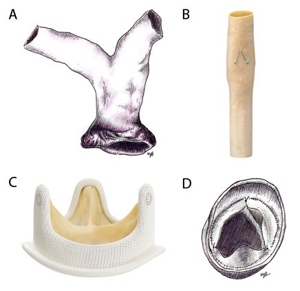
Figure 1
Surgical RV-PA conduits and prosthetic valves for PVR in ACHD: (A) Pulmonary homograft, (B) Contegra™ pulmonary valved conduit, (C) Hancock™ II bioprosthesis, (D) ePTFE valved conduit. Contegra pulmonary valved conduit and Hancock II bioprosthesis images reprinted with permission from Medtronic.
Bioprosthetic valves and homografts are most used because they avoid lifelong anticoagulation, provide favorable hemodynamics. and seem to show the best durability (mean lifespan of 10-20 years). Surgical outcomes are generally excellent in experienced centers, with low perioperative mortality (approximately 1-4%) and consistent reductions in RV volumes and symptomatic improvement.25,26,27,28,29,30,31,32,33,34,35
However, durability limitations are intrinsic. Structural valve degeneration (SVD), calcification, and conduit obstruction occur over time, particularly in younger patients. RV-PA conduits eventually fail due to tissue degeneration, calcification, fixed size mismatch, and mechanical distortion leading to PS and/or PR, RV pressure/volume overload and subsequently need repeat intervention. Furthermore, pulmonary homografts, which seem to show the best durability and freedom from reintervention (freedom from reintervention up to roughly 98.6% at 5 and 10 years), have obvious limited availability.28,30,34 Comparative and single-center outcome literature show device-specific failure phenotypes (eg, BJV conduit neointimal hyperplasia/stenosis and variable calcification profile; porcine-valved conduits/Hancock porcine-valved Dacron classic bioprosthetic SVD with progressive calcification and fibrosis), with differences in freedom from reintervention across conduit types and ages/sizes.25,29,32 Finally, infective endocarditis (IE) is a possible complication that usually accelerates conduit deterioration and need for reintervention. Conduit types showed significant differences in the cumulative incidence of IE, suggesting pulmonary homografts as the safest choice (< 1%) and BJV conduits to have much more propensity to this complication (~ 9-11%).27,31,33 Therefore, surgical intervention should be planned with the entire lifetime strategy in mind, especially considering that even if there is still heterogeneity in the choice of pulmonary valve prostheses, future valve-in-valve transcatheter procedures increasingly reduce the number of repeat sternotomies required across the lifespan.36
Transcatheter Therapeutics of RVOT Dysfunction
Transcatheter PVR (TPVR) has transformed the management paradigm for RVOT disease. Initially developed for dysfunctional conduits, TPVR offers reduced procedural morbidity, shorter hospital stays, and faster recovery compared with surgery.37,38 The contemporary transcatheter toolbox aims to restore RVOT competence and reduce gradients while minimizing cumulative morbidity from repeat sternotomies. Adults after the Ross procedure, truncus arteriosus repair, ToF with pulmonary atresia, and many complex RVOT reconstructions typically develop SVD and/or conduit obstruction, often amenable to catheter-based therapy because the landing zone is cylindrical and constrained. Native/patched RVOTs are frequently dilated and noncylindrical, historically limiting catheter valves. Dedicated devices and “landing-zone creation” strategies have expanded transcatheter candidacy.
Nowadays TPVR is becoming the cornerstone therapy for RVOT dysfunction when anatomy is suitable. Its goals are: eliminate/reduce PR, lower RVOT gradients, promote RV reverse remodeling, defer/avoid repeat surgery.
Two main types of transcatheter valves are available in current practice: (1) The balloon-expandable valves, which include Melody™ valve (Medtronic), made of a bovine jugular vein sewn into a Cheatham-platinum stent with three sizes available (18, 20, and 22 mm), and (2) the Sapien™ transcatheter heart valve (Edwards Lifesciences), made of bovine pericardium, chemically treated and sewn into a cobalt-chromium stainless steel stent (last version, Sapien S3, available in sizes 20, 23, 26, and 29 mm). Another balloon-expandable valve is Myval transcatheter heart valve (Meril Life Sciences Pvt Ltd.), made of bovine pericardial leaflets sewn into a nickel-cobalt stent frame (numerous sizes from 20 mm up to 32 mm). This valve is not currently available in the United States and approved only in India and Europe. The second valve type are self-expanding valves and pre-stent specifically designed to address the dilated, compliant RVOT typical of rToF. The Venus P-Valve™ (Venus MedTech) is a porcine pericardial valve within a nitinol frame that is covered with pericardial tissue except at the distal end, available in 18 mm to 36 mm diameter and 20 mm to 35 mm length. The Harmony™ valve (Medtronic) is a porcine pericardial valve within a nitinol frame, covered with polyester fabric and available in two sizes (TPV 22 and TPV 25) and length from 51 to 55 mm. The Alterra™ Adaptive Prestent (Edwards Lifesciences), designed to create a rigid landing zone in native/patched RVOT for the 29 mm Sapien S3, is a nitinol frame partially covered with PTFE (length of 49 mm and an inflow/outflow diameter of 40 mm). Other self-expanding valve platforms include the Med-Zenith PT-valve (Med-Zenith) and the Pulsta™ valve (TaeWoong Medical).39
Early feasibility studies established the safety and effectiveness of balloon-expandable valves in conduits and surgical bioprostheses.40,41,42,43 Subsequent multicenter trials and registries demonstrated durable hemodynamic improvement and high freedom from early reintervention. Patients treated with the Melody valve in the long-term outcome US Investigational Device Exemption Trial showed a cumulative survival of 90% at 10 years of follow-up, 79% freedom from reoperation, 60% freedom from any intervention, 53% freedom from valve dysfunction, and 81% freedom from IE.44 Furthermore, a large multicenter European and Canadian post-approval Melody Valve registry showed an incidence rate of 4.2% per person per year of a composite end point of death, reoperation, and reintervention and an incidence rate of 2.3% per person per year of IE after Melody valve implantation. The same registry found a strong correlation between final invasive RV-PA gradient and the risk of the composite end point and IE.45 On the other side, patients treated with the Sapien valve showed a survival rate of 98%, 93.7%, freedom from reintervention, and 97.1% freedom from IE at 3-year follow-up in the COMPASSION trial,46 while the last-generation Sapien 3 valve showed acute promising results with no mortality, IE, thrombosis, or stent fracture and a cumulative incidence of 4.3% of SVD at 1 year in the COMPASSION S3 Trial.47 Furthermore, two recent multicenter registries confirmed the excellent long-term outcome of both types of balloon-expandable valves.48,49
Technical considerations, typically for the balloon-expandable valves, include coronary artery compression testing (incidence of coronary compression ≈5%, especially in case of coronary artery anomalies and after Ross procedure), pre-stenting to reduce stent fracture risk, and careful imaging assessment of RVOT dimensions.50,51 As experience has grown, outcomes have continued to improve, making TPVR the preferred strategy for many patients with prior conduits or bioprosthetic valves.1,3
More recently, device innovation has expanded treatment to patients with large or native/patched RVOTs previously unsuitable for catheter therapy. Self-expanding systems and adaptive pre-stent platforms provide conformable solutions for complex geometries, broadening candidacy and further reducing surgical burden.52,53 For the Harmony valve, freedom from reintervention or valve dysfunction was 96% at 2 years, freedom from IE was 98% at 1 year, freedom from all-cause mortality was 98% at 1 year, and procedural serious adverse events occurred in 4% of cases (no device embolization or death), while 19% of patients required treatment for post-implant VA in a recent multicentric registry study.54 The 3-to-5-year outcome study confirmed those good results of sustained valve function, positive cardiac remodeling, and improved quality of life; of note, early cases of VT resolved, and there were no new arrhythmias.55 In Europe, a recent 3-year CE (Conformité Européenne) study showed that the Venus P-valve is a promising self-expandable platform with good clinical and hemodynamic results and less arrhythmic complication compared to other systems (no early procedure-related or late mortality, one case of IE, one case of valve thrombosis, two cases of VA).56 Finally, the Alterra Adaptive Prestent in combination with the SAPIEN 3 valve showed excellent outcomes at 2 years, with no significant valve dysfunction and no cases of IE, although approximately 34.4% of patients developed early VA.57
Despite these advances, TPVR is not without complications. IE remains a significant long-term concern, with nontrivial incidence across devices and studies (annualized incidence of 2.2 per 100 patient-years in the whole cohort of balloon expandable valves and up to 2.4 per 100 patient-years for the Melody valve). Thus, awareness, preventive measures, and prompt evaluation of febrile illness remain essential components of lifelong care.58,59,60 Finally, valve longevity also remains a concern. Several risk factors have been described to be correlated to progressive valve dysfunction: residual gradient (especially when invasive RV-PA is > 15 mm Hg, which increases the risk of IE), mechanical compression (especially without pre-stenting and conduit rehabilitation), IE, thrombosis, and leaflet dysfunction.39,61,62
Currently, numerous designs of transcatheter pulmonary valves are available for the treatment of RVOT dysfunction (Figure 2), and the choice should consider patient anatomy, comorbidities, somatic growth, and life expectancy.
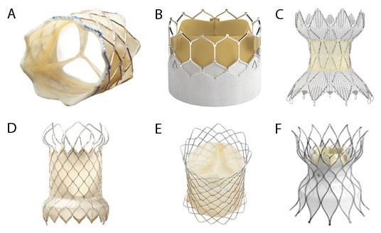
Figure 2
Transcatheter pulmonary valves for right ventricular outflow tract (RVOT) dysfunction in adult congenital heart disease: (A) Melody™ transcatheter pulmonary valve, (B) Sapien™ 3 Ultra-Resilia valve, (C) Harmony™ transcatheter pulmonary valve, (D) Venus P-Valve™ System, (E) Pulsta™ valve, (F) Alterra™ Adaptive Prestent. Reprinted with permission from Medtronic (Melody, Harmony valves), Edwards Lifesciences (Sapien 3 Ultra Resilia, Alterra Adaptive Prestent), Venus MedTech (VenusP-valve) and Taewoong Medical (Pulsta valve).
Overall, surgical and transcatheter therapies should be viewed as complementary tools within a staged, lifetime treatment algorithm rather than competing approaches.
Results and Complications After Reintervention
Both surgical and transcatheter interventions typically produce rapid hemodynamic improvement, reduction in RV volumes, and enhanced exercise capacity. Reverse remodeling is most pronounced when intervention occurs before advanced dilation or dysfunction, reinforcing the importance of timely referral.11,12,13,14
Nevertheless, reintervention does not eliminate all risks. VAs may persist because the underlying scar substrate remains. Consequently, longitudinal rhythm surveillance is required even after technically successful valve replacement. Selected patients may benefit from electrophysiologic evaluation, catheter ablation, or implantable cardioverter-defibrillator therapy.18,19,20,22 Furthermore, TPVR can cover critical areas of the RVOT and infundibular septum, hindering future electrophysiological studies.63 Concerns regarding post-implant VAs were raised with new generation percutaneous self-expandable systems, where the arrhythmic risk seems to be increased because of device mechanical myocardial irritation.54,57,64 Pre- and post-procedural expert electrophysiologic evaluation and anatomic isthmus ablation in these cases is an active field of research.65
Endocarditis is another major determinant of long-term morbidity. Both conduits and transcatheter valves are susceptible. Infection often necessitates repeat intervention and may adversely affect survival. Structured education, dental hygiene, and rapid treatment of bacteremia are therefore integral to management.27,33,60
Finally, the inevitability of prosthetic degeneration means that many patients will undergo multiple procedures over their lifetime. Planning must account for future access, preservation of vascular routes, and cumulative surgical risk. Multidisciplinary ACHD teams are uniquely positioned to coordinate this complex, longitudinal care.
Surgical and Transcatheter Management of RVOT Dysfunction: Competition Versus Integration
Comparative data regarding surgical versus transcatheter RVOT dysfunction therapeutics are limited because no randomized controlled trial has ever been (and likely will ever be) performed in this field.66,67 Clear superiority of one strategy over another is not reported, and background confounding by indication makes direct comparison analysis challenging. The general perception that TPVR can be performed safely with reasonable early/mid-term results is a strong argument in favor of this strategy.48 However, it must be emphasized that many ACHD patients with RVOT failure do present low surgical risk, and surgical PVR is safe and effective when performed in expert centers.35 In addition, the excellent longevity of surgical pulmonary homograft and the possibility that longitudinal surgical pulmonary homograft failure may be treated with TPVR are key players in this decision-making.
In the future, large multicenter studies should be specifically designed to address the important topic of differential indication to transcatheter versus surgical PVR, dissecting patient-level and anatomic-level risk factors for early failure of one modality over the other, with the long-term goal of reducing periprocedural risk while limiting the longitudinal need for multiple reinterventions.
It is time to consider a convenient integration of both modalities in the lifelong management of RVOT dysfunction in the ACHD population.
Conclusion
RVOT dysfunction in ACHD is a lifelong, progressive condition rather than an isolated postoperative issue. Chronic hemodynamic lesions drive structural remodeling and electrical vulnerability that together influence symptoms, functional capacity, and survival. Contemporary management emphasizes proactive surveillance using CMR, functional assessment, and arrhythmic risk stratification to guide timely intervention.
Surgical and transcatheter therapies each play essential roles and should be deployed within a lifetime strategy aimed at minimizing cumulative morbidity while preserving future options. Persistent risks, including arrhythmias, IE, and prosthetic degeneration, necessitate lifelong follow-up within specialized centers.
Adopting this comprehensive, anticipatory framework allows clinicians to shift from reactive treatment toward structured lifelong management, ultimately improving quality of life and long-term outcomes for ACHD patients with RVOT dysfunction.
Key Points
Right ventricular outflow tract dysfunction in adults with congenital heart disease is a progressive condition driven by chronic pulmonary regurgitation and/or stenosis that leads to right ventricular remodeling, dysfunction, and arrhythmic risk rather than representing an isolated valvular problem.
Timing of reintervention should be proactive and imaging-guided, with cardiac magnetic resonance–derived right ventricular volumes, functional capacity, and electrical markers preferred over symptoms alone to prevent irreversible myocardial damage.
Surgical and transcatheter pulmonary valve replacement are complementary strategies: surgery remains essential for complex anatomies and concomitant repairs, whereas transcatheter therapy is increasingly first-line for suitable conduits and bioprostheses, reducing cumulative procedural morbidity.
Valve replacement improves hemodynamics but does not eliminate late risks, as arrhythmias, infective endocarditis, and prosthetic degeneration persist and frequently necessitate repeat interventions.
Optimal outcomes require lifelong follow-up within specialized ACHD programs, with anticipatory planning that integrates multimodality imaging, electrophysiologic surveillance, and staged lifetime treatment strategies.