
Table 1
Clinical, echocardiographic, and multimodality imaging features of constrictive pericarditis, effusive-constrictive pericarditis, and transient constrictive pericarditis. HF: heart failure; JVP: jugular venous pressure; RA: right atrial; CRP/ESR: C-reactive protein/ erythrocyte sedimentation rate; TB: tuberculosis; IVC: inferior vena cava; CT: computed tomography; CMR: cardiac magnetic resonance
| FEATURE CATEGORY | CHRONIC CONSTRICTIVE PERICARDITIS (CP) | EFFUSIVE–CONSTRICTIVE PERICARDITIS (ECP) | TRANSIENT CONSTRICTIVE PERICARDITIS (TCP) |
|---|---|---|---|
| Clinical Presentation | Chronic fatigue, dyspnea, peripheral edema; signs of right-sided HF Elevated JVP with prominent x and y descents Kussmaul’s sign; symptoms due to fixed fibrotic/calcific disease | Presents with large effusion with or without tamponade Persistent HF symptoms after drainage RA pressure remains elevated Mixed effusive and constrictive physiology | Similar to CP initially but reversible Often idiopathic, viral, autoimmune, or post-surgical Improves with anti-inflammatory therapy |
| Biomarkers | CRP/ESR usually normal (non-inflammatory) | Elevated if inflammatory or infectious (eg, TB) | CRP/ESR frequently elevated and normalize with treatment |
| Echocardiography | Respirophasic septal shift Respiratory variation > 25% in mitral E wave velocity Late diastolic hepatic vein flow reversal with expiratory end-diastolic reversal ratio > 0.79 Medial e′ ≥ 9 cm/s Annulus reversus | Before drainage: effusion with or without tamponade features After drainage: mixed effusion/constriction signs; septal bounce; hepatic vein reversal; medial e′ > 8 cm/s; persistent IVC dilation | Constrictive pattern identical to CP initially E′ velocities, strain, and septal motion improve with therapy |
| CMR Findings | Pericardial thickening Minimal LGE/T2 edema Evaluates myocardial fibrosis/infiltration | Effusion + constrictive findings Inflammatory changes vary by etiology Visceral pericardial involvement may be subtle | Strong inflammatory signal (pericardial T2 edema, LGE) Predictors of reversibility include LGE thickness ≥ 3 mm and higher quantitative DHE Improves with therapy |
| CT Findings | Pericardial thickening and/or calcification common; helpful for surgical planning | Effusion ± pericardial thickening; calcification etiology dependent | Mild or normal pericardial thickening; calcification absent |
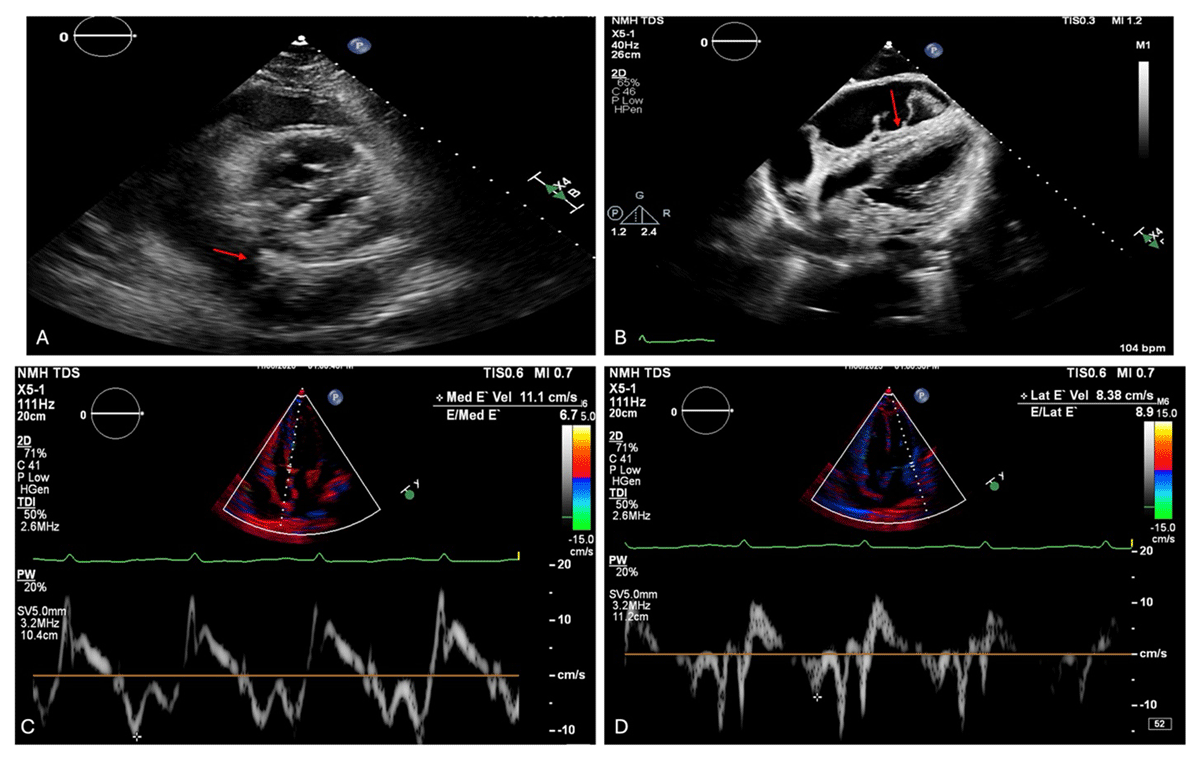
Figure 1
Echocardiographic features of effusive-constrictive pericarditis at presentation. Transthoracic echocardiography demonstrates concurrent features of pericardial effusion, tamponade physiology, and constrictive hemodynamics. (A) Parasternal short-axis view with a large circumferential pericardial effusion containing fibrinous strands (arrow). Video 1 also shows the respirophasic septal shift. (B) Diastolic right ventricular free wall collapse in the subcostal view, consistent with tamponade physiology (arrow; Video 2). Tissue Doppler imaging reveals annulus reversus, with (C) accentuated medial mitral annular e′ velocity (11.1 cm/s) exceeding the (D) lateral e′ velocity (8.38 cm/s). The respirophasic septal shift and annulus reversus are suggestive of concomitant constrictive pathophysiology in a patient who has pericardial tamponade.
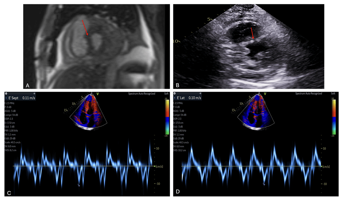
Figure 2
Persistent constrictive physiology following pericardial drainage. Cardiac magnetic resonance (CMR) imaging and transthoracic echocardiography obtained after therapeutic pericardiocentesis (600 mL drained) demonstrate persistent features of constrictive physiology. Panel A shows a CMR image with respirophasic interventricular septal shift (arrow; corresponding motion shown in Video 3). Panel B demonstrates residual pericardial effusion with persistent interventricular septal flattening on post-drainage echocardiography (arrow; corresponding motion shown in Video 4). Panels C and D show tissue Doppler imaging of the mitral annulus, with accentuated medial e′ velocity exceeding the lateral e′ velocity, consistent with persistent annulus reversus after relief of tamponade.
Video 1
Parasternal short-axis view demonstrating a large circumferential pericardial effusion with fibrinous strands and respirophasic interventricular septal shift, consistent with constrictive physiology; also see at https://vimeo.com/1167807972.
Video 2
Subcostal view demonstrating diastolic right ventricular free wall collapse, consistent with pericardial tamponade physiology; see also at https://vimeo.com/1167809971.
Video 3
Cardiac magnetic resonance cine imaging demonstrating respirophasic interventricular septal shift after therapeutic pericardiocentesis, consistent with persistent constrictive physiology; see also at https://vimeo.com/1167810637.
Video 4
Transthoracic echocardiography demonstrating residual pericardial effusion with persistent interventricular septal flattening following pericardiocentesis, consistent with persistent constrictive physiology; see also at https://vimeo.com/1167814267.
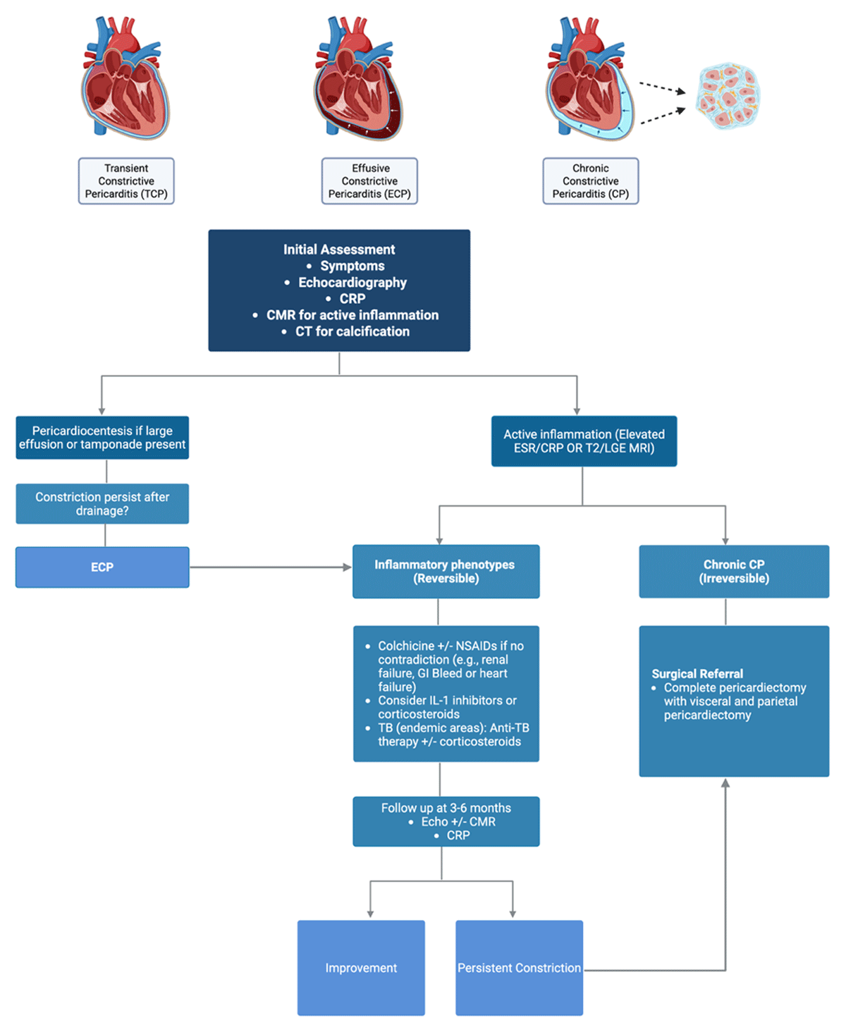
Figure 3
Schematic summary of management across the pericardial constriction spectrum (CP–TCP–ECP). (Created in BioRender with permission. Harake L, 2026, https://BioRender.com/m89rbq2). CP: constrictive pericarditis; TCP: transient constrictive pericarditis; ECP: effusive–constrictive pericarditis; CMR: cardiac magnetic resonance; CT: computed tomography; CRP: C-reactive protein; ESR: erythrocyte sedimentation rate; LGE: late gadolinium enhancement; NSAIDs: nonsteroidal anti-inflammatory drugs; IL-1: interleukin-1; TB: tuberculosis