
Introduction
Pericardial diseases encompass a broad spectrum of disorders affecting the pericardium, including acute and recurrent pericarditis, pericardial effusion, cardiac tamponade, constrictive pericarditis (CP), and effusive–constrictive pericarditis (ECP).1 These entities vary in etiology, pathophysiology, and clinical presentation, ranging from self-limited inflammatory syndromes to a chronic fibrotic process.2,3 Over recent years, major advances in the understanding of pericardial pathophysiology, along with developments in multimodality imaging, have refined both diagnosis and management.1
Constrictive pericarditis represents a complication of pericardial inflammation caused by an inelastic pericardium that inhibits cardiac filling.4,5 The resulting constrictive pathophysiology impairs diastolic ventricular filling and produces a heart failure syndrome with preserved ejection fraction.5 CP can develop as a consequence of chronic fibrosis and/or calcification, termed chronic constrictive pericarditis, or as a potentially reversible inflammatory process, termed transient constrictive pericarditis.6 Recognizing this distinction is crucial, as transient forms may resolve spontaneously or improve with anti-inflammatory therapy.
ECP is a distinct clinical entity characterized by the coexistence of pericardial effusion under pressure and constriction of the heart by an inflamed, noncompliant visceral pericardium.7, 8, 9 It was first described in the 1950s and formally defined in 1971 as persistent elevation in right atrial pressure after pericardiocentesis, indicating ongoing constrictive pathophysiology despite normalization of intrapericardial pressure.10 Increasingly, echocardiography has been the central modality in assessing for features of constrictive pathophysiology before and after drainage of a pericardial effusion.11
Inflammation plays a central role in both CP and ECP. Active pericardial inflammation can promote transient constriction, which may respond to anti-inflammatory therapy, whereas chronic fibrotic disease tends to be unresponsive.12 Similarly, in ECP, marked visceral pericardial inflammation underlies persistent constrictive physiology despite drainage of effusion. Advances in echocardiography and cardiac magnetic resonance have enhanced the ability to detect inflammation, guiding therapeutic decisions and identifying patients who may benefit from medical therapy.1,7 Understanding the timing, duration, and patient selection for such therapy is increasingly important, as radical pericardiectomy is reserved for advanced and irreversible cases.12,13
This review summarizes current evidence on the role of medical therapy in CP and ECP, emphasizing how advances in imaging and understanding of disease pathophysiology inform treatment decisions.
Pathophysiology and Etiology
CP arises when pericardial inflammation leads to loss of compliance, impaired diastolic filling, and exaggerated ventricular interdependence, with chronic disease reflecting fibroblast activation, collagen deposition, and calcification.4,5 This results in abrupt cessation of ventricular filling once the intrapericardial volume limit is reached, causing elevated venous pressures, reduced stroke volume, and heart failure syndrome with preserved ejection fraction.5 The ventricles fill only within the constraint of the inelastic pericardium and, as a result, inspiratory right ventricular expansion occurs at the expense of left ventricular filling. Dissociation between intrathoracic and intracardiac pressures further impairs left-sided filling. Specifically, during inspiration, the negative intrathoracic pressure leads to preserved filling of extrapericardial pulmonary veins but not the intrapericardial left atrium. Accordingly, the lack of transmission of this negative intrathoracic pressure into the heart leads to relatively reduced left-sided filling during inspiration. On echocardiography, the result is the characteristic septal shift to the left during inspiration. This enhanced interventricular interaction also leads to the exaggerated respirophasic variation with Doppler assessment of mitral and tricuspid inflows.14,15
CP encompasses transient, effusive, and chronic forms that often overlap in clinical practice.2 Neither the prevalence nor overall incidence of CP has been well-established, but it remains uncommon. Approximately 1.8% of patients with acute pericarditis develop chronic constrictive physiology, and it is more common with bacterial and tuberculous pericarditis. After cardiac surgery, chronic CP rarely occurs (0.2-2.4%).16,17 Transient CP represents a potentially reversible inflammatory variant, typically following viral or idiopathic pericarditis or cardiac surgery; in transient CP, pericardial edema and fibrin deposition predominate more than dense fibrosis.6,18 Constriction in these cases results from active pericardial inflammation rather than fibrosis, and it typically resolves following effective anti-inflammatory therapy.19,20
ECP represents a distinct clinical syndrome form of CP characterized by the coexistence of a pericardial effusion and persistent constrictive physiology after fluid removal.8-10 The hallmark feature is sustained elevation in right atrial pressure following pericardiocentesis, reflecting constriction that primarily involves the visceral pericardium. ECP occurs in about 1% of pericarditis cases but up to 50% in tuberculous cohorts.9,21,22 Some patients present with tamponade, and constrictive physiology becomes evident only after fluid drainage; however, overt tamponade is not required for diagnosis.23 The additive effects of elevated pericardial pressure and reduced visceral compliance define its hemodynamic profile.
The hemodynamic profile of ECP incorporates features of both tamponade and constriction.10,24 In tamponade, uniformly elevated intrapericardial pressure restricts filling throughout diastole with equalization of diastolic pressures in the cardiac chambers. In constriction, early rapid filling is preserved with an abrupt mid- to late-diastolic halt once the pericardial limit is reached. These divergent patterns of restrictive filling underlie their distinct respiratory interactions. In tamponade, inspiratory venous return increases and ventricular interdependence is accentuated because pleural pressure changes are transmitted directly to the heart. Conversely, in constriction, the noncompliant pericardium prevents this transmission of intrathoracic pressure, leading to reduced left-sided filling during inspiration and the characteristic respirophasic septal shift.25 Accordingly, constriction demonstrates discordant respiratory changes in ventricular pressures. In addition, there is a preserved or increased right atrial pressure with inspiration, termed a Kussmaul’s sign, with an associated inspiratory decrease in pulmonary capillary wedge pressure.4,26
Inflammatory mechanisms play a central role in ECP.12 Ntsekhe et al. demonstrated that patients with tuberculous ECP had markedly elevated levels of interleukin-10 (IL-10), interferon gamma, and transforming growth factor beta in both serum and pericardial fluid compared with those with nonconstrictive effusions, suggesting that these cytokines sustain inflammation and fibrosis.23 In non-tuberculous ECP, Kim et al. reported that pericardial fluid contained a higher proportion of neutrophils despite similar total white cell counts, consistent with more intense inflammation.11
Etiologies vary by region and disease pattern.27 In high-income countries, CP is most commonly idiopathic or post-surgical, with radiation-associated cardiac disease as a less common cause, whereas tuberculosis remains the leading cause in low- and middle-income regions and may be influenced by HIV coinfection.1,3,5 Autoimmune, post-infectious, and malignant causes, particularly in ECP, also contribute to the spectrum, with iatrogenic and procedure-related cases increasingly reported.8,9 Contemporary frameworks integrate histopathology, hemodynamics, and multimodality imaging, viewing CP as a dynamic continuum from inflammatory and fibroelastic stages to fibrotic and calcific remodeling.27 Within this spectrum, transient and effusive constrictive syndromes represent the inflammation-dominant reversible phase, underscoring the importance of early identification and targeted medical therapy before irreversible calcification.2,27
Clinical Presentation and Diagnostic Approach
The diagnostic evaluation of CP and ECP centers on recognizing constrictive pathophysiology and determining the degree of active inflammation since this distinction directly informs the likelihood of response to medical therapy.1,12 Clinically, patients with CP often present with fatigue, dyspnea, and peripheral edema, reflecting right-sided heart failure, while some may show pleuritic pain or fever due to ongoing pericardial inflammation.4 A careful physical examination remains invaluable; elevated jugular venous pressure with prominent x and y descents and a positive Kussmaul’s sign (an inspiratory increase in jugular venous pressure) are classic though not universal findings. In patients presenting with pericardial effusion or tamponade, persistence of heart failure symptoms after drainage should raise suspicion for effusive–constrictive physiology.9 Inflammatory biomarkers complement clinical evaluation and serve as important tools for patient stratification. Elevated C-reactive protein (CRP) levels identify an active, potentially reversible phase of disease.5 Similarly, higher baseline inflammatory markers predicted reversibility and guided treatment duration, as normalization of CRP and ESR can parallel clinical improvement.19 Key clinical features and distinguishing echocardiographic and multimodality imaging findings across constrictive pericarditis phenotypes are summarized in Table 1.
Table 1
Clinical, echocardiographic, and multimodality imaging features of constrictive pericarditis, effusive-constrictive pericarditis, and transient constrictive pericarditis. HF: heart failure; JVP: jugular venous pressure; RA: right atrial; CRP/ESR: C-reactive protein/ erythrocyte sedimentation rate; TB: tuberculosis; IVC: inferior vena cava; CT: computed tomography; CMR: cardiac magnetic resonance
| FEATURE CATEGORY | CHRONIC CONSTRICTIVE PERICARDITIS (CP) | EFFUSIVE–CONSTRICTIVE PERICARDITIS (ECP) | TRANSIENT CONSTRICTIVE PERICARDITIS (TCP) |
|---|---|---|---|
| Clinical Presentation | Chronic fatigue, dyspnea, peripheral edema; signs of right-sided HF Elevated JVP with prominent x and y descents Kussmaul’s sign; symptoms due to fixed fibrotic/calcific disease | Presents with large effusion with or without tamponade Persistent HF symptoms after drainage RA pressure remains elevated Mixed effusive and constrictive physiology | Similar to CP initially but reversible Often idiopathic, viral, autoimmune, or post-surgical Improves with anti-inflammatory therapy |
| Biomarkers | CRP/ESR usually normal (non-inflammatory) | Elevated if inflammatory or infectious (eg, TB) | CRP/ESR frequently elevated and normalize with treatment |
| Echocardiography | Respirophasic septal shift Respiratory variation > 25% in mitral E wave velocity Late diastolic hepatic vein flow reversal with expiratory end-diastolic reversal ratio > 0.79 Medial e′ ≥ 9 cm/s Annulus reversus | Before drainage: effusion with or without tamponade features After drainage: mixed effusion/constriction signs; septal bounce; hepatic vein reversal; medial e′ > 8 cm/s; persistent IVC dilation | Constrictive pattern identical to CP initially E′ velocities, strain, and septal motion improve with therapy |
| CMR Findings | Pericardial thickening Minimal LGE/T2 edema Evaluates myocardial fibrosis/infiltration | Effusion + constrictive findings Inflammatory changes vary by etiology Visceral pericardial involvement may be subtle | Strong inflammatory signal (pericardial T2 edema, LGE) Predictors of reversibility include LGE thickness ≥ 3 mm and higher quantitative DHE Improves with therapy |
| CT Findings | Pericardial thickening and/or calcification common; helpful for surgical planning | Effusion ± pericardial thickening; calcification etiology dependent | Mild or normal pericardial thickening; calcification absent |
Multimodality imaging forms the cornerstone of diagnosis and therapeutic decision-making.2,27 As noted, the two hallmark hemodynamic features that define constrictive pathophysiology are dissociation between intrathoracic and intracardiac pressures during respiration and enhanced interventricular dependence with accentuated early diastolic filling pressures in both ventricles. These physiologic changes are best appreciated on echocardiography, which provides real-time assessment through characteristic Doppler and 2-dimensional patterns.1,5 The Mayo Clinic echocardiographic diagnostic criteria incorporate four parameters that reflect these hemodynamic alterations: (1) respirophasic ventricular septal shift; (2) predominant early mitral inflow with exaggerated respiratory variation; (3) expiratory end-diastolic hepatic-vein flow reversals; and (4) elevated medial mitral e′ velocities. The presence of ventricular septal shift in combination with either a medial e′ ≥ 9 cm/s or a hepatic-vein expiratory diastolic reversal ratio ≥ 0.79 provides excellent diagnostic accuracy.28 Strain imaging adds sensitivity for detecting subtle diastolic constraint through reduced longitudinal motion of the left ventricular free wall.5 In transient CP, improvement in tissue Doppler velocities (lateral/septal e′) and longitudinal strain over serial follow-up correlates with clinical recovery during anti-inflammatory therapy.20 Echocardiographic features can also identify constrictive pathophysiology in patients with large pericardial effusions or pericardial tamponade.8 In a series of 205 patients undergoing pericardiocentesis, ECP was reported in 16%, characterized by respirophasic septal shift, pronounced mitral inflow variation, expiratory hepatic vein flow reversal, and elevated medial e′ velocities (8.9 vs 6.9 cm/s).11 Miranda et al. confirmed similar findings, with ECP demonstrating higher medial and lateral e′ than tamponade and intermediate respiratory variation in E:A ratios.29 Figures 1 and 2 illustrate a representative case of ECP, highlighting the complementary roles of echocardiography and cardiac magnetic resonance imaging. Figure 1 demonstrates the coexistence of tamponade and constrictive physiology at presentation, while Figure 2 shows persistent constrictive features following therapeutic pericardiocentesis. Videos 1, 2, 3, 4 show corresponding motion.
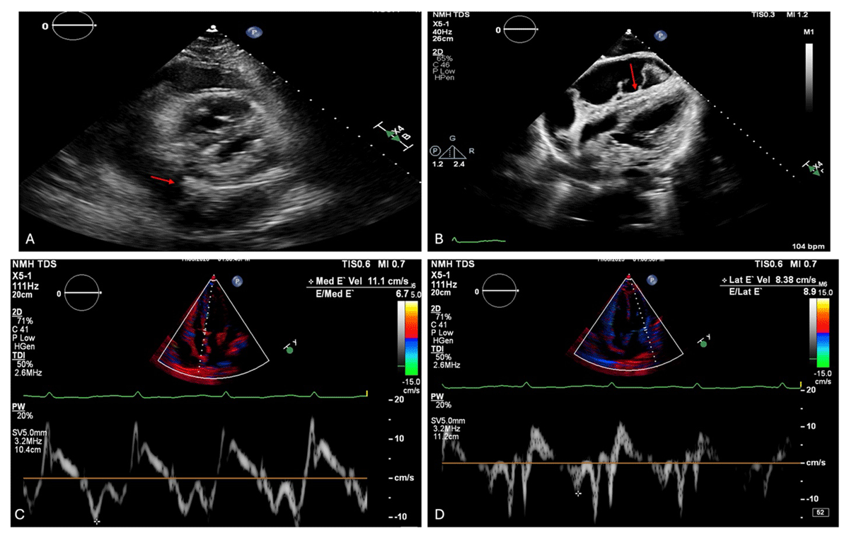
Figure 1
Echocardiographic features of effusive-constrictive pericarditis at presentation. Transthoracic echocardiography demonstrates concurrent features of pericardial effusion, tamponade physiology, and constrictive hemodynamics. (A) Parasternal short-axis view with a large circumferential pericardial effusion containing fibrinous strands (arrow). Video 1 also shows the respirophasic septal shift. (B) Diastolic right ventricular free wall collapse in the subcostal view, consistent with tamponade physiology (arrow; Video 2). Tissue Doppler imaging reveals annulus reversus, with (C) accentuated medial mitral annular e′ velocity (11.1 cm/s) exceeding the (D) lateral e′ velocity (8.38 cm/s). The respirophasic septal shift and annulus reversus are suggestive of concomitant constrictive pathophysiology in a patient who has pericardial tamponade.
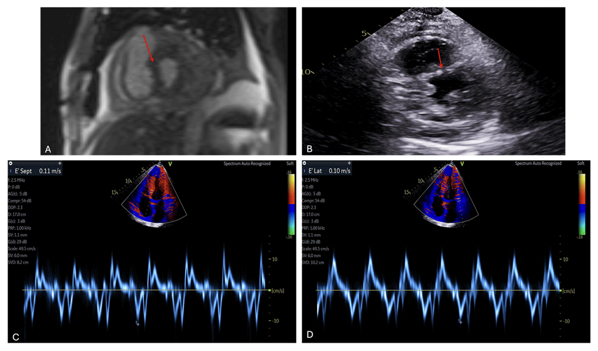
Figure 2
Persistent constrictive physiology following pericardial drainage. Cardiac magnetic resonance (CMR) imaging and transthoracic echocardiography obtained after therapeutic pericardiocentesis (600 mL drained) demonstrate persistent features of constrictive physiology. Panel A shows a CMR image with respirophasic interventricular septal shift (arrow; corresponding motion shown in Video 3). Panel B demonstrates residual pericardial effusion with persistent interventricular septal flattening on post-drainage echocardiography (arrow; corresponding motion shown in Video 4). Panels C and D show tissue Doppler imaging of the mitral annulus, with accentuated medial e′ velocity exceeding the lateral e′ velocity, consistent with persistent annulus reversus after relief of tamponade.
Video 1
Parasternal short-axis view demonstrating a large circumferential pericardial effusion with fibrinous strands and respirophasic interventricular septal shift, consistent with constrictive physiology; also see at https://vimeo.com/1167807972.
Video 2
Subcostal view demonstrating diastolic right ventricular free wall collapse, consistent with pericardial tamponade physiology; see also at https://vimeo.com/1167809971.
Video 3
Cardiac magnetic resonance cine imaging demonstrating respirophasic interventricular septal shift after therapeutic pericardiocentesis, consistent with persistent constrictive physiology; see also at https://vimeo.com/1167810637.
Video 4
Transthoracic echocardiography demonstrating residual pericardial effusion with persistent interventricular septal flattening following pericardiocentesis, consistent with persistent constrictive physiology; see also at https://vimeo.com/1167814267.
Cardiac magnetic resonance (CMR) provides complementary anatomic and tissue-level assessment, crucial for identifying active inflammation.2 Increased pericardial signal on T2-weighted imaging signifies edema, while late gadolinium enhancement (LGE) reflects neovascularization and fibroblast proliferation.1,15,27 The presence and thickness of LGE have strong prognostic implications; in Feng et al.’s cohort, LGE pericardial thickness ≥ 3 mm predicted reversibility with 86% sensitivity and 80% specificity, confirming that inflammation, rather than fibrosis, drives constriction in such patients.19 Quantitative assessment of pericardial LGE further refines this prognostic value. Greater degrees of delayed hyperenhancement independently predicted clinical improvement with anti-inflammatory therapy in patients with CP and provided incremental discrimination beyond symptoms and inflammatory markers. Collectively, these findings confirm that CMR not only identifies an inflammatory constrictive phenotype but also guides therapeutic decision-making in patients with potentially reversible disease.13 CMR also allows evaluation for concomitant myocardial fibrosis or infiltration using LGE and mapping sequences, providing incremental prognostic information and aiding surgical planning.2 Cardiac CT serves as a supplementary tool to evaluate pericardial calcification and fluid to assist with preoperative planning.30
When noninvasive findings are inconclusive, cardiac catheterization provides hemodynamic confirmation. Classic features include elevated and near-equalized diastolic filling pressures and discordant respiratory variation between right and left ventricular systolic pressures. Enhanced ventricular interdependence can be quantified using the systolic area index, defined as the ratio of right ventricular to left ventricular systolic pressure-time area during inspiration versus expiration; an inspiratory-to-expiratory systolic area index ≥ 1.1 has been shown to be highly sensitive (97%), with excellent predictive accuracy for CP.31 In effusive–constrictive disease, the hallmark is a right atrial pressure that fails to fall below 10 mm Hg or by at least 50% following pericardiocentesis, reflecting ongoing visceral constriction.10,22,24 Persistent elevation of right atrial pressure with prominent y descent and a dip-and-plateau right ventricular contour further confirm the diagnosis.24 Taken together, the integration of inflammatory markers, echocardiographic and CMR indicators of active inflammation, and hemodynamic assessment allows clinicians to identify a potentially reversible, inflammation-driven subset of constrictive disease, one in which timely medical therapy may obviate the need for surgery.
Management Strategies in Constrictive and Effusive–Constrictive Pericarditis Medical Therapy
For patients with evidence of active pericardial inflammation detected by elevated inflammatory markers or pericardial LGE on CMR, medical therapy should precede surgical intervention.1,3,13 This approach targets the reversible, inflammation-dominant phase of CP and ECP, aiming to relieve symptoms and, in some cases, restore pericardial compliance. These features are most commonly seen in idiopathic, viral, post-surgical, or autoimmune etiologies, whereas calcific or fibrotic disease is unresponsive to anti-inflammatory therapy.4,5 The stepwise management approach across transient, effusive-constrictive, and chronic constrictive pericarditis is summarized in Figure 3.
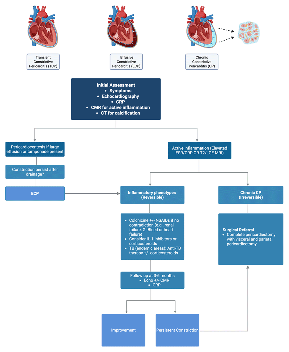
Figure 3
Schematic summary of management across the pericardial constriction spectrum (CP–TCP–ECP). (Created in BioRender with permission. Harake L, 2026, https://BioRender.com/m89rbq2). CP: constrictive pericarditis; TCP: transient constrictive pericarditis; ECP: effusive–constrictive pericarditis; CMR: cardiac magnetic resonance; CT: computed tomography; CRP: C-reactive protein; ESR: erythrocyte sedimentation rate; LGE: late gadolinium enhancement; NSAIDs: nonsteroidal anti-inflammatory drugs; IL-1: interleukin-1; TB: tuberculosis
Nonsteroidal anti-inflammatory drugs (NSAIDs) and colchicine form the first-line treatment with gradual tapering of NSAIDs guided by symptom resolution and normalization of C-reactive protein. NSAIDs are typically given for a few weeks. NSAIDs should be avoided in patients with heart failure as it can lead to further decompensation and renal insufficiency. Colchicine is typically prescribed for at least 3 to 6 months.2,12,18 Corticosteroids are used when NSAIDSs and colchicine are contraindicated or ineffective, typically at 0.25 to 0.5 mg/kg/day with a slow taper.2 In a cohort of 29 patients, 48% demonstrated complete resolution after anti-inflammatory treatment, and corticosteroids were used in two-thirds.19 Similarly, Sato et al. reported clinical and echocardiographic resolution in 57% of patients treated with anti-inflammatory therapy, with recovery in tissue Doppler and strain parameters paralleling improvement in symptoms.20 For transient or early CP, a treatment course of 3 to 6 months is generally recommended, with follow-up imaging at 8 to 12 weeks to assess reversibility using echocardiography with weekly CRP until normalization.5
More recently, targeted cytokine inhibition has emerged as an adjunctive option in inflammation-driven constrictive pericarditis.2 Specifically, IL-1 receptor inhibition with anakinra has shown promise in refractory pericarditis complicated by constrictive pathophysiology. In a prospective cohort, Andreis et al. observed complete reversal in 63% of patients within 1.2 months, whereas 37% progressed to chronic constriction requiring pericardiectomy.32 A case report similarly described improvement with IL-1 blockade in inflammatory CP, suggesting that early intervention may prevent transition to fibrosis,33 IL-1 inhibitors may thus serve as second- or third-line options in corticosteroid-dependent or colchicine-resistant cases, although prospective randomized data are lacking.
Tuberculous CP and ECP require etiology-specific therapy. Standard anti-tuberculous treatment leads to resolution of constrictive physiology in up to 75% of patients within 6 months.21,22,34 Adjunct corticosteroids may hasten improvement but have not been shown to improve survival. In the Investigation of the Management of Pericarditis in Africa (IMPI) trial, which enrolled 1,400 patients with tuberculous pericarditis, prednisolone reduced the incidence of constrictive pericarditis but did not decrease mortality and was associated with higher risk of HIV-related malignancies.35 Corticosteroids are thus reserved for HIV-negative patients with active inflammation or early constrictive physiology, while early pericardiectomy is avoided unless there is hemodynamic compromise or failure of medical therapy after 4 to 8 weeks.35
In chronic fibrotic or noninflammatory CP, medical therapy is primarily supportive. Loop diuretics and mineralocorticoid receptor antagonists help control volume overload but do not alter the disease course.1,5,6 Therefore, a prolonged medical therapy should not delay definitive surgery once imaging and biomarkers indicate a transition to irreversible fibrosis in symptomatic patients (ie, New York Heart Association [NYHA] class II-IV) or in patients with evidence of end-organ dysfunction (ie, liver or renal dysfunction).12 Ultimately, the integration of clinical findings, inflammatory markers, and multimodality imaging is essential to identify patients most likely to benefit from anti-inflammatory therapy and to determine when definitive pericardiectomy becomes necessary.
Percutaneous Interventions and Surgical Management
In ECP, the initial management step is pericardiocentesis to relieve tamponade and confirm the diagnosis.10 Drainage often leads to rapid symptomatic improvement; however, persistence of elevated right atrial pressure or constrictive physiology after fluid removal indicates ongoing visceral pericardial constriction.24,36 Surgical pericardial window is considered when percutaneous drainage is unsafe or in cases of recurrent effusions, particularly in malignant or purulent etiologies.12 Despite adequate drainage, progression to chronic constriction may occur, emphasizing the importance of close hemodynamic and imaging follow-up.
Pericardiectomy remains the definitive therapy for chronic constrictive and refractory effusive-constrictive pericarditis. Surgery is indicated for patients with symptomatic heart failure (NYHA class II-IV) with either (1) persistent constrictive physiology despite anti-inflammatory therapy, or (2) chronic constriction with extensive pericardial fibrosis and/or calcification.1,3,12 Radical pericardiectomy is preferred and involves removal of the pericardium between—including posterior to the phrenic nerves, plus diaphragmatic and posterior segments—and is often performed on cardiopulmonary bypass to ensure complete resection.37 If the visceral pericardium contributes to constriction, epicardiectomy or the waffle procedure may be required to relieve epicardial tethering, but this approach should not be necessary if a patient has received appropriate anti-inflammatory treatment prior surgery.38 Partial pericardiectomy carries a high risk of recurrence and incomplete symptom relief.39
When feasible, surgery should be delayed until inflammatory activity has subsided considering that operating in the presence of active pericardial inflammation is challenging and associated with higher perioperative morbidity.1 Given the complexity of radical pericardiectomy, referral to experienced high-volume centers with specialized pericardial expertise is recommended to optimize outcomes.3 Persistent echocardiographic features of constriction are common postoperatively but often resolve with time and should not be mistaken for recurrence in asymptomatic patients.4
Prognosis
The prognosis of CP and ECP is determined by the underlying etiology, inflammatory activity, and timing of intervention. Patients with inflammatory or transient forms, particularly those with evidence of pericardial LGE and/or elevated inflammatory markers, often experience full recovery with timely anti-inflammatory therapy. In contrast, chronic fibrotic disease and advanced presentations carry a less-favorable course. In the review by Ntsekhe et al., pericardiectomy was required in 65% of effusive–constrictive pericarditis cases, with higher rates among idiopathic etiologies compared with tuberculous or postinfectious disease.40 Conversely, Kim et al. observed that only 2 of 33 patients required surgery over nearly 4 years of follow-up, highlighting that early recognition and anti-inflammatory treatment may prevent progression and improve outcomes.11 The true risk of progression remains unclear, as current data are limited to observational findings from small cohorts.
Operative mortality ranges between 6% and 12%, largely dependent on etiology and comorbidities.41 Idiopathic constrictive pericarditis carries the most favorable prognosis, with operative mortality below 1.5% and 5- to 7-year survival exceeding 80%.42 Postsurgical constrictive pericarditis demonstrates intermediate outcomes, with operative mortality around 4% to 10% and 5-year survival between 50% and 66%.43,44 In contrast, post-radiation constriction carries the highest risk, with operative mortality of approximately 10% to 27% and survival rates of 53% and 32% at 5 and 10 years, respectively.42,45 Radiation-related heart disease often extends beyond the pericardium, involving the myocardium, valves, and coronary arteries. In particular, a concomitant cardiomyopathy, which is often restrictive, contributes to a poor outcome.3,43
Inoperable or “end-stage” constrictive pericarditis, characterized by cachexia, atrial fibrillation, advanced hepatic congestion and cirrhosis, and low cardiac output, carries a poor prognosis even with supportive therapy.3 In contrast, patients with inflammatory or transient forms have excellent outcomes when identified early and treated with appropriate anti-inflammatory therapy, underscoring the importance of multimodality imaging in prognostication and management.
Conclusion
CP and ECP represent overlapping syndromes within a continuum of pericardial disease, ranging from transient inflammatory forms to chronic calcific constriction. Many cases of transient constrictive pericarditis and effusive–constrictive pericarditis resolve with timely recognition and anti-inflammatory therapy, thereby avoiding the need for surgery. Treatment should be guided by the underlying etiology, with anti-inflammatory therapy as first-line management in cases showing active inflammation and radical pericardiectomy reserved for refractory or chronic disease. Multimodality imaging remains essential for identifying reversible inflammation and directing therapy. Close clinical and imaging follow-up is critical to monitor for resolution or progression toward chronic constriction. As diagnostic criteria and imaging techniques continue to evolve, early detection, targeted medical therapy, and individualized management will be key to improving outcomes across the spectrum of constrictive pericardial disease.
Key Points
Constrictive pericarditis (CP) and effusive–constrictive pericarditis (ECP) exist along a spectrum of pericardial inflammation and fibrosis, with transient CP representing an early, reversible stage.
Identification of pericardial inflammation through clinical, biomarker, and multimodality imaging assessment is critical to guide therapy and distinguish reversible from chronic constriction.
Anti-inflammatory therapy including nonsteroidal anti-inflammatory drugs, colchicine, corticosteroids, and interleukin-1 inhibitors remains the cornerstone of treatment in inflammatory and transient forms, with close follow-up to assess resolution.
Radical pericardiectomy should be reserved for patients with persistent constrictive physiology, advanced fibrosis, or refractory symptoms despite optimal medical therapy and is best performed at experienced referral centers with multidisciplinary perioperative care; outcomes depend largely on etiology and comorbidity.
Early recognition, targeted medical therapy, and individualized, imaging-guided management can improve outcomes and may prevent progression to irreversible chronic constriction.
Competing Interests
Dr. Al-Kazaz has received research grants from Kiniksa Pharmaceuticals, Ventyx BioSciences, and Cardiol Therapeutics, is on the Speakers Bureau for Kiniksa Pharmaceuticals, and is a consultant for Edwards Lifesciences. Dr. Cremer is a consultant for Kiniksa Pharmaceuticals, CardiolRx, Ventyx Biosciences, Monte Rosa Therapuetics, Pfizer, Boston Scientific, and General Electric. Dr. El Harake has no conflicts to declare.