
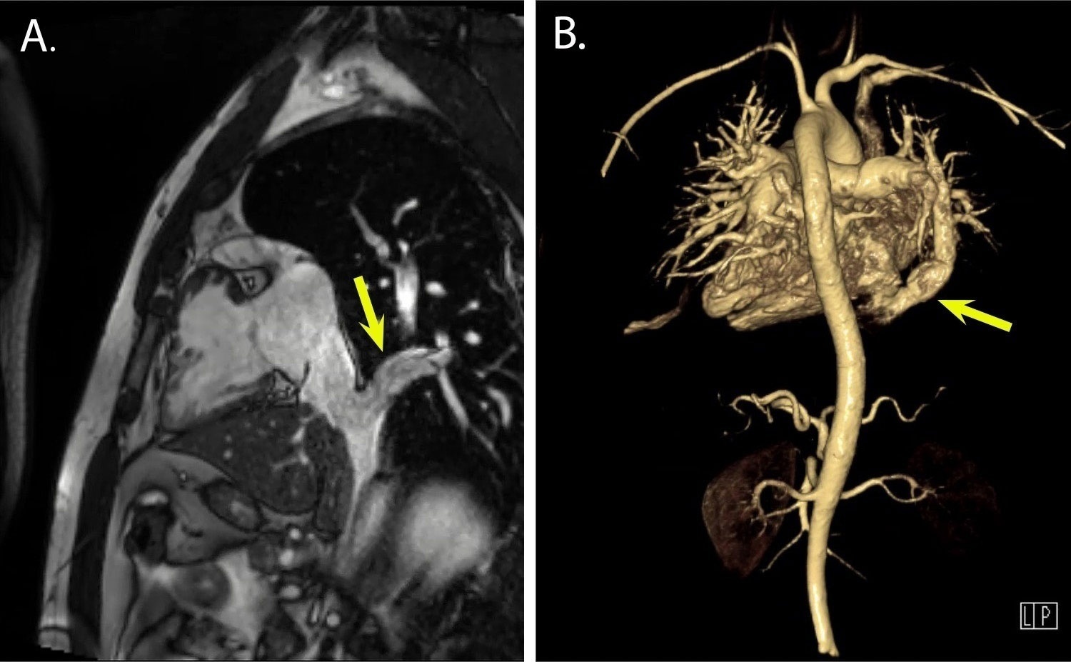
Figure 1
Magnetic resonance imaging of the scimitar vein from a (A) lateral and (B) posterior 3-dimensional-reconstruction view. The yellow arrows point at the scimitar vein.
Video 1
Intra-atrial baffling of the scimitar vein and inferior vena cava enlargement without circulatory arrest; see also at https://vimeo.com/1168242844.
Video 2
Postoperative computed tomography scan showing a patent baffle inflow from the scimitar vein at the inferior vena cava site and a patent outflow to the left atrium via the atrial septostomy on axial and coronal planes. The red arrows point at the inflow and outflow of the baffle; see also at https://vimeo.com/1174555320.