
Introduction
Scimitar syndrome is a rare congenital defect in which an anomalous right-sided pulmonary vein drains into the inferior vena cava (IVC), causing left-to-right shunt, and is associated with right lung hypoplasia and other congenital defects. In some cases, the scimitar vein (SV) can be reimplanted directly to the left atrium (LA); however, if the anatomy is not favorable, intra-atrial baffling is preferred, typically by implementing deep hypothermic circulatory arrest.1
Here we report the case of a 47-year-old male patient with scimitar syndrome who underwent successful intra-atrial baffling of an infradiaphragmatic SV without using circulatory arrest. An Institutional Review Board review was waived per institutional policy, and informed patient consent was obtained.
Case Report
A 47-year-old male patient with progressive dyspnea on exertion presented at the emergency department with additional stroke-like symptoms. Although evaluation ruled out stroke, computed tomography (CT) angiography of the chest and later cardiac magnetic resonance imaging (MRI) revealed a hypoplastic right lung with an anomalous partial right pulmonary venous return to the IVC, below the diaphragm (Figure 1). Shunt evaluation by cardiac MRI and right heart catheterization showed a significant left-to-right shunt (QP/QS = 1.7) and a severely dilated right ventricle and atrium. The interatrial septum was intact, and the patient was deemed a candidate for surgical correction.
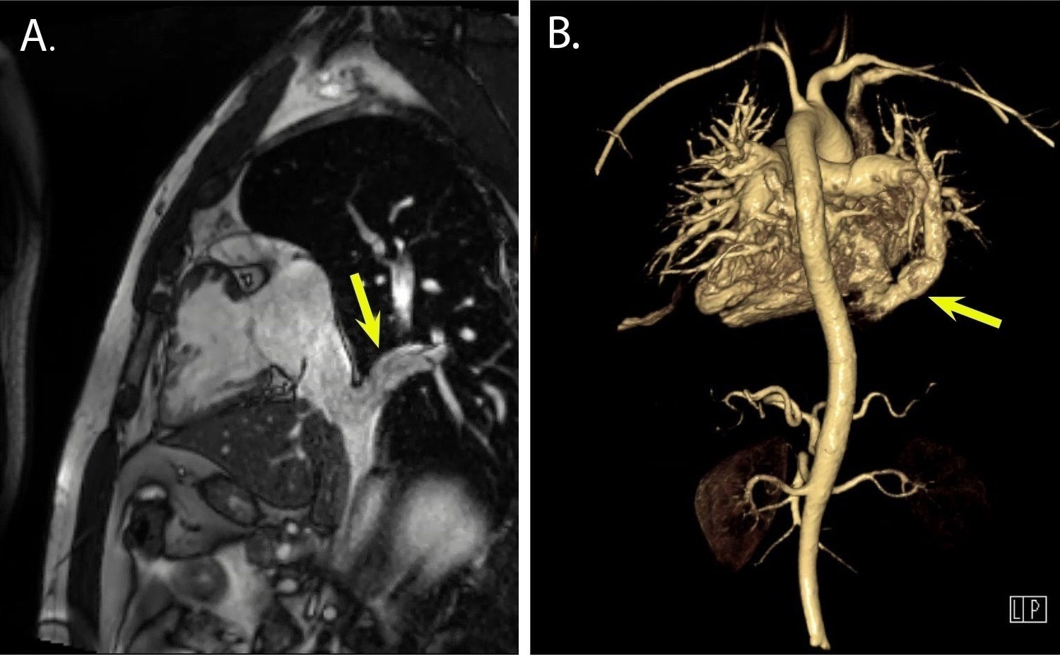
Figure 1
Magnetic resonance imaging of the scimitar vein from a (A) lateral and (B) posterior 3-dimensional-reconstruction view. The yellow arrows point at the scimitar vein.
Surgical Technique
After median sternotomy, cardiopulmonary bypass was established with bicaval cannulation using a direct superior vena cava (SVC) and percutaneous right femoral vein access. The SVC and IVC were snared, and cooling was set to 28°C. Once the right pleural space was opened, the SV was identified crossing the diaphragm. The heart was arrested using del Nido cardioplegia in the aortic root, and the right atrium was longitudinally opened. An atrial septostomy of 2 cm was created into the fossa ovalis, and the free edges were reinforced with a running suture. The IVC snare was released, and the inferior venous cannula was pulled to the level of the renal veins. The hepatic veins were continuously vented throughout the procedure, and vacuum-assisted venous drainage was employed to prevent air lock. The SV was identified from the IVC ostium. A GORE-TEX baffle was then sutured from the ostium of the SV across the right atrium to the atrial septostomy. The IVC and cavoatrial junction were enlarged using an autologous pericardial patch; then, a 4-mm fenestration on the baffle was created using a punch—allowing the unloading of the LA and left ventricle with a left-to-right shunt, which acts as a valve in case of late baffle obstruction. The heart was carefully de-aired and the patient was weaned from cardiopulmonary bypass uneventfully (Video 1).
Video 1
Intra-atrial baffling of the scimitar vein and inferior vena cava enlargement without circulatory arrest; see also at https://vimeo.com/1168242844.
Transesophageal echocardiography showed regular laminar drainage from the SV to the LA, with a peak gradient of 1 mm Hg. Total cardiopulmonary bypass time was 210 minutes, while cross-clamp time was 152 minutes. The patient was extubated 16 hours postoperatively and discharged from the hospital on postoperative day 4 without complications. After 1-year follow-up, the patient reported improved symptoms, and postoperative CT confirmed a patent baffle (Video 2).
Video 2
Postoperative computed tomography scan showing a patent baffle inflow from the scimitar vein at the inferior vena cava site and a patent outflow to the left atrium via the atrial septostomy on axial and coronal planes. The red arrows point at the inflow and outflow of the baffle; see also at https://vimeo.com/1174555320.
Discussion
We describe the case of a patient with scimitar syndrome who successfully underwent intra-atrial baffling of the SV into the LA without needing circulatory arrest, instead using vacuum-assisted venous drainage. There are several cases in which direct reimplantation of the SV to the LA was described either via sternotomy or a minimally invasive/robotic approach; however, there are certain anatomic situations when intra-atrial baffling is inevitable.1, 2 In most cases, to obtain a bloodless surgical field, deep hypothermic circulatory arrest is required, exposing patients to an increased risk of neurological events.3 Surgical techniques to avoid circulatory arrest of scimitar syndrome patients were previously reported, especially if SVs were draining into the IVC at a supradiaphragmatic level.4 Regarding our case, the SV ostium level was infradiaphragmatic; therefore, the distal suture line of the baffle was challenging while keeping the field bloodless. For inferior venous body drainage, we used femoral access with vacuum-assisted venous drainage (40 mm Hg), and the cannula was withdrawn to the level of the renal veins to avoid air lock. With short periods of flow reduction, a clearly visible work field could be achieved with continuous venting of the hepatic veins. Pump-flow was uncompromised during the procedure.
Conclusion
An intra-atrial baffle correction technique can be applied to patients with scimitar syndrome without the need of circulatory arrest when using vacuum-assisted venous drainage, even in cases where the SV is below the diaphragm, to reduce the risk and morbidity of the repair.
Competing Interests
The authors have no competing interests to declare.