
Introduction
Post-cardiac injury syndrome (PCIS) was first recognized in patients after cardiac surgery and myocardial infarction (MI), and it is characterized by pericarditis with possible pleural effusions and systemic inflammatory features.1, 2, 3 In contemporary practice, PCIS is increasingly encountered due to the growing number of cardiac surgical and catheter-based procedures, even as classic post-MI pericarditis has become rare with modern reperfusion therapies.4
The clinical importance of PCIS lies in its impact on patient recovery and healthcare resources. Post-pericardiotomy syndrome (PPS) can prolong hospital stay or lead to readmissions and, in rare cases, may cause cardiac tamponade or progression to constrictive pericarditis if inadequately managed.5, 6 Recognizing the syndrome and instituting appropriate therapy is therefore crucial for improving patient outcomes after cardiac surgery or other cardiac injuries. This review discusses the evaluation and management of PCIS, incorporating evidence from published studies and the new 2025 European Society of Cardiology (ESC) guidelines on pericardial disease.1 We review the epidemiology and risk factors, pathophysiological mechanisms, clinical presentation, diagnostic criteria, and both preventive and therapeutic strategies for PCIS. The aim is to provide clinicians with a comprehensive understanding of this syndrome and current best practices for its management.
Epidemiology
The incidence of PCIS varies significantly depending on the specific subtype. While PCIS serves as an umbrella term for various forms of cardiac damage, the epidemiology differs between surgical and post-ischemic causes. Because of aging populations, changes in lifestyle, and the growing number of indications for percutaneous cardiac procedures and operations, PCIS is often more common in developed countries.7, 8 PCIS is considered a common complication after cardiac surgery,9 thus the real incidence is unknown.8
Post-MI pericarditis presents with two distinct phenotypes: early post-MI pericarditis (typically within days, related to transmural necrosis) and late post-MI syndrome, properly termed Dressler syndrome (occurring weeks to months later with an immune mediated mechanism). Due mainly to early detection and repair of heart injuries, Dressler syndrome has grown less common over time. Its incidence has dropped from up to 3% to less than 1% because of emergency coronary artery bypass surgery and percutaneous coronary intervention.10 One study estimated that 3% of acute MI patients had PCIS in the pre-reperfusion era; however, another study found none in its cohort.7, 8
The incidence of post-pericardiotomy syndrome varies with patient populations and the nature of cardiac injury. The volume of PPS is rising alongside the increase in cardiac procedures. While retrospective data varies widely up to 40%, recent prospective studies place the incidence at up to 29%.2, 3 The risk correlates with the extent of direct pericardial trauma and bleeding. An extensive cohort study identified the hierarchy of risk as ascending aortic surgery, aortic or mitral valve replacements, and coronary artery bypass grafting.11 Another factor that may contribute includes multiple procedures during a single operation, which increases risk.5 While less studied, PPS has been documented in case reports following transcatheter interventions such as transcatheter aortic valve implantation (TAVI), MitraClip, and lead fixation.12, 13, 14, 15
Iatrogenic pericarditis is a complication of cardiac procedures, with a reported incidence of 0.5% to 5%. It is associated with myocardial injury caused by percutaneous coronary interventions (PCI), pacemaker lead insertion, and radiofrequency ablation.4 For instance, pericarditis occurs in an estimated 10% of cases after atrial fibrillation ablation and a smaller percentage (1-2%) of patients after pacemaker or other device implantation. Even diagnostic or therapeutic cardiac catheterization carries a very low risk of PCIS (around 0.2%).4, 16 These trends reflect the shifting epidemiology—as invasive cardiac procedures become more common, the overall prevalence of PCIS is increasing even though the post-MI subset has waned.
Pediatric patients, particularly children with congenital heart defects, tend to have at least similar if not higher rates of PCIS after surgery (reported in the 10-28% range), potentially due to more robust immune responses.11
Risk Factors
Younger patient age has been associated with a higher risk of PCIS, as have certain types of surgery (eg, valve surgery, interatrial defect correction and procedures involving pericardial opening). A history of prior pericarditis or a history of autoimmune disorders might predispose individuals to an exaggerated inflammatory response.
Several risk factors for post-cardiac injury syndrome have been identified.5 In adults, female sex and pleural incisions are recognized independent predictors. Regarding body habitus, evidence suggests an inverse relationship between body mass index (BMI) and disease risk: patients with a lower preoperative BMI appear to have a higher susceptibility to PPS, whereas a higher BMI has been associated with a protective effect in large surgical cohorts. Conversely, perioperative steroid use has not consistently demonstrated preventive benefit. Surgical variables, including the use of cardiopulmonary bypass, the extent of pericardial trauma, and significant bleeding, are also critical determinants of risk (Table 1).9
Table 1
Risk factors for developing post-cardiac injury syndrome.
| CATEGORY | RISK FACTOR | PATHOPHYSIOLOGY & NOTES | KEY REFERENCES |
|---|---|---|---|
| Patient Characteristics | Younger age | Younger patients (typically < 60 years) mount a more vigorous immune and inflammatory response to surgical trauma compared to older patients. | 49 5 |
| Female sex | Females have a statistically higher incidence in many cohorts, likely due to hormonal factors or a general predisposition to autoimmune-like reactions. | 5 50 | |
| History of pericarditis | A prior history of pericarditis creates a “primed” immune system, increasing susceptibility to recurrence after surgical triggering. | 51 | |
| Surgical Factors | Pleural incision (pleurotomy) | Opening the pleura allows blood and inflammatory mediators to spread from the pericardium into the pleural space, significantly increasing post-pericardiotomy syndrome (PPS) risk. | 5 51 |
| Type of surgery (valve vs CABG) | Valve surgery (mitral/aortic) and aortic root surgery carry a higher risk than simple coronary artery bypass grafting (CABG), likely due to greater tissue manipulation. | 49 50 | |
| Blood transfusions | The receipt of red blood cell units is associated with increased systemic inflammation and higher PPS rates. | 50 | |
| Postoperative | Retained blood / effusions | Accumulation of blood in the pericardial or pleural spaces acts as a substrate for local inflammation and oxidative stress. | 2 |
Pathophysiology
Although the exact pathophysiology of PCIS remains incompletely understood, it is primarily believed to be an immune-mediated inflammatory reaction triggered by injury to the heart or pericardium (Figure 1).4, 17, 18
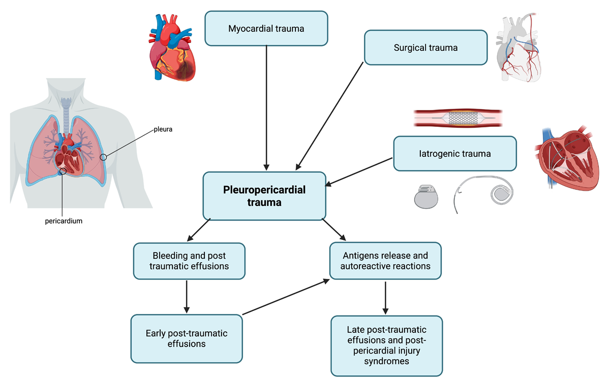
Figure 1
Flowchart illustrating the pathogenesis of post-cardiac injury syndrome.
The pathophysiology of pericardial damage involves distinct phases. In the first phase, damage-associated molecular patterns (DAMPs) trigger pattern recognition receptors. In the second, NF-κB activation leads to NOD-like receptor protein 3 (NLRP3) inflammasome assembly and the subsequent production of interleukin-1β (IL-1β), IL-6, and IL-18. IL-1β signaling via the IL-1 receptor (IL-1R) sustains pericardial inflammation.19 These molecular pathways provide the rationale for the therapeutic targets currently used in clinical practice.
The leading hypothesis is that cardiac injury (surgical incision, MI, necrosis, or trauma) releases intracellular cardiac antigens, which, in some patients, incite an inflammatory and immune response.7, 8 Circulating antimyocardial antibodies may arise even if their pathogenetic role is debatable, being probably an epiphenomen, or they form and create immune complexes that deposit in the pericardium and pleural membranes, leading to inflammatory pericarditis and pleuritis. This autoimmune mechanism is supported by the characteristic latency period of a few days to several weeks between the inciting event and onset of symptoms as well as the frequent elevation of inflammatory markers (eg, C-reactive protein) during episodes.7, 8 The syndrome also tends to respond well to anti-inflammatory therapies and often shows a relapsing or remitting course, further suggesting an immune basis similar to what is hypothesized in classical “idiopathic” recurrent pericarditis.
Evidence for the immune pathogenesis includes studies demonstrating higher titers of antibodies against cardiac antigens (such as anti-actin or anti-myosin antibodies) in patients who develop PCIS. However, the precise role of these antibodies remains incompletely defined, and not all aspects of PCIS can be explained solely by activation of the immune system. Other contributing mechanisms may include a nonspecific inflammatory response to surgical trauma and, in some cases, viral infection triggers. Notably, cases of PCIS have been observed even in immunosuppressed patients (for example, after heart transplantation), leading some researchers to question the purely autoimmune origin and consider additional factors such as viral infections or other immune triggers. Some reports note seasonal peaks and concurrent viral illness in PCIS patients, hinting that viral myocarditis/pericarditis could sometimes mimic or contribute to the syndrome.7, 8, 20
Overall, the pathophysiology of PCIS involves a complex interplay among immune activation, pericardial and pleural inflammation, and patient-specific susceptibility. The understanding continues to evolve, but the autoimmune pericarditis model currently guides most therapeutic approaches (eg, anti-inflammatory and immunosuppressive treatments).
Clinical Presentation and Diagnosis
Patients with PCIS typically present within 1 to 6 weeks after the precipitating cardiac injury, with most cases occurring in the first month post-surgery or infarction (Table 2).1, 4, 5 The clinical presentation closely resembles that of acute pericarditis. Key symptoms include:
Pleuritic chest pain: classically sharp chest pain that may improve on sitting up and worsen when lying down, indicative of pericardial inflammation.
Fever: low-grade fever is common, reflecting systemic inflammation, and in some cases, patients appear febrile without other infectious cause.
Dyspnea or fatigue: often related to the presence of pericardial effusion or pleural effusions, causing discomfort and breathing difficulty.
Pericardial effusion: fluid accumulation in the pericardial space is frequent, sometimes large enough to cause an enlarged cardiac silhouette on chest X-ray or attenuated heart sounds.
Pleural effusions: inflammation often extends to the pleura; many patients have an accompanying pleural effusion, which contributes to respiratory symptoms.
Pericardial friction rub: on auscultation, a scratchy triphasic rub may be heard due to inflamed pericardial layers rubbing together, though this is present in only around one-third of cases.1, 4, 5
Table 2
Diagnostic criteria for post-cardiac injury syndrome. Post-cardiac injury syndrome is diagnosed in patients with recent cardiac surgery or other cardiac injury when acute pericarditis is present, defined by >1 of the 4 primary pericarditis criteria, in the appropriate clinical context, after exclusion of alternative diagnoses. EKG: electrocardiogram; CT: computed tomography
| CATEGORY | CRITERION | DESCRIPTION (2025 ESC-ALIGNED) |
|---|---|---|
| Symptoms | Clinical presentation | Typical sharp, pleuritic chest pain, often positional (worse supine, relieved by sitting forward) or dyspnea |
| Major Diagnostic Criteria | Pericardial friction rub | Superficial, scratchy sound on auscultation, reflecting pericardial inflammation |
| Typical EKG changes | New diffuse ST-segment elevation and/or PR-segment depression | |
| Systemic inflammation | Elevated C-reactive protein and/or erythrocyte sedimentation rate | |
| New or worsening pericardial effusion | Detected by echocardiography or other cardiac imaging | |
| Imaging evidence of pericardial inflammation | Pericardial edema or late gadolinium enhancement on cardiac magnetic resonance or pericardial thickening on CT | |
| Clinical Context | Recent cardiac injury | Cardiac surgery (post-pericardiotomy), myocardial infarction, or iatrogenic cardiac trauma |
| Supporting Findings | Pleural effusion | |
Diagnostic Definition:
| ||
Clinical examination and history of recent cardiac surgery or myocardial infarction are usually the first clues. These criteria underscore that PCIS is essentially a diagnosis of pericarditis (with or without pleuropericardial effusions) temporally associated with a cardiac injury after excluding other causes of symptoms. Infectious causes, pulmonary embolism, and postoperative pneumonia, for instance, must be ruled out as alternate explanations for fever or chest pain in the post-surgery setting.1
Fever is frequently the first symptom of PCIS in children, and it is commonly accompanied by exhaustion and appetite loss. The clinical signs of PCIS in children are comparable to those in adults. However, once the illness manifests, children’s symptoms may be more severe than those of adults due to their more active immune systems.7
Laboratory tests typically show elevated markers of inflammation, such as high C-reactive protein or erythrocyte sedimentation rate in most patients. Modest leukocytosis can be present. Cardiac biomarkers, such as troponin, may be mildly elevated in the presence of concomitant myocardial inflammation but typically not to the extent seen in MI. Importantly, these tests are nonspecific; their primary utility is to support the diagnosis (when no other source of inflammation is found) and to track disease activity.1, 5
Electrocardiography (EKG) may reveal changes characteristic of pericarditis, such as diffuse ST-segment elevations with associated PR-segment depressions across multiple leads or pseudo-infarct-like patterns. These changes, if present, bolster the diagnosis of PCIS (similar to idiopathic pericarditis). However, postsurgical patients may have abnormal EKGs due to the surgery itself or underlying disease, so EKGs are interpreted in the context.1, 4, 5, 7, 8, 20
Imaging is crucial in the evaluation. A chest X-ray can show cardiomegaly if a significant pericardial effusion has developed and may also demonstrate pleural effusions or atelectasis. Transthoracic echocardiography is the first-line imaging modality to assess for pericardial effusion in suspected PCIS. Echocardiography can detect even small effusions and is essential for evaluating signs of cardiac tamponade (eg, diastolic chamber collapse or inspiratory variation in flows).1, 4, 5, 7, 8 In PCIS, echo findings commonly include a new moderate pericardial effusion; in the absence of hemodynamic compromise, this is managed medically, but any signs of tamponade would necessitate urgent pericardial drainage.
In cases where pericardial thickening or constriction is a concern or the diagnosis is uncertain, advanced imaging such as cardiac magnetic resonance imaging (CMR) or cardiac computed tomography can be employed.1, 4, 5, 6, 7, 8, 20 CMR represents the gold standard for noninvasive myocardial tissue characterization, and its use is underlined in the ESC 2025 guidelines. Unlike other imaging modalities, it provides a multiparametric assessment of myocardial injury, achieved primarily through the synergistic application of T2-weighted imaging and late gadolinium enhancement (LGE). The first is used to detect edema, the second to detect fibrosis and persistent injury. T2-weighted short-tau inversion recovery sequences are sensitive to increases in tissue water content. Notably, inflammation cascade induces interstitial edema, typical of an acute process. LGE imaging relies on extracellular contrast agents (gadolinium chelates). In healthy myocardium, gadolinium washes out rapidly, but in a damaged heart with fibrosis, necrosis, or severe inflammation, the washout is delayed. In order to lower the risk of recurrence, persistent LGE or edema on CMR may justify the maintenance of anti-inflammatory medication even in the absence of symptoms or increased C-reactive protein.
Cardiac CT offers an alternative for assessing pericardial effusions. This imaging technique is able to quantify fluid density using Hounsfield Units (HU). This “virtual biopsy” allows for the differentiation of fluid subtypes based on specific density: transudate (< 10 HU), exudate (20-60 HU) and hemorrhagic (< 60 HU).1 This synergistic approach allows clinicians to distinguish acute inflammatory processes from chronic scarring, providing definitive evidence of pericardial and myocardial involvement that guides targeted anti-inflammatory management.
CMR and CT have a limited role in hemodynamically unstable patients.1, 8, 20 It should be noted that routine pericardial fluid sampling or biopsy is not necessary in typical PCIS cases since the etiology (postsurgical or postinjury inflammation) is usually evident. In atypical or severe cases, however, one might perform diagnostic pericardiocentesis to rule out infection (eg, purulent pericarditis) or hemopericardium, especially if the presentation is unusual or the patient fails to respond to conventional therapy. The 2025 ESC guidelines advise reserving invasive diagnostic procedures for cases where the diagnosis is unclear or the effusion behaves unexpectedly.1
Management
Reducing pericardial inflammation is the primary goal of PCIS treatment to alleviate patients’ symptoms until the illness resolves (Figure 2). PCIS is treated similarly to acute pericarditis, with an emphasis on anti-inflammatory medications to reduce immune-mediated inflammation. Throughout PCIS management, patients are advised to limit physical activity because exertion can exacerbate pericardial inflammation. Athletes are generally restricted from competitive sports for at least 3 months (and often up to 6 months) after an episode of pericarditis to allow full recovery.1
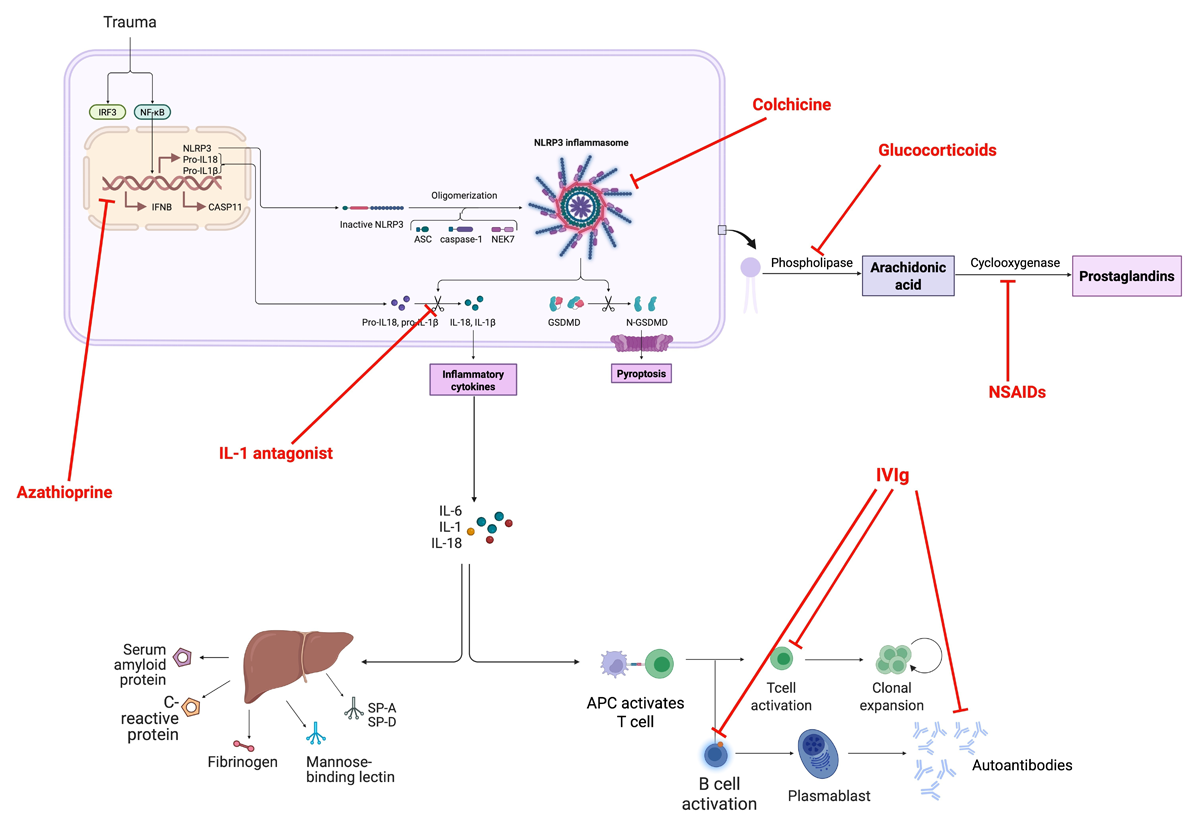
Figure 2
Pathophysiology and molecular targets in post-cardiac injury syndrome.
First-Line Therapy: Anti-inflammatory Medications and Colchicine
According to the 2025 ESC guidelines, nonsteroidal anti-inflammatory drugs (NSAIDs) together with colchicine are the first-line treatment for PCIS.1, 3, 21 This approach hastens symptom resolution and reduces the risk of recurrence. High-dose aspirin is often recommended as the NSAID of choice in post-MI cases (Dressler syndrome) or in patients who require antiplatelet therapy. In post-pericardiotomy cases without a recent MI, common NSAIDs used include ibuprofen or indomethacin, although aspirin is also effective. A typical regimen might be ibuprofen 600 to 800 mg every 6 to 8 hours, or aspirin on the order of 750 to 1000 mg every 6 to 8 hours, with doses tapered gradually over weeks. The course of NSAIDs generally continues for several weeks (often 2-4 weeks at full dose) until patients have no symptoms and inflammatory markers have normalized, followed by a taper. It is crucial to ensure gastric protection (proton-pump inhibitor) in patients on high-dose NSAIDs, primarily when used for an extended period.1
Colchicine is a key adjunct in therapy. It is usually given in addition to NSAIDs for acute PCIS, as it has been proven to reduce the risk of both initial nonresponse and future recurrences of pericarditis. Colchicine dosing for adults is typically 0.5 once daily (for body weight < 70 kg) or twice daily (for body weight ≥ 70 kilograms), continued for at least 3 months (some guidelines suggest 3-6 months). Colchicine therapy in PCIS is extrapolated from extensive evidence in idiopathic and recurrent pericarditis: a meta-analysis of clinical trials confirmed that colchicine added to anti-inflammatories significantly improves remission rates and reduces recurrences.1, 22 In the context PCIS, the COlchicine for the Prevention of the Post-Pericardiotomy Syndrome (COPPS) trial and subsequent studies demonstrated that colchicine administration can halve the incidence of PPS compared with placebo.2, 21 Consequently, the ESC guidelines recommend colchicine for PCIS treatment and even as prophylaxis in surgical patients (see below) if there are no contraindications (eg, severe renal impairment).1, 3
Patients usually experience improvement in fever and chest pain within a few days of starting NSAIDs and colchicine. If a large pericardial effusion is present, serial echocardiography is advisable to ensure the effusion is not progressing; typically, with anti-inflammatory therapy, the effusion will stabilize and then regress. Therapeutic pericardiocentesis is not routinely needed unless there is hemodynamic compromise (tamponade) or diagnostic uncertainty. In cases of cardiac tamponade physiology, pericardial drainage is indicated emergently, even as medical therapy is initiated.1, 2, 3, 21
Compared with placebo, perioperative colchicine use decreased the incidence of postpericardiotomy syndrome but not of postoperative atrial fibrillation (AF) or postoperative pericardial/pleural effusion in individuals following cardiac surgery.3
Corticosteroids and Immunotherapy for Refractory Cases
Most patients respond well to NSAIDs plus colchicine, but a subset with refractory or recurrent PCIS may require additional therapy. Corticosteroids can be very effective anti-inflammatories in this context. Still, they are generally reserved for second-line treatment due to concerns that high-dose steroids might increase recurrence rates if tapered too quickly. If needed for symptom control (for instance, if NSAIDs are contraindicated or ineffective), a low-to-moderate dose of corticosteroid such as prednisone (eg, 0.25-0.5 mg/kg/day) can be used. It is vital to use the lowest effective steroid dose and taper slowly. Studies in recurrent pericarditis have shown that using high initial steroid doses (≈ 1 mg/kg) leads to more frequent relapses than using moderate doses (≤ 0.5 mg/kg). Therefore, current practice is to avoid high-dose steroids in PCIS unless necessary and to attempt a slow taper (over several weeks or even months) once the patient’s condition improves.1
For patients who are corticosteroid-dependent or have multiple recurrences despite NSAIDs, colchicine, and a steroid, the introduction of immunomodulatory therapy is recommended. Notably, IL-1 blockade has emerged as an effective strategy in recurrent pericarditis, including post-cardiac injury cases.1 The IL-1 receptor antagonist anakinra has been shown to induce remission in refractory pericarditis and to prevent progression to constrictive pericarditis in cases with incessant inflammation. The 2025 ESC guidelines give a Class I recommendation for IL-1 antagonists (eg, anakinra, and, by extension, newer agents such as rilonacept) in patients with refractory PCIS to avert recurrences and avoid chronic constriction. Typically, anakinra is initiated at 100 mg daily subcutaneously (with dose adjustments for pediatric patients’ weight or renal function as needed), and a clinical response, including symptom relief and C-reactive protein reduction, is often seen within days to a couple of weeks. Therapy is usually continued for at least 6 months or longer in recurrent pericarditis to ensure sustained remission, with colchicine often maintained concurrently.1, 6
Other immunosuppressive agents have been used in severe cases, including azathioprine or IV immunoglobulin (IVIG) for recurrent pericarditis that is intolerant or unresponsive to anakinra and steroids (Figure 3).7 These therapies are considered third-line treatment and tailored to individual cases, often in consultation with a tertiary care center. Their evidence in PCIS specifically is limited to case series, but they are part of the armamentarium for complex cases (Table 3).1
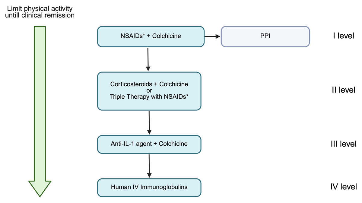
Figure 3
Step-by-step suggested management algorithm for post-cardiac injury syndrome. PPI: proton pump inhibitors; NSAIDs: nonsteroidal anti-inflammatory drugs; IL-1: interleukin-1; IV: intravenous
*NSAIDs or aspirin
Table 3
Drugs for post-cardiac injury syndrome according to 2025 European Society of Cardiology guidelines. CRP: C-reactive protein; ESC: European Society of Cardiology
| DRUG CLASS | DRUG | RECOMMENDED DOSE | DURATION | ESC RECOMMENDATION (2025) | COMMENTS/PRECAUTIONS |
|---|---|---|---|---|---|
| Nonsteroidal Anti-Inflammatory Drugs (NSAIDs) | Aspirin | 750-1000 mg every 6-8 h | 1-2 weeks, then gradual taper guided by symptoms and CRP | Class I, Level B | Preferred after myocardial infarction or when antiplatelet therapy is required |
| Ibuprofen | 600-800 mg every 6-8 h | 1-2 weeks, then taper | Class I, Level B | Common first-line agent after cardiac surgery; use gastroprotection | |
| Indomethacin | 25-50 mg every 8 h | Short-term | Class IIb, Level C | Less commonly used; higher risk of adverse effects | |
| Colchicine | Colchicine | 0.5 mg once daily (< 70 kg) or twice daily (≥ 70 kg) | 3 months | Class I, Level A | Reduces symptom duration and recurrence; dose adjusted for renal impairment |
| Corticosteroids | Prednisone | 0.25-0.5 mg/kg/day | Short course with slow taper | Class IIa, Level C | Reserved for NSAID/colchicine intolerance or refractory disease; avoid high doses |
| Interleukin-1 Inhibitors | Anakinra | 100 mg subcutaneously daily | ≥ 6 months, individualized | Class I, Level B | For recurrent or refractory PCS, rapid symptom and CRP response |
| Rilonacept | Loading 320 mg, then 160 mg weekly | Long-term, not known | Class IIa, Level B | Alternative IL-1 blockade in recurrent pericarditis | |
| Adjunctive Therapy | Proton-pump inhibitor | Standard dose | During NSAID therapy | Good clinical practice | Gastrointestinal protection |
| Preventive Therapy | Colchicine (prophylaxis) | 0.5 mg once or twice daily | 1 month | Class IIa, Level A | Start 48-72 h before cardiac surgery to prevent PCS |
[i] Notes: Tapering should be guided by clinical resolution and normalization of inflammatory markers, not by fixed time intervals. Antibiotics are not indicated unless the infection is proven.
Nonpharmacological Therapy
Pericardiectomy is a nonpharmacological therapy option that may be considered when medication treatment becomes unsatisfactory.1
Prevention of Post-Cardiac Injury Syndrome
Preventive strategies have been studied, especially for patients undergoing cardiac surgery who are at risk for PPS. The most robust evidence for prevention is with colchicine prophylaxis. The COPPS trial (2010) and COPPS-2 trial (2014) found that giving colchicine around the time of cardiac surgery significantly reduced the incidence of PPS in the postoperative period. In COPPS, colchicine was started on postoperative day 3 and continued for 1 month, resulting in a reduction in PPS from 21% (placebo group) to 11% in the colchicine group. COPPS-2 specifically examined starting colchicine preoperatively (48-72 hours before surgery) to improve tolerability and found a reduction in PPS occurrence.2, 3 Based on these findings, the latest ESC guidelines recommend in Class IIa level of evidence A to use colchicine starting 48 to 72 hours prior to cardiac surgery and continued for a month to prevent PCIS in patients who were not contraindicated.1 This practice is increasingly adopted, particularly in higher-risk patients, unless there are contraindications such as significant liver disease, renal failure, or intolerance.1, 2, 3, 21
In contrast, corticosteroid prophylaxis has not shown clear benefit and is not recommended for routine use to prevent PPS.7, 9, 23 Some earlier studies in pediatric cardiac surgery suggested a possible reduction in PPS with a short course of steroids, but larger analyses (including a 2014 Dexamethasone for Cardiac Surgery substudy) did not find a significant preventive effect.24 Similarly, attempts to use NSAIDs prophylactically (eg, high-dose aspirin started postoperatively) have not conclusively prevented PPS in clinical trials, and they carry risks such as bleeding.25 One exception is a small study that indicated indomethacin given before surgery might reduce PPS incidence, but this finding requires further confirmation and has not become standard practice. Therefore, colchicine is the leading evidence-based prophylactic agent for PPS.26
All patients who have experienced PCIS should be followed clinically for some time after recovery. The prognosis of PCIS is generally good since most patients have complete resolution with appropriate therapy. However, recurrences occur in an estimated 10% to 15% of cases despite initial treatment.4, 5 These recurrences are usually manageable with repeat anti-inflammatory therapy, often with more extended tapering periods and adjuncts as described above. A tiny minority (around 0.5% of PPS patients) may develop constrictive pericarditis due to chronic inflammation and scarring. Consequently, current guidelines advise careful follow-up of PCIS patients, especially those with recurrent episodes, to detect any progression towards constrictive physiology. If constrictive pericarditis is suspected (eg, signs of diastolic heart failure, pericardial knock on exam, or septal bounce on echo), timely evaluation with imaging and pericardial biopsy may be warranted, and some patients could ultimately require surgical pericardiectomy.1 Fortunately, such outcomes are rare in the era of early and aggressive anti-inflammatory treatment (Table 4).
Table 4
Clinical trials on the treatment and prevention of the post-cardiac injury syndrome. AF: atrial fibrillation; CPB:, cardiopulmonary bypass; GI:, gastrointestinal; NNH: number needed to harm; NNT: number needed to treat; PCS: post-cardiac injury syndrome; PPS: post-pericardiotomy syndrome
| STUDY (YEAR) | DESIGN/SETTING | POPULATION | INTERVENTION vs PLACEBO | PRIMARY ENDPOINT | MAIN RESULTS(PCS/PPS-RELATED) | KEY SAFETY FINDINGS |
|---|---|---|---|---|---|---|
| Horneffer et al. (1990) | Randomized, double-blind, placebo-controlled trial | Adults with established PPS after cardiac surgery (149 enrolled) | Ibuprofen or indomethacin (treatment) | Resolution ≥ 2 of fever, chest pain, rub within 48 h | Ibuprofen 90.2% and indomethacin 88.7% effective vs placebo 62.5% (P = .003) | Side effects were low and similar across groups; no difference in length of stay or significant effusions reported |
| COPPS (Imazio et al., 2010) | Multicenter, randomized, double-blind, placebo-controlled | Adults after cardiac surgery (colchicine started post-op day 3) | Colchicine | PPS incidence at 12 months | PPS reduced: 8.9% vs 21.1% (P = .002; NNT = 8). Secondary composite (hospitalization/tamponade/constriction/relapse) also decreased (0.6% vs 5.0%, P = .024) | GI intolerance similar: 8.9% vs 5.0% (P = .212) |
| COPPS-2 (Imazio et al., 2014) | Multicenter, randomized, placebo-controlled | Adults undergoing cardiac surgery (colchicine started 48–72 h pre-op, continued 1 month) | Colchicine 0.5 mg OD (< 70 kg) or BD (≥ 70 kg) | PPS within 3 months | PPS reduced: 19.4% vs 29.4% (absolute difference 10.0%; NNT = 10). No significant difference for effusions; AF reduction only in on-treatment analysis | More adverse events with colchicine (20.0% vs 11.7%; NNH = 12), mainly GI; no serious adverse events |
| DECS Substudy (Bunge et al., 2014) | Substudy of RCT (valve surgery cohort), placebo-controlled | Adults undergoing valvular cardiac surgery (n = 822) | High-dose intra-op dexamethasone 1 mg/kg | PPS occurrence | No protective effect: PPS 13.5% vs 15.5% (RR 0.88; 95% CI, 0.63-1.22). Complicated PPS has also not been reduced | No PPS benefit; trial-level safety not PPS-specific here |
| Wilson et al. (1994) | Randomized, double-blind, placebo-controlled | Children with established PPS (n = 21) | Prednisone 2 mg/kg/day, taper to zero over 14 days | Remission at 72 h and 1 week | No difference at 72 h; higher remission at 1 week with prednisone (10/12 vs 3/9, P = .03) | Pericardial effusion enlargement was noted in two steroid-treated children |
| Mott et al. (2001) | Randomized, double-blind, placebo-controlled | Children undergoing cardiac surgery with CPB (analyzed n = 246) | Methylprednisolone 1 mg/kg pre-CPB + 4 doses over 24 h | PPS incidence/severity | No reduction in PPS incidence (overall PPS 16%; no inter-group difference, P = .73). Marginal increase in complicated PPS in steroid arm (P = .05) | Short-course steroid prophylaxis did not prevent PPS and may worsen complicated PPS |
Management in Special Populations
Pregnancy
Management of PCIS during pregnancy requires careful selection of pharmacological agents to ensure fetal safety. During pregnancy, until the 20th week of gestation, NSAIDs can be administered at high dosages.27, 28, 29 Among NSAIDs, ibuprofen is preferred due to its favorable pharmacokinetic profile and limited cross-placental transfer. After 20 weeks of gestation, only a low-dose aspirin (100 mg daily) may be continued until the third trimester.27, 28, 30, 31 Colchicine is considered safe in pregnancy, is highly effective in preventing recurrence of pericarditis, and is permitted throughout all trimesters of pregnancy.27, 32, 33 Corticosteroids can be used but only at the lowest effective dose, and low-to-moderate doses (typically ≤ 20 mg daily) are considered safe throughout pregnancy.27, 28, 31
While real-world data on anakinra during pregnancy are evolving, emerging evidence from case reports suggests it is effective in preventing recurrence, with no adverse fetal outcomes reported to date. According to a recently published case study, anakinra was effectively used during pregnancy and lactation in women with recurrent pericarditis, showing clinical efficacy—sustained remission of pericarditis symptoms and clearance of inflammatory markers—with no adverse pregnancy or fetal outcomes.34, 35, 36, 37
IVIG may be used throughout pregnancy for severe refractory disease and should be taken into consideration for the induction of remission in individuals with severe disease prior to conception.38
Pediatric Population
In the pediatric population, therapeutic management calls for particular concerns. Aspirin is not advised for children under the age of 12 due to Reye’s syndrome and hepatotoxicity, although NSAIDs and colchicine are advised as first-line therapy. Corticosteroids may be administered to individuals who are refractory, have contraindications, or are unable to take first-line medications, but chronic corticosteroid therapy is contraindicated in pediatric patients, and IL-1 drugs may be taken into consideration.39
Geriatric Population
Therapeutic management in geriatric population demands a tailored approach focused on drug safety and comorbidity management. NSAIDs have risks in elderly patients, including gastrointestinal bleeding and fluid retention that may exacerbate underlying heart failure. It is necessary to adjust the dose according to the eventual renal impairment. Consequently, the ESC guidelines emphasize the mandatory use of gastroprotection (proton-pump inhibitors) and careful tapering in these high-risk individuals. Colchicine must be used at low dose and adjusted by weight and renal function to avoid toxicity.27
Future Drug Directions
New drugs for pericarditis are now under development that specifically target the inflammasome acivation at different levels.40 For example, VTX2735 is an oral small-molecule inhibitor currently in a phase II trial. It functions by preventing the assembly of the NLRP3 inflammasome, thereby stopping IL-1 production at the source. This offers the potential for controlling inflammation without requiring biologic injections.41 CardiolRx (CT-100) is a cannabidiol derivative that modulates the NLRP3 inflammasome. Following positive phase II results showing reduced flares, it has moved to a pivotal phase III trial (MAVERIC). It is notable for being an oral, non-immunosuppressive agent.42
The IL-1 blockade has become a cornerstone in the management of pericarditis; in fact, different molecules are being studied. Anakinra, a recombinant IL-1 receptor antagonist, is administered daily by subcutaneous injection. The Anakinra-Treatment of Recurrent Idiopathic Pericarditis (AIRTRIP) trial showed that it drastically reduced recurrence (18% vs 90% on placebo) in steroid-dependent patients.43 Rilonacept is a soluble IL-1 receptor “trap” that neutralizes both IL-1α and IL-1β. It is the first therapy approved by the US Food and Drug Adminstration for recurrent pericarditis following the RHAPSODY (Study to Assess the Efficacy and Safety of Rilonacept Treatment in Participants With Recurrent Pericarditis) trial, where it nearly eliminated recurrences (7% vs 74% on placebo).44 Goflikicept, a fusion protein similar to rilonacept (IL-1α/β trap), is currently in mid-stage development and has shown promise in recent phase II/III trials.45 Canakinumab, a monoclonal antibody targeting IL-1β, is presently supported only by case reports.46 KPL-387, a long-acting anti-IL-1 receptor antibody, is slated for phase II/III trials in 2025; it aims to offer a monthly dosing alternative to daily anakinra, pending safety and cost-benefit evaluations.47
While the pipeline is dominated by IL-1/inflammasome inhibitors, other mechanisms, such as IL-6 blockade (eg, tocilizumab), are being explored theoretically, though they currently lack formal trial data.48
Conclusion
Post-cardiac injury syndrome is an essential postoperative and post-infarction complication characterized by pericardial inflammation due to an immune response to cardiac injury. It encompasses post-pericardiotomy, post-MI (Dressler syndrome), and other post-traumatic pericarditis syndromes. Timely recognition of the syndrome is facilitated by its classic presentation of fever, pleuritic chest pain, pericardial friction rub, and effusions arising a few weeks after a cardiac event. The evaluation should confirm the diagnosis of inflammatory pericarditis and exclude other causes of the symptoms.
The cornerstone of management is anti-inflammatory therapy (high-dose aspirin/NSAIDs and colchicine), which is highly effective at relieving symptoms and preventing recurrences in most patients. The addition of colchicine has substantially improved outcomes and is also helpful as a preventive measure in high-risk surgical patients. In cases of refractory or recurrent PCIS, advanced therapies such as IL-1 inhibitors have shown excellent results in achieving remission. With appropriate treatment, serious complications like tamponade or constrictive pericarditis are uncommon, but careful follow-up is advised. Ongoing research and the latest guidelines continue to refine diagnostic criteria and treatments, promising even better prevention and management of post-cardiac injury syndrome in the future.
Key Points
Post-cardiac injury syndrome (PCIS) is an immune-mediated pericarditis that occurs after cardiac surgery, myocardial infarction (MI), or other cardiac injuries, presenting with fever, chest pain, and pericardial/pleural effusions.
Incidence of post-pericardiotomy syndrome ranges from ~10% to 30% after cardiac surgery, with higher rates in specific surgeries and pediatric patients. Dressler’s post-MI pericarditis is now rare (< 1% of MIs) in the reperfusion era.
Diagnosis is clinical, requiring at least two of the following: fever without other cause, pericarditic chest pain, pericardial/pleural rub, new effusion, and elevated inflammatory markers. electrocardiography, echocardiography, and inflammatory labs help confirm pericarditis and an effusion.
First-line treatment is aspirin or nonsteroidal anti-inflammatory drugs (NSAIDs) plus colchicine, which hasten symptom resolution and reduce recurrences. High-dose aspirin is preferred after MI, while ibuprofen/NSAID is used post-surgery, with colchicine given for 3 months.
Colchicine prophylaxis around cardiac surgery (starting 2-3 days preoperatively) significantly lowers the risk of PCIS and is recommended in the 2025 European Society of Cardiology guidelines (Class IIa).
Refractory or recurrent PCIS is managed with corticosteroids (low-moderate dose) and, if needed, interleukin-1 blockers like anakinra to achieve remission and prevent progression to constrictive pericarditis. Follow-up is essential, as a small percentage of patients can develop constrictive pericarditis over time.
Competing Interests
The authors have no competing interests to declare.