
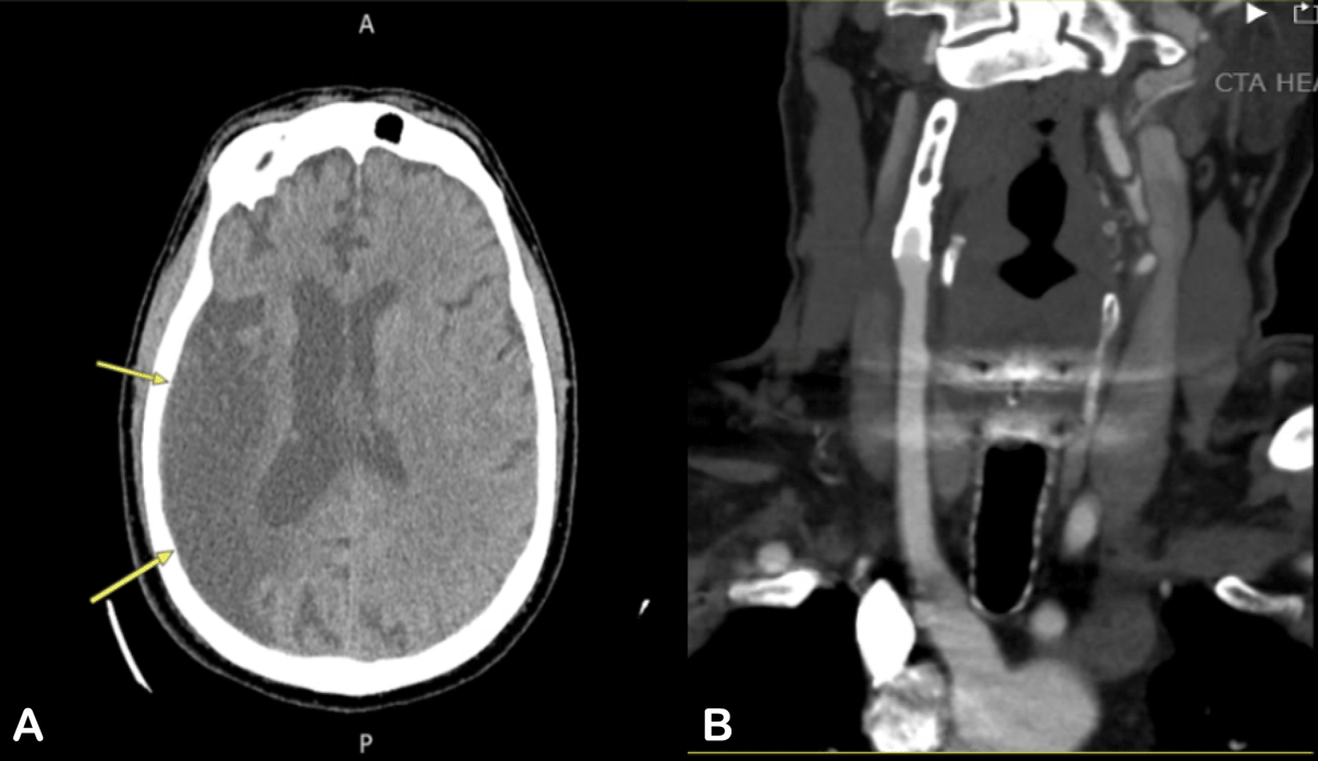
Figure 1
(A) Computed tomography (CT) of the head without contrast (yellow arrows) showing previous right middle cerebral artery territory infarct. (B) CT angiography of the head and neck show right internal carotid artery with stent in-situ and no distal flow, suggestive of chronic occlusion.
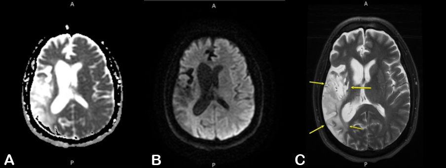
Figure 2
Magnetic resonance imaging T2 sequences (left to right). (A) diffusion coefficient map, (B) fluid-attenuated inversion recovery and (C) axial T2 (arrows demarcating previously infarcted middle cerebral artery territory).