
Introduction
Left atrial appendage occlusion (LAAO) with the Watchman device (Boston Scientific) is increasingly used as an alternative strategy for stroke prevention in patients with atrial fibrillation who are unable to tolerate long-term anticoagulation. Although large trials and real-world registries have demonstrated its efficacy and safety, periprocedural neurologic complications such as stroke, transient ischemic attack, or device-related thromboembolism—while uncommon—can still occur.1,2 Acute visual loss following structural cardiac procedures is particularly challenging because potential etiologies include retinal ischemia, ophthalmic artery occlusion, posterior circulation stroke, posterior reversible encephalopathy syndrome, and contrast-induced cortical blindness (CICB).3
CICB is a rare but recognized phenomenon characterized by transient bilateral visual loss after exposure to iodinated contrast, often with preserved pupillary responses and normal fundoscopic examination. Patients with preexisting cerebrovascular disease or chronic large-vessel occlusion may be more susceptible to transient cortical dysfunction due to impaired autoregulation or increased blood–brain barrier permeability.4,5 Given the rise in LAAO procedures and the diagnostic complexity of periprocedural neurologic deficits, it is essential to distinguish ischemic causes from reversible contrast-related neurotoxicity. We present a case of acute cortical blindness and hemispatial neglect following Watchman implantation, ultimately consistent with contrast-induced cortical blindness, underscoring the importance of early multidisciplinary assessment and advanced neuroimaging.
History of Presenting Illness and Medical History
A 66-year-old male with a history of ischemic right middle cerebral artery (MCA) stroke (March 2024) complicated by residual left-sided deficits (contracture of left upper extremity, weakness in left lower extremity), chronic right internal carotid artery (ICA) occlusion, long-standing persistent atrial fibrillation (questionable Xarelto adherence), hypertension, and depression underwent a planned Watchman LAAO procedure.
Immediately post-procedure, it was noted that the patient appeared confused, unable to visually track, persistently looked leftward, and reported severe headache with acute bilateral vision loss. Pre-procedure, he was highly functional and independent of activities of daily living.
A stroke code was called, and the patient was assessed with the following vitals: temperature of 98.5°F, heart rate of 71 beats/min, blood pressure of 160/78 mm Hg, respiratory rate of 18, and oxygen saturation of 95% on room air. A neurological exam showed left gaze preference with inability to cross midline, right hemi spatial neglect, and worsened left-sided weakness.
Further Evaluation, Management, and Differential Diagnosis
Ophthalmology and Vascular Neurology were consulted for further evaluation, and a stroke protocol was initiated.
On examination, the patient’s visual acuity was limited to light perception bilaterally, with previously documented 20/20 vision during a recent outpatient visit. Pupils were equal, round, and reactive to light, although a trace relative afferent pupillary defect was noted on the left. Intraocular pressures were within normal limits, and fundoscopic examination revealed healthy-appearing optic nerves without evidence of retinal whitening, ischemia, or visible emboli. A left gaze preference was observed, although the patient was unable to cooperate fully with extraocular movement testing.
Computed tomography (CT) of the head showed no acute infarct or hemorrhage, and CT angiography with brain perfusion revealed chronic right ICA occlusion with diminished right MCA and anterior cerebral artery flow with no new occlusion (Figure 1).
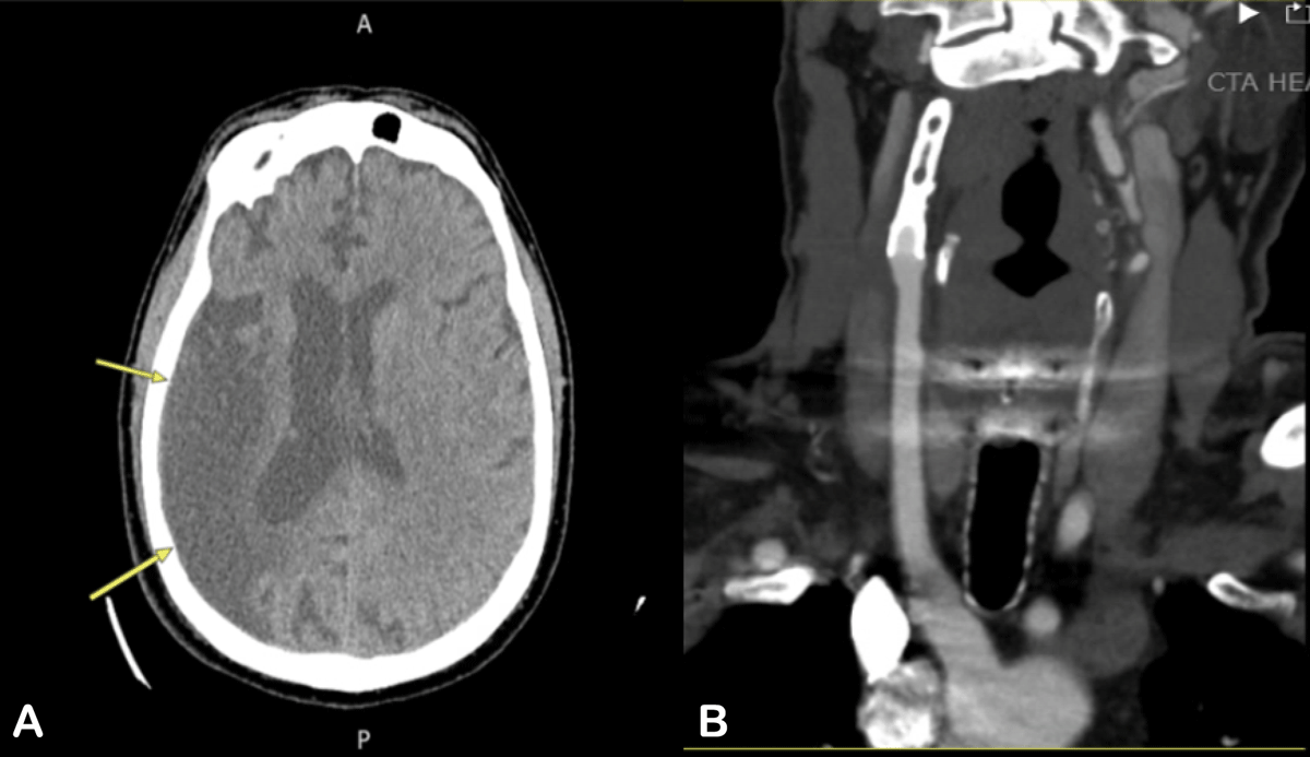
Figure 1
(A) Computed tomography (CT) of the head without contrast (yellow arrows) showing previous right middle cerebral artery territory infarct. (B) CT angiography of the head and neck show right internal carotid artery with stent in-situ and no distal flow, suggestive of chronic occlusion.
Given clinical findings and negative imaging, the vascular neurology consult determined that the patient was not a candidate for mechanical thrombectomy due to his known chronic right internal carotid artery (ICA) occlusion and no new major vessel occlusion. The use of tenecteplase was also contraindicated due to intraprocedural heparin administration. The neurology team recommended initiation of low-intensity heparin therapy for anticoagulation and advised further diagnostic evaluation to identify the etiology of acute visual loss.
Broad differentials were considered at this point, including bilateral occipital lobe infarctions resulting in cortical blindness, bilateral retinal or ophthalmic artery occlusions, bilateral ischemic optic neuropathy, cerebral air embolism, post-stroke recrudescence (possibly secondary to peri-procedural hypotension or hypoxia), and posterior reversible encephalopathy syndrome. Additionally, CICB was considered since this entity can present with transient bilateral vision loss following exposure to iodinated contrast, often with normal pupillary responses and fundus examination, and has the potential for spontaneous visual recovery.
Follow-up
Magnetic resonance imaging of the brain to further evaluate occipital pathology was obtained, demonstrating chronic occlusion of the right ICA with a large area of encephalomalacia in the right ACA and MCA territories and no acute restricted diffusion or intracranial hemorrhage (Figure 2). Therefore, recent ischemic or hemorrhagic lesions in this region were excluded.
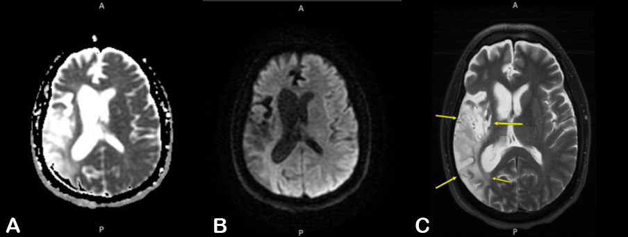
Figure 2
Magnetic resonance imaging T2 sequences (left to right). (A) diffusion coefficient map, (B) fluid-attenuated inversion recovery and (C) axial T2 (arrows demarcating previously infarcted middle cerebral artery territory).
Additionally, as per multispecialty recommendations, the patient was continued on secondary stroke prevention, anticoagulation, and high-flow oxygen therapy to support cerebral oxygenation. No specific ophthalmic intervention was indicated at that time. The patient woke up the next day with improved vision to finger counting in both eyes and no gaze preference, and he was completely back to baseline on day two after the procedure.
Discussion
There was no discernible objective pathology identified that could account for the patient’s presentation. An embolism originating during the procedure was highly improbable because it would be exceedingly unlikely for embolic material to selectively migrate into both ophthalmic arteries, particularly in the presence of a chronic right ICA occlusion and negative pre-procedure transesophageal echocardiogram immediately prior to device implantation for left atrial appendage or intracardiac thrombus. In addition, the patient had a normal ophthalmologic examination, which showed no evidence of retinal or optic nerve ischemia. Similarly, an embolic event involving the bilateral distal posterior cerebral artery (PCA) territories is also an unlikely explanation since such a process would typically produce additional neurological deficits and would not be expected to result in complete bilateral vision loss with otherwise intact neurological function. This diagnosis is further negated by the normal CTA, CT perfusion, and magnetic resonance imaging findings, which demonstrated no perfusion deficits or vascular occlusions in the posterior circulation. The procedure was performed under moderate sedation without general anesthesia. There were no documented episodes of periprocedural hypotension or hypoxia, which could explain recrudescence. This makes CICB the most probable diagnosis.5
The patient’s rapid improvement regaining finger-counting vision by the next morning and returning to baseline within 48 hours further supports CICB, which is typically self-limited, with spontaneous resolution reported in most cases.6 High-flow oxygen therapy and supportive neurologic monitoring were appropriately pursued while ischemia was being ruled out.
This case underscores the importance of considering nonischemic cortical processes, such as contrast neurotoxicity, in the differential diagnosis of acute bilateral visual loss following structural cardiac interventions. Early multidisciplinary involvement, particularly neurology, ophthalmology, and radiology, are critical in distinguishing CICB from posterior circulation stroke or embolic complications, thereby preventing unnecessary interventions and guiding appropriate management.
Competing Interests
The authors have no competing interests to declare.