
Introduction
Transthoracic echocardiography (TTE) remains the standard technique for assessing left ventricular function and evaluating myocardial structure.1 Routine monitoring of ejection fraction (EF) is critical in monitoring for cardiotoxic effects of chemotherapy.1 Additionally, global longitudinal strain (GLS) by speckle-tracking echocardiography (STE) is highly sensitive in detecting early myocardial dysfunction, even before a decline in EF becomes apparent.1
Two-dimensional (2D) echocardiography is essential for visualizing intracardiac masses, which are most commonly attributable to thrombi, vegetations, or myxoma.2 Myocardial metastases are less common, with an incidence ranging from 2.3% to 18.3% depending on cancer subtype.3,4 A multimodal imaging approach using ultrasound enhancing agents, transesophageal echocardiography (TEE), or cardiac magnetic resonance (CMR) imaging is recommended for better characterization and diagnosis of unknown cardiac mass.5
There are no published reports of using GLS to localize intramyocardial metastases prior to cardiac magnetic resonance (CMR) imaging confirmation. We present a case in which a cardiac metastasis was highly suspected and localized on GLS imaging on TTE and subsequently confirmed by CMR imaging.
Case Presentation
A 60-year-old man with a history of renal cell carcinoma with metastatic disease to the lung and brain—status post primary resection, stereotactic radiosurgery, and immunotherapy—was noted to have findings concerning for subacute embolic left parietal stroke on routine restaging imaging. Non-oncologic chronic comorbidities included hypertension, hyperlipidemia, diabetes mellitus, and class I obesity. He had no known preexisting cardiovascular disease.
This index diagnosis of stroke triggered an additional panel of imaging tests, including carotid artery duplex ultrasonography, TTE with agitated saline, and upper- and lower-extremity venous duplex ultrasonography. Echocardiography with agitated saline contrast was positive for interatrial shunt only with Valsalva maneuver. Interatrial shunting compatible with patent foramen ovale (PFO) was confirmed on TEE. Notably, echocardiography revealed focal thickening of the mid inferior and inferoseptal walls of the left ventricle, suspicious for hypertrophy of the posteromedial papillary muscle, although the differential included metastatic infiltration of the myocardium or, less likely, adherent thrombus (Figure 1). Contrast echocardiography with an echo enhancing agent was not performed. Tissue deformation of this region was markedly reduced relative to the rest of the myocardium, favoring infiltration (Figure 2). Lower- and upper-extremity ultrasonography was negative for deep vein thrombi.
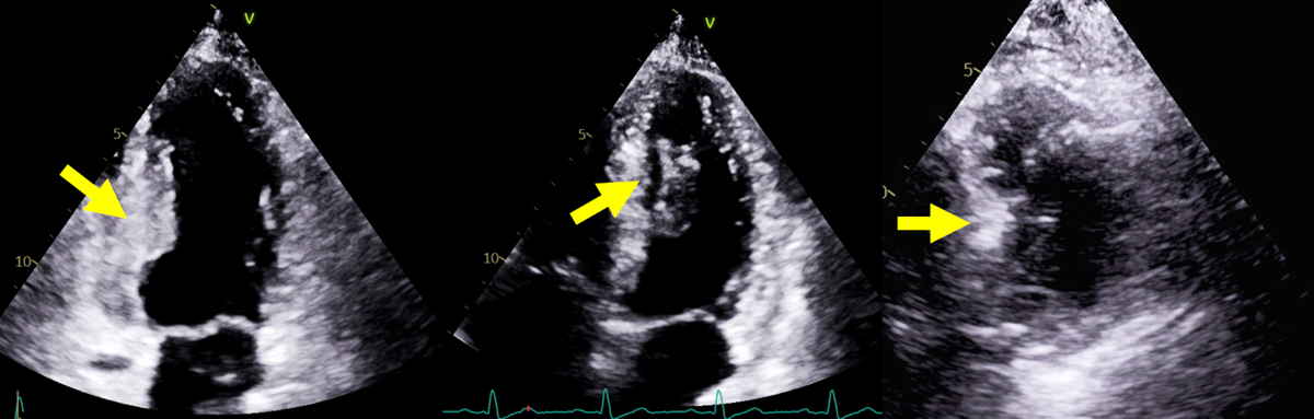
Figure 1
Transthoracic echocardiography (TTE) images. 2- and 4-chamber TTE images with significant thickening (1.4 cm) within the mid inferior and inferoseptal walls (yellow arrows). Short axis imaging on echocardiography was technically limited, however there was no visible mass. Contrast echocardiography was not performed.
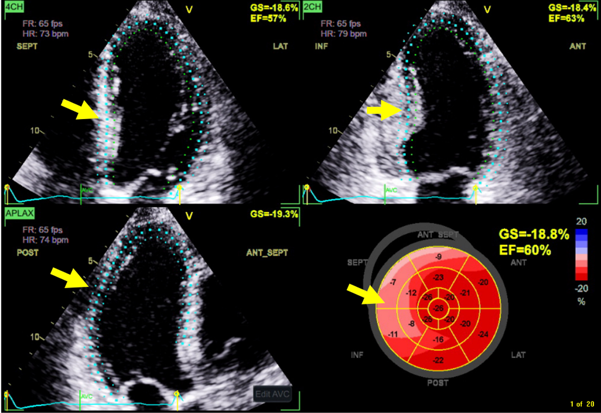
Figure 2
Despite overall preservation of GLS, the inferior and inferoseptal segments had significant reduction in regional GLS. Yellow arrows refer to location of mass. GLS: global longitudinal strain
CMR imaging was obtained and revealed a 2.7 × 2.3 cm mass located in the inferior and inferoseptal walls, hyperintense on both T1- and T2-weighted sequences, mild peripheral contrast uptake on early perfusion imaging, and marked contrast uptake on late gadolinium enhancement images (Figure 3). The CMR findings were consistent with intracardiac metastasis.
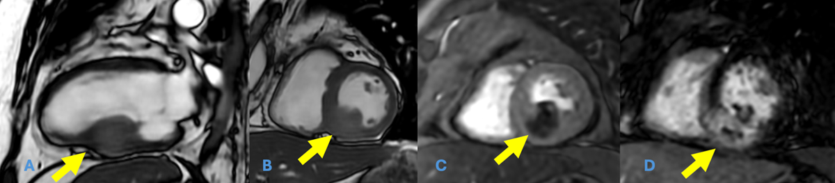
Figure 3
(A, B) Steady state free precession cine images reveal a 2.7 × 2.3 cm mass within the mid inferior and inferoseptal wall (yellow arrows). (C) Perfusion imaging with mild contrast uptake. (D) Diffuse late gadolinium enhancement throughout the mass.
In the absence of venous thrombi of the upper and lower extremity and without expansion of the tumor into the inferior vena cava, the cerebrovascular infarct was most likely related to the intracardiac masses as opposed to paradoxical embolism from venous sites. Additionally, routine hospital telemetry detected no episodes of atrial fibrillation, and magnetic resonance angiography of the head showed no large vessel occlusion. Antithrombotic therapy was escalated from aspirin to therapeutic anticoagulation with apixaban for presumed secondary prophylaxis of cardioembolic events. PFO closure was not indicated in this setting of an intracardiac source requiring prolonged anticoagulation. The patient was then discharged home on hospital day 6 with outpatient oncology follow-up to discuss systemic chemotherapy, and he was ultimately switched to targeted oral therapy with belzutifan given his progressive oncologic disease. Despite treatment, his cancer continued to progress, and the patient passed away at home 3 months later.
Discussion
The focal thickening of the inferior and inferoseptal myocardium along with reduced GLS in the associated segments on echocardiography were later confirmed by CMR tissue characteristics to be consistent with intracardiac metastasis. This was crucial for changing the long-term antithrombotic strategy from single antiplatelet monotherapy to therapeutic anticoagulation and a comprehensive oncologic reevaluation of treatment strategy.
Although TTE and wall thickness evaluation are standard tools to assess cardiac structure, localized abnormalities with GLS offer additional valuable information and may prompt further targeted imaging and multimodality confirmation for proper diagnosis.
Conclusion
In addition to focal hypertrophy, reduced GLS on TTE should prompt targeted multimodality imaging for diagnostic confirmation.
Competing Interests
Dr. Deswal is cochairman of the executive committee, clinical heart failure trial sponsored by Bayer. The other authors have no competing interests to declare.