
A 40-year-old asymptomatic male patient was incidentally detected to have an echogenic mass measuring 5.1 × 4.2 cm in the region of the basal interventricular septum on transthoracic echocardiogram (TTE) and transesophageal echocardiogram (TEE) during the workup for renal transplant donation (Figure 1). The valves were normal with normal biventricular function. Cardiac magnetic resonance was considered for further characterization and exact location of the tumor (Figure 2A–G). It revealed a well-defined intramyocardial lesion with the epicenter in the basal inferior, inferoseptal, and inferolateral walls. The myocardium was splayed by the tumor with intact myocardium all around. The lesion was hyperintense on T1- and T2-weighted images, with signal drop on fat suppression sequences. Post-contrast images demonstrated no contrast enhancement. Biventricular functions were preserved and no regional wall motion abnormality was seen. Computed tomography (CT) also confirmed the diffuse fatty attenuation of the lesion with no intralesional calcification (Figure 2H). These findings supported the diagnosis of intramyocardial lipoma arising from the left ventricular wall. Since the patient was asymptomatic, a conservative approach with regular follow-up was adopted. At 1-year clinical and echocardiographic follow-up, there was no change in the tumor size and the patient remains asymptomatic.
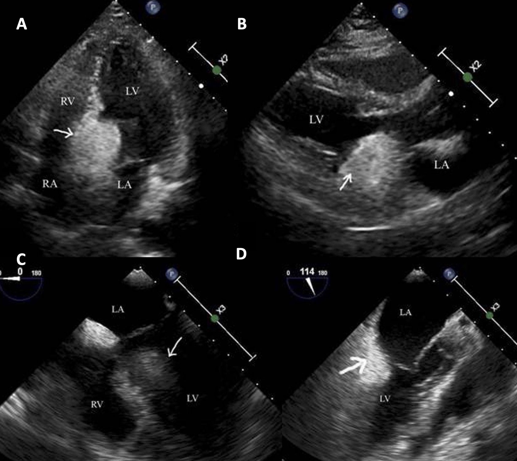
Figure 1
(A) Transthoracic echocardiogram in apical four-chamber view and (B) parasternal long-axis view showing a large echogenic mass in the region of the basal interventricular septum (depicted by white arrow) transesophageal echocardiogram in (C) four-chamber and (D) three-chamber views demonstrate the echodense mass. RV: right ventricle; LV: left ventricle; RA: right atrial; LA: left atrial
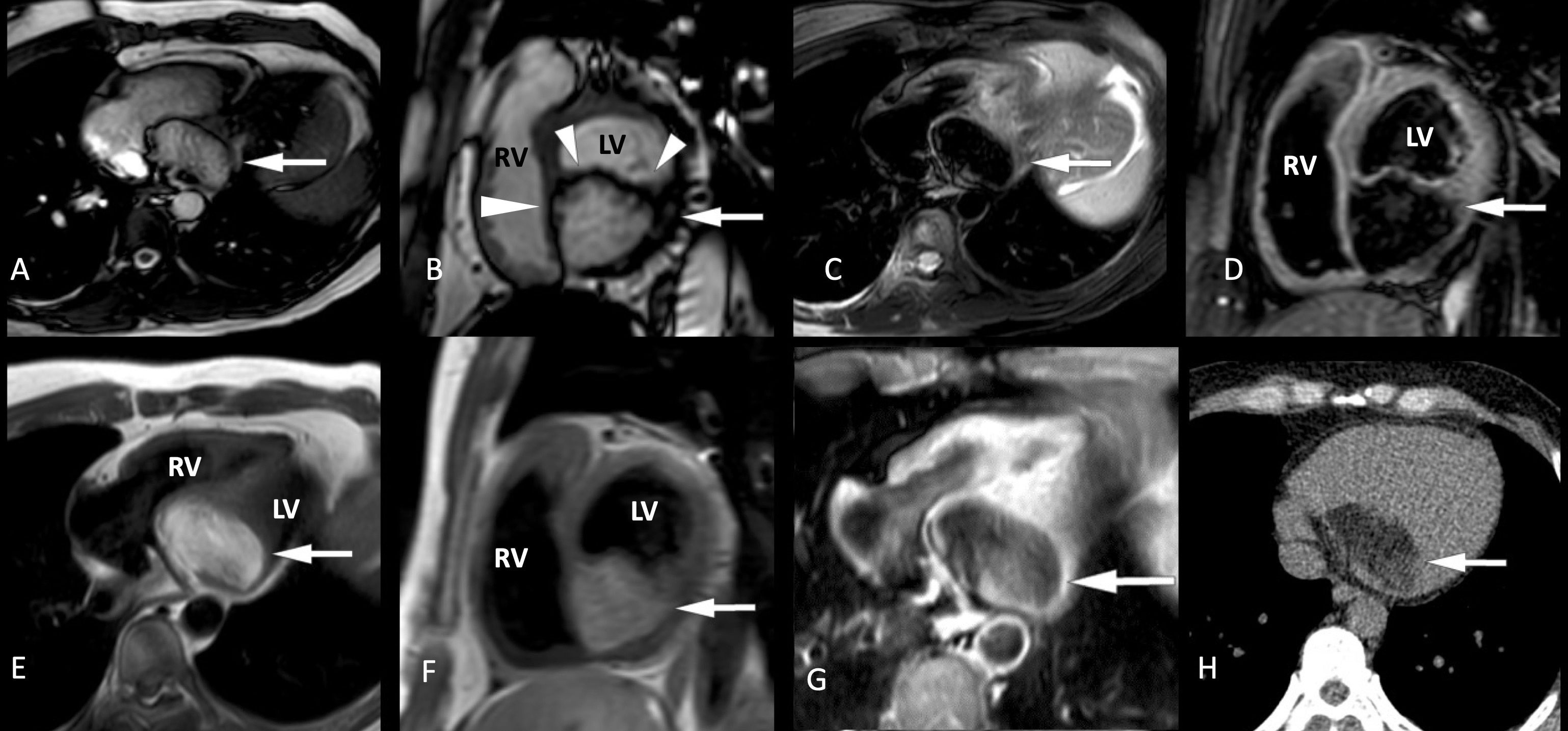
Figure 2
(A) Steady state free precession sequence in horizontal long-axis (HLA) and (B) short-axis (SA) view demonstrating a hyperintense lesion (depicted by white arrow) arising in the intramyocardial location from the basal inferior, inferoseptum, and inferolateral left ventricular wall. The lesion is (C) hypointense on T2 fat saturated (FS) HLA and (D) SA images and hyperintense on (E) T1 non-fat saturated HLA and (F) SA images. (G) Post-contrast T1 FS HLA shows no contrast enhancement. (H) Non-contrast-ECG gated CT scan of the chest confirmed the diffuse fatty attenuation of the lesion, with no intralesional calcification. ECG: electrocardiography; CT: computed tomography; RV: right ventricle; LV: left ventricle
Cardiac lipomas account for 8.4% of primary heart tumors and are most commonly diagnosed in the 40- to 60-year age group, with equal gender distribution.1 Most patients remain asymptomatic, and the lesion is detected incidentally. However, some may become symptomatic and present with shortness of breath, obstruction-related symptoms, and arrhythmias when the tumor is near the vital structures.2 Most cardiac lipomas are located in the left ventricular epicardial surface, interventricular septum, and right atrium.3 The occurrence of lipomas in the intramyocardial location, as seen in our case, is rare, with only a few cases reported in literature.4 Intramyocardial masses have the potential to affect the conduction system and therefore can cause arrythmias. TTE is usually the first-line investigation because it provides information regarding the shape, location, and any secondary changes in cardiac hemodynamics, but it may be inadequate for tissue characterization. MRI and CT together can provide an unequivocal diagnosis due to their ability to identify fat with a high degree of specificity. Asymptomatic cardiac lipomas may be managed conservatively with regular follow-up imaging. In symptomatic patients, however, surgical resection is curative.5
Competing Interests
The authors have no competing interests to declare.