
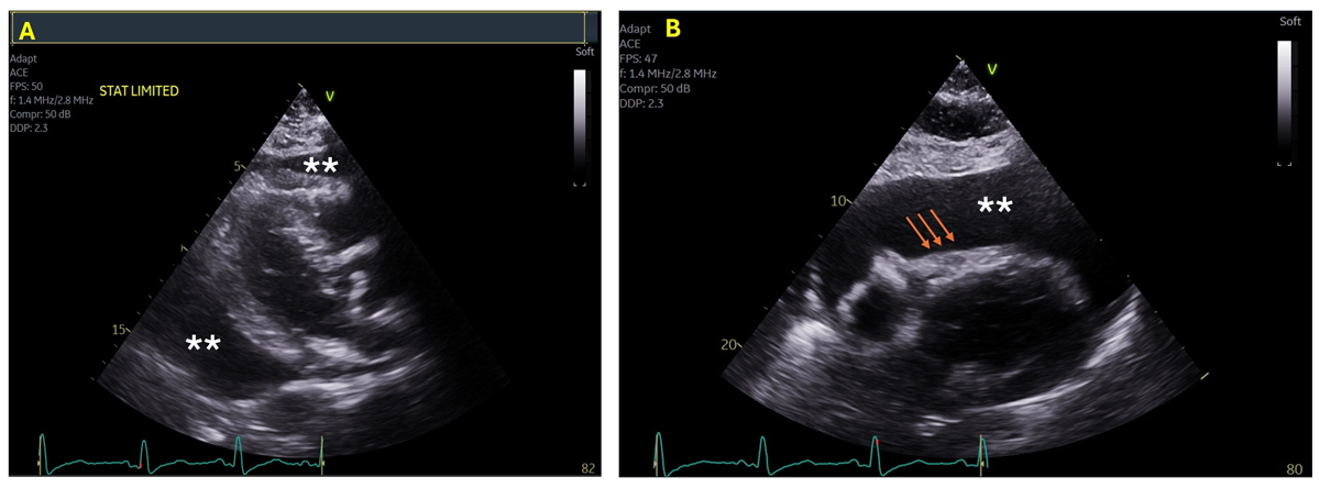
Figure 1
Transthoracic echocardiogram, parasternal long-axis view (Panel A), and subcostal view (Panel B) showing large circumferential pericardial effusion (asterisks) with right ventricular diastolic collapse (arrows).
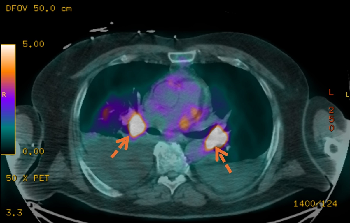
Figure 2
F-18 labeled fluorodeoxyglucose (FDG)-positron emission tomography showing extensive FDG avid disease in the chest (arrows). The primary malignancy appears to be a medial right lower lobe lung mass.