
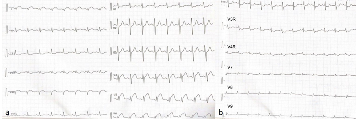
Figure 1
Electrocardiogram showing ST-segment elevation in (A) leads I, aVL and (B) V4-V9.
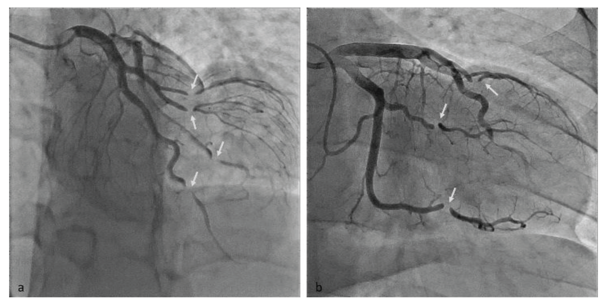
Figure 2
Coronary angiography showing (A) marked narrowing of the left anterior descending artery, left circumflex artery, and obtuse marginal branches; (B) subtotal stenoses of the left circumflex artery, obtuse marginal artery, and left anterior descending artery (arrows).
Video 1
Echocardiography showing akinesis and thickening of the apical left ventricular walls; see also at https://vimeo.com/1161503628/a15b7bf09f.
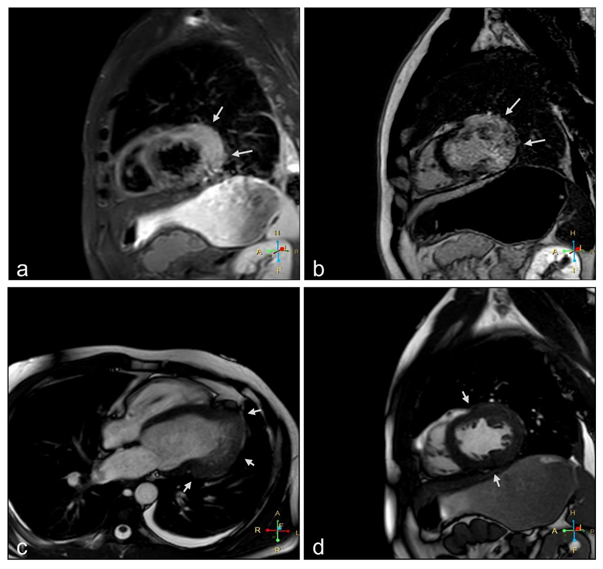
Figure 3
Cardiac magnetic resonance imaging (a) short-axis view shows myocardial edema and (b) late gadolinium enhancement in the left ventricle lateral wall; (c) 4-chamber apical view and (d) short-axis view showing a partial absence of the pericardium and myocardial indentation (arrows).