
Background
Congenital partial pericardial defects are rare and typically discovered incidentally during autopsy or thoracic surgery.1 While most pericardial defects are asymptomatic, some patients present with chest pain, dyspnea, palpitations, or syncope.2 Pericardial defects are known to cause heart murmurs on auscultation, cardiomegaly and heart displacement on the chest x-ray, arrhythmias, and pneumopericardium associated with the development of spontaneous pneumothorax.3 Extremely rare cases of fatal complications have been described, such as sudden cardiac arrest or myocardial infarction due to cardiac herniation through the defect.3,4
Given the rarity of these complications, partial pericardial defects are often not considered in the differential diagnosis of acute coronary syndrome. We present the case of a 33-year-old man with a previously undiagnosed congenital partial pericardial defect who presented with acute myocardial infarction (MI). Strangulation of the heart occurred due to spontaneous herniation of the left ventricle (LV) through the pericardial defect that caused compression of the coronary arteries, which led to the MI.
Case Presentation
A 33-year-old man with no significant medical history was admitted to the intensive care unit with acute-onset severe chest pain. An electrocardiogram demonstrated ST-segment elevation in leads I, aVL, and V4-V9 (Figure 1). He had received aspirin and clopidogrel before arrival.
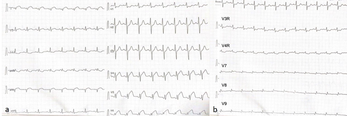
Figure 1
Electrocardiogram showing ST-segment elevation in (A) leads I, aVL and (B) V4-V9.
Emergency coronary angiography revealed subtotal narrowing of the left anterior descending artery, left circumflex artery, diagonal branches, and obtuse marginal artery (Figure 2). The lesions lacked characteristics of atherosclerotic plaque, thrombus, or embolic obstruction. Intracoronary nitroglycerin injection had no effect, arguing against vasospasm. The angiographic pattern suggested the possibility of extrinsic coronary arterial compression, and percutaneous coronary intervention was deferred.
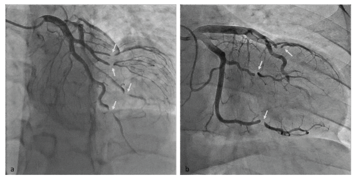
Figure 2
Coronary angiography showing (A) marked narrowing of the left anterior descending artery, left circumflex artery, and obtuse marginal branches; (B) subtotal stenoses of the left circumflex artery, obtuse marginal artery, and left anterior descending artery (arrows).
Laboratory tests showed significant elevation in troponin levels. Transthoracic echocardiography revealed a distorted LV shape with marked thickening and akinesis of the apical LV segments. The maximal apical LV wall thickness was approximately 17 mm. The LV ejection fraction was moderately reduced at 42% (Video 1). There was no pericardial effusion.
Video 1
Echocardiography showing akinesis and thickening of the apical left ventricular walls; see also at https://vimeo.com/1161503628/a15b7bf09f.
Alternative causes of acute coronary syndrome were considered. Based on the thickening of the LV apical segments observed on echocardiography, a cardiac tumor was suspected, and cardiac magnetic resonance (CMR) was performed. CMR revealed signs of myocardial edema on T2-weighted imaging and transmural late gadolinium enhancement (LGE) in the anterior and lateral midventricular segments and the entire apex, which was consistent with a large acute MI. No myocardial mass was identified, and no thrombus was present in the LV cavity. Instead, CMR identified a left-sided partial pericardial defect with focal indentation and herniation of the LV apex into the defect (Figure 3).
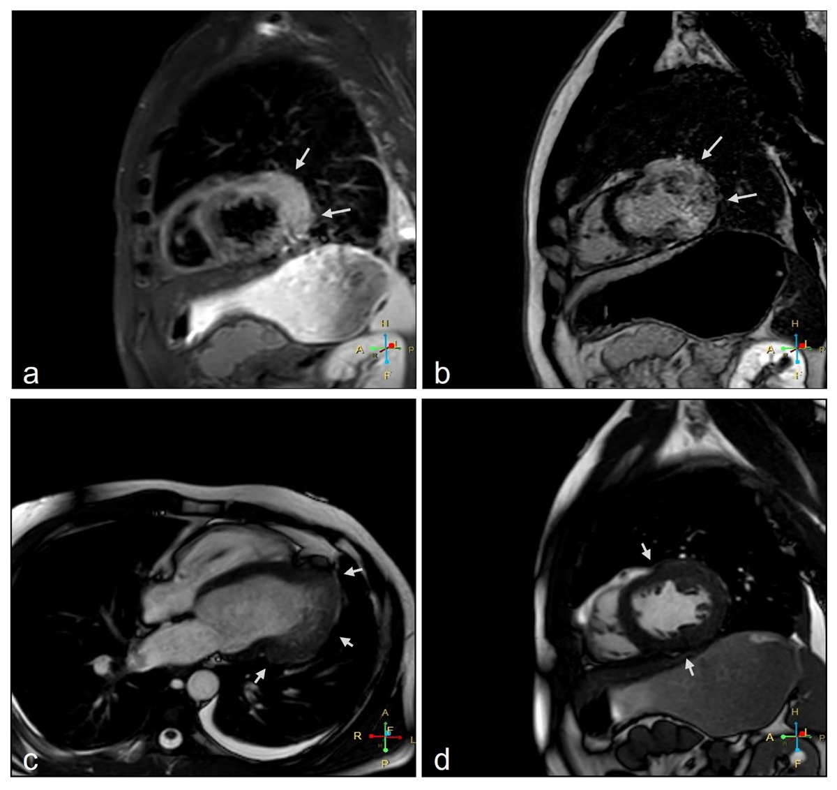
Figure 3
Cardiac magnetic resonance imaging (a) short-axis view shows myocardial edema and (b) late gadolinium enhancement in the left ventricle lateral wall; (c) 4-chamber apical view and (d) short-axis view showing a partial absence of the pericardium and myocardial indentation (arrows).
Based on multimodal imaging findings, the patient was diagnosed with congenital partial pericardial defect and concomitant LV herniation. Although surgical repair was strongly recommended, the patient refused further hospitalization and was discharged. At 3 months, he was asymptomatic and declined to return for follow-up.
Discussion
Cardiac strangulation is a rare complication with several etiologies. This complication has been reported to be secondary to epicardial pacemaker leads, pericardial rupture after trauma, or congenital pericardial absence.4,5,6,7 In partial pericardial defects, dynamic cardiac motion can allow parts of the heart, more likely the LV, to invaginate through the defect. The causes of left atrial appendage or right ventricle herniation have also been described.8,9 The symptoms of cardiac herniation can vary from intermittent chest pain to MI or hemodynamic collapse. In this case, an LV herniation through a partial pericardial defect led to coronary artery compression, which caused an acute MI. A timely and correct diagnosis is critical for appropriate management but can be challenging when the pericardial defect is previously unknown.
Despite a typical presentation of ST-elevation MI, the patient’s echocardiography and angiography results were highly unusual. A markedly increased apical LV thickness and abnormal LV morphology may be observed in a variety of pathologies, including hypertrophic and infiltrative cardiomyopathies, cardiac tumors, or thrombus. However, very few processes can cause such abrupt changes in coronary arteries.2 The differential diagnosis included multi-vessel spasm, embolization, spontaneous coronary artery dissection (SCAD), or external compression.
Although SCAD was considered given the patient’s young age, the angiographic pattern was not typical. Coronary angiography revealed focal, subtotal stenoses involving multiple coronary arteries of different calibers and their branches, with abrupt transitions from a normal segment to severe narrowing and back to normal distally. These stenoses appeared to align along a common anatomical plane, which was inconsistent with intrinsic coronary disease. There were no features of coronary spasm or embolic occlusion.
Given both echocardiographic and angiographic findings together, suspicion for external coronary artery compression, such as by a tumor or, more rarely, a partial pericardial defect, should be raised.2 Cardiac computed tomography or magnetic resonance imaging is a possible next step to identify the cause of the coronary artery obstruction as well as LV thickening and dysfunction.10,11 In this case, CMR defined the extent of the pericardial defect and confirmed the diagnosis of cardiac strangulation by revealing signs of myocardial herniation.
The thickened and akinetic LV segments were located adjacent to the pericardial strangulation line and were a consequence of acute edema due to MI and extrinsic compression of the coronary vessels. A tumor diagnosis was unlikely due to several factors, including the acute onset, the specific strangulation pattern on angiography, the absence of an additional mass and vascularization within the “tumor-like” area, and the large homogeneous area of edema and LGE on CMR.
Surgical intervention is recommended for symptomatic cardiac herniation through a pericardial defect. Pericardiectomy is the most commonly reported treatment for this condition.12,13 Closure of the defect using pericardioplasty has also been described.14 The choice of surgical approach depends on intraoperative findings, including the size and location of the defect and the degree of cardiac herniation.
Conclusion
Partial absence of the pericardium is rarely included in the differential diagnosis of patients presenting with acute chest pain; however, considering such rare etiologies is crucial for selecting appropriate imaging and management strategies. Coronary angiography and multimodality imaging (computed tomography or CMR) are essential for diagnosing cardiac strangulation. Surgical intervention remains the definitive treatment for this life-threatening condition.
Competing Interests
The authors have no competing interests to declare.