
Introduction
Advances in pediatric cardiology and cardiac surgery have transformed the prognosis of congenital heart disease (CHD), such that more than 90% of affected infants now survive into adulthood.1 As a result, there are currently more adults than children living with CHD in developed nations, creating an expanding cohort of patients who require lifelong surveillance and specialized care.2 Rhythm disorders are among the most common complications in this population, contributing significantly to morbidity, hospitalizations, and premature mortality.3 Indeed, sudden cardiac death (SCD) and progressive arrhythmia burden have supplanted many surgical and hemodynamic complications as leading causes of late adverse outcomes in adult CHD (ACHD).4,5
The arrhythmogenic substrate in ACHD is shaped by a convergence of developmental, surgical, and acquired factors. Surgical scars, prosthetic material, atriotomies, baffles, conduits, and ventriculotomies create zones of conduction block and slow conduction that provides the anatomic milieu for macroreentrant circuits.6 Chronic pressure and volume overload, chamber dilation, and adverse remodeling exacerbate the electrical vulnerability of atrial and ventricular myocardium.7 Congenital conduction system anomalies predispose to bradyarrhythmias and progressive conduction disease.8 Consequently, the arrhythmia spectrum in ACHD is both diverse and lesion-specific, encompassing intra-atrial reentrant tachycardia (IART), atrial fibrillation (AF), monomorphic ventricular tachycardia (VT), polymorphic VT, and conduction disturbances necessitating pacing.3
Traditional management strategies for arrhythmias in acquired heart disease cannot be directly extrapolated to ACHD because of fundamental differences in anatomy, substrate, and clinical context. For example, catheter ablation of IART in a Fontan patient demands different access routes, mapping strategies, anesthesia and hemodynamic management, techniques to avoid inadvertent atrioventricular (AV) block, and considerations of the nature and severity of potential complications compared to ablation of cavotricuspid isthmus-dependent flutter in structurally normal hearts.9 Similarly, substrate-guided VT ablation in repaired tetralogy of Fallot (rTOF) must account for slow-conducting anatomical isthmuses between surgical patches and annuli that are not encountered in acquired heart disease.10 Device therapy also presents unique challenges, with distorted venous pathways, systemic right ventricles, and young patient age complicating decisions regarding defibrillators, resynchronization, and pacing systems.11
The past decade has witnessed rapid technological progress that is transforming arrhythmia management in ACHD. Three-dimensional electroanatomic mapping and high-density multielectrode catheters have enhanced visualization of atrial and ventricular circuits.12 Robotic magnetic navigation overcomes catheter access challenges while providing stable catheter control across various complex anatomies.13 Pulsed field ablation (PFA), a nonthermal energy source, has demonstrated superior safety in atrial fibrillation ablation and is being explored in ACHD.14 Intraoperative conduction mapping has emerged as a preventive strategy to reduce iatrogenic AV block during complex repairs.15 Device therapy has evolved with the advent of subcutaneous implantable cardioverter-defibrillators (ICDs), leadless pacemakers, and conduction system pacing techniques that preserve physiologic activation.16,17 Beyond interventional advances, the application of artificial intelligence (AI) to electrocardiograms (ECGs) and imaging, together with personalized “digital twin” models, are enabling individualized risk prediction and virtual testing of therapeutic strategies.18,19
The purpose of this review is to synthesize contemporary innovations in the management of rhythm disorders in ACHD organized across mechanistic mapping, ablation technologies, device therapies, and predictive tools. Emphasis is placed on developments that have immediate clinical relevance, are supported by emerging data, and are poised to influence future guidelines. By contextualizing these advances within lesion-specific substrates and lifelong ACHD care, we aim to provide a roadmap for integrating novel approaches into routine practice while highlighting critical knowledge gaps that warrant prospective investigation.
Arrhythmia Mechanisms and Mapping
Atrial Arrhythmias
Atrial arrhythmias are the most prevalent rhythm disorders in ACHD, with IART being the predominant arrhythmia followed by AF.20 Both contribute to heart failure, thromboembolism, and impaired quality of life.21,22 The underlying substrate is heterogeneous, comprising atriotomy scars, suture lines from baffles or conduits, atrial dilation, and areas of low-voltage myocardium that anchor reentry circuits.23 Electrophysiologists specialized in CHD generally favor the label IART over atrial flutter. Unlike “typical” or “atypical” flutter, IART avoids presupposing the existence of a standard cavotricuspid isthmus. In the ACHD population, the anatomy is often unconventional: the tricuspid valve may be rudimentary, absent, displaced into the pulmonary venous atrium, or even split between atria. In AV septal defects, the right-sided valve itself is not truly tricuspid, rendering “cavotricuspid” a misnomer. On top of this structural variability, electrocardiographic signatures are often less clear-cut, and reentry frequently involves complex double-loop or figure-of-eight circuits traversing the AV valve to vena cava region. High-density mapping catheters now permit rapid identification of low-voltage corridors and fractionated potentials, enabling tailored ablation of non-cavotricuspid–dependent circuits with improved acute success.24
Fontan patients represent a particularly vulnerable group. Surgical modifications of the atria, incisions, baffles, elevated venous pressures, and progressive atrial fibrosis create an arrhythmogenic milieu that evolves over decades.25 In these patients, arrhythmias are not only symptomatic but may be poorly hemodynamically tolerated and precipitate thromboembolism and Fontan circulatory failure. Mapping challenges include catheter stability in markedly dilated right atria among patients with classic modified Fontan procedures and access to the pulmonary venous atrium in those with total cavopulmonary connections.9 Integration of imaging modalities, such as intracardiac echocardiography (ICE) and preprocedural computed tomography (CT) or cardiac magnetic resonance (CMR) imaging, with electroanatomic mapping, facilitates access via transcaval, transbaffle, or robotic magnetic-guided retrograde aortic approaches.26,27
Ventricular Arrhythmias
Patients with CHD who face an elevated risk of ventricular arrhythmias and SCD can be broadly categorized into two conceptual groups: those in whom reentrant monomorphic VT arises from well-defined anatomic circuits, as in rTOF, and those with more diffuse arrhythmogenic substrates, exemplified by patients with systemic right ventricles.28 Reentrant monomorphic VT was first recognized decades ago in patients with rTOF, with inducible VT identified as an important marker of risk.29 More recently, the critical anatomic isthmuses underlying these reentrant circuits have been more precisely delineated.30 The pathophysiologic substrate is defined by so-called slow-conducting anatomical isthmuses (SCAIs) that are bordered by patches, valve annuli, or surgical scars. Among the four principal isthmuses that have been characterized, the isthmus between the ventricular septal defect patch and pulmonary anulus is most commonly implicated in reentrant VT (Figure 1).10
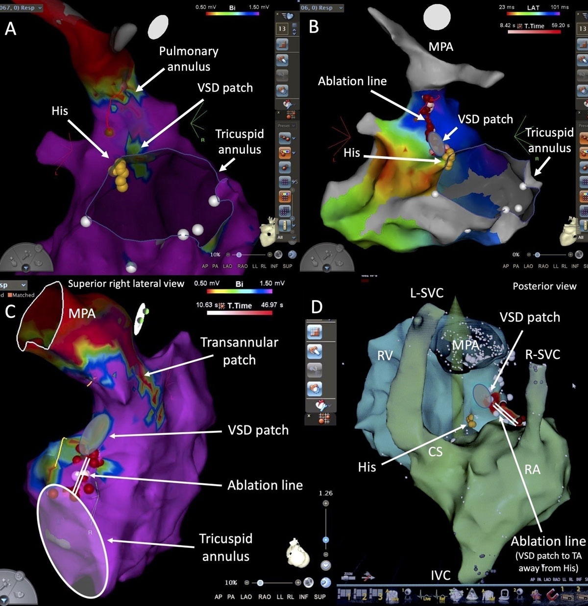
Figure 1
Ventricular tachycardia (VT) ablation in tetralogy of Fallot (TOF). (A) Patient 1 with repaired TOF and VT in which the critical isthmus targeted for ablation was between the ventricular septal defect (VSD) patch and pulmonary annulus, identified by voltage mapping. (B) The ablation line connected the two electrically inert structures (VSD patch and pulmonary annulus). (C) Patient 2 with repaired TOF and VT in which the critical isthmus was between the VSD patch and tricuspid annulus (TA), as identified by voltage mapping. The ablation line connected these two electrically inert structures (VSD patch and TA). (D) The ablation line was anchored on the VSD patch and directed away from the His bundle. Note that this patient had bilateral superior vena cavae (SVC), with a left SVC (L-SVC) draining into the coronary sinus (CS). Reprinted with permission from Wolters Kluwer Health, Inc. IVC: inferior vena cava; MPA: main pulmonary artery; RA: right atrium; R-SVC: right SVC; RV: right ventricle
Traditional risk factors such as QRS duration, nonsustained VT, and extensive ventricular fibrosis provide modest predictive power for VT and SCD in rTOF. By contrast, inclusion of electrophysiological studies with inducibility of VT and identification of SCAIs appears to provide additional prognostic value.10,31 Three-dimensional late gadolinium enhancement CMR imaging (3D-LGE-CMR) has emerged as a potential tool to noninvasively identify high-risk substrates.32,33 Electrocardiographic markers also provide valuable insights. Fragmented low-amplitude R′ wave in lead V1 and terminal S waves in inferior leads have been attributed to conduction abnormalities across the septal isthmus, establishing a mechanistic link between right ventricular activation and these common electrocardiographic patterns.34 Preventive strategies are under active investigation. The “Catheter Ablation of Ventricular Tachycardia Before Transcatheter Pulmonary Valve Replacement in Repaired Tetralogy of Fallot” (CATAPULT-TOF) study is testing whether ablation of critical isthmuses involved in inducible VT together with prophylactic ablation of SCAIs prior to transcatheter pulmonary valve replacement reduces the risk of subsequent clinical VT and SCD.35 This approach embodies a shift toward mechanism-based prevention, integrating hemodynamic and arrhythmic interventions.
Intraoperative Conduction Mapping
Conduction disturbances, including postoperative AV block and sinus node dysfunction, remain major challenges in ACHD. Anatomical variability of the conduction system, such as inferior displacement of the AV node in AV septal defect or anterior positioning in congenitally corrected transposition of the great arteries, increases susceptibility to spontaneous AV block and iatrogenic injury during catheter-based and surgical procedures.3
High-density intraoperative conduction mapping allows real-time localization of the His-Purkinje system on the beating heart. By applying multielectrode grids intraoperatively, the conduction system can be localized and assist with planning incision sites and patch placements. Early studies have shown that this strategy significantly reduces postoperative pacemaker implantation rates in complex repairs, including AV septal defects and heterotaxy syndromes.36 In a prospective cohort, integration of intraoperative mapping into surgical planning halved the incidence of postoperative complete AV block, underscoring its preventive potential.15 Intraoperative mapping also provides unique insights into the developmental variability of conduction pathways, generating datasets that may refine preoperative imaging predictions and computational models. As these approaches mature, they may become integral to multidisciplinary surgical-electrophysiological collaborations in CHD surgery.
Innovations in Catheter Ablation
Catheter ablation has become an indispensable tool for arrhythmia management in ACHD. Nevertheless, procedural success rates historically lagged behind those in structurally normal hearts, due in part to complex anatomy, multiple coexisting arrhythmias, limited catheter maneuverability, atypical arrhythmogenic substrates, and progressive arrhythmogenic remodeling.37 Technological advances have transformed this field, improving safety, efficacy, and accessibility for increasingly complex patient populations.
High-density Mapping and Imaging Integration
Conventional point-by-point mapping is often long and cumbersome in ACHD, where circuits traverse nontraditional isthmuses and scar corridors. High-density mapping catheters, with closely spaced multipolar electrodes, generate rapid, high-resolution activation and voltage maps, unveiling slow-conduction channels and functional lines of block that were previously difficult to delineate. For atrial arrhythmias, high-density mapping has improved recognition of non-cavotricuspid isthmus-dependent IART circuits in atrial switch or Fontan patients, thereby increasing acute ablation success (Figure 2).12 Integration of imaging modalities has further enhanced substrate definition. Fusion of CMR or CT data into electroanatomic maps facilitates catheter navigation around complex postsurgical geometries. ICE has become an essential adjunct, guiding transbaffle punctures, confirming tissue contact, and detecting complications in real time.38
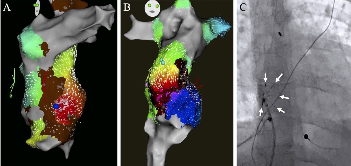
Figure 2
High-density mapping of atrial arrhythmias in patients with Fontan surgery. (A) A high-density electroanatomic map of a nonautomatic focal atrial tachyardia along a scar border in a 41-year-old patient with an intracardiac lateral tunnel Fontan. Earliest electrical activation (red) is along the inferomedial aspect of the Fontan baffle. (B) A high-density map of an intra-atrial re-entrant tachycardia in a 28-year-old patient with a lateral tunnel Fontan. Earliest electrical activation is denoted in red with arrows depicting the wavefront propagating clockwise around dense scar (brown and grey areas) to the latest points in purple. (C) A fluoroscopic view of a multielectrode mapping catheter (PentaRay, Biosense Webster,) used to create these high-density maps. A total of 20 electrodes are distributed among five soft radiating splines indicated by the white arrows. Reprinted with permission from Elsevier.
Robotic Magnetic Navigation
Robotic magnetic navigation represents a major advance in catheter stability and maneuverability, particularly in scenarios where manual techniques are limited, such as vascular constraints that preclude reaching the arrhythmia, high-risk or non-feasible trans-septal, transbaffle, or transconduit access to the pulmonary venous atrium, challenges in manipulating a manual catheter within the target chamber, or failure to maintain stable tissue contact needed to deliver effective and durable ablation lesions.13 By remotely controlling a magnetically responsive catheter with externally applied magnetic fields, operators can achieve consistent tissue contact and access to regions that are otherwise difficult to reach. Multicenter experiences, albeit limited, have demonstrated the safety and efficacy of robotic magnetic-guided ablation in ACHD in complex anatomies, including in patients with atrial switch baffles, systemic venous anomalies, and total cavopulmonary connection Fontan surgeries.39 Beyond improving acute success, robotic magnetic navigation minimizes radiation exposure for patients and operators while alleviating operator fatigue during prolonged interventions (Figure 3).
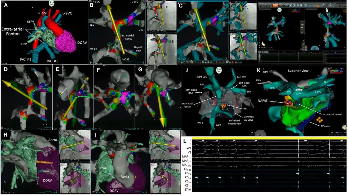
Figure 3
Robotic magnetic nativation-guided ablation in a patient with intracardiac tunnel Fontan. (A) CT scan images segmented and imported into a 3D-electroanatomic mapping system. (B) Electroanatomic mapping of the systemic venous atrium. The yellow arrow designates the direction of the magnetic field and the starburst at the tip of the ablation catheter indicates good tissue contact. (C) Merging of electroanatomic mapping and CT scan images of the systemic venous atrium in multiple views. Shown is the operator view of CT scan and 3D mapping images (left) during the merging process, overlaying of these images on orthogonal fluoroscopic views (middle), and views from the 3D electroanatomic mapping system. The yellow arrows indicate the direction of the magnetic field. (D-G) Robotic magnetic navigation-guided retrograde aortic access. (H-I) Crossing of the common AV valve of the double-outlet right ventricle (DORV) to enter the pulmonanry venous atrium (PVA). The green arrows represent the direction of the magnetic field set by the operator whereas the yellow arrows indicate the actual direction of the magnetic field, with a brief lag time between the two. (J-K) 3D view of electroanatomic maps of nonautomatic focal atrial tachycardia (NAFAT). (L) Surface ECG leads I, II, aVF, and V2; intracardiac recordings from the distal (MAP1-2) to proximal (MAP3-4) and unipolar (MAPUni) mapping catheter. Reprinted with permission from Elsevier.27 STIM: stimulation channel; R-SVC: right superior vena cava; L-SVC: left superior vena cavae; RSVP: right superior pulmonary vein; RPA: right pulmonary artery; RAA: right atrial appendage; RIPV: right inferior pulmonary vein; PVA: pulmonary venous atrium; DORV: double-outlet right ventricle; IVC: inferior vena cava; LPA: pulmonary artery; AV: denotes atrioventricular
Pulsed Field Ablation
While radiofrequency ablation remains the predominant energy source for catheter ablation in ACHD, cryoablation has primarily been used in selected patients with perinodal arrhythmias40 and atrial fibrillation.41,42 The introduction of PFA is a paradigm shift in energy delivery in that it employs nonthermal electrical fields to induce irreversible electroporation of cardiomyocytes, sparing adjacent structures such as the esophagus and phrenic nerve.43 Large randomized and registry-based studies in paroxysmal and persistent AF have shown PFA to be noninferior in efficacy and superior in safety compared with conventional thermal energy sources.44 The MANIFEST-17K (Multi-National Survey on the Safety of the Post-Approval Clinical Use of Pulsed Field Ablation in 17,000+ Patients) registry extended these findings to more than 17,000 patients and confirmed low complication rates, including an absence of atrioesophageal fistula.45
In adults with CHD, PFA may offer an advantage in anatomically constrained regions because its nonthermal, electroporative mechanism can generate lesions without bulk heating, potentially reducing thermal injury to adjacent viable myocardium or delicate structures.14,46 However, its performance in regions adjacent to surgical patches or dense scar tissue remains uncertain and requires careful study, especially given risks such as coronary spasm and variable lesion formation in heterogeneous substrates. Early feasibility studies suggest that PFA may be effective in ablating outflow tract ectopy and even scar-related VT.47 However, long-term durability in heavily scarred ventricular substrates remains uncertain, complications are not negligible, and ACHD-specific PFA studies are lacking.
Innovations in Cardiac Implantable Electronic Devices
Implantable Cardioverter-Defibrillators
Implantable cardioverter-defibrillators remain the cornerstone of SCD prevention in ACHD, but their application is challenged by the heterogeneous population with diverse mechanisms leading to SCD, limitations of existing risk models, and uncertainties inherent to SCD prediction.48 Vascular access limitations and complex anatomy may preclude transvenous leads, resulting in an important role for the subcutaneous ICD.48 First implanted in 2008, the subcutaneous ICD has since evolved into a system with improved testing, sensing algorithms, programming, and MRI compatibility. Although initial studies included only a small number of patients with CHD, later series have confirmed feasibility, provided careful screening is undertaken to limit inappropriate shocks, particularly in those with wide QRS complexes such as in rTOF.49,50 Limitations include the inability to provide antitachycardia pacing (ATP) or chronic bradycardia support, making patient selection crucial. Modern algorithms (eg, Smart Pass) and dual-zone programming have reduced inappropriate shock rates from 14% to 3.5%.51 With the exception of unipolar leads, compatibility with most pacemakers is achievable, which is relevant to the ACHD cohort given the high prevalence of coexisting bradyarrhythmias (Figure 4).52
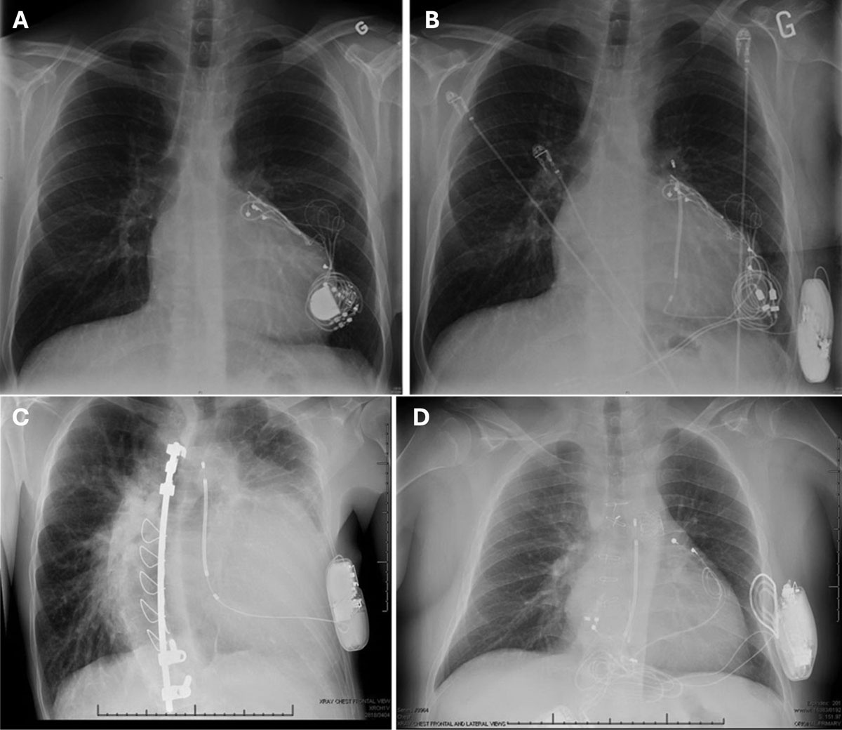
Figure 4
Subcutaneous implantable cardioverter defibrillator in congenital heart disease. (A) A 26-year-old male after lateral tunnel fontan had undergone epicardial pacemaker for symptomatic sinus node dysfunction. (B) The patient underwent generator replacement with a dedicated bipolar device to avoid future interdevice interaction as well as relocation to the right upper abdomen to exclude the device from the future subcutaneous implantable cardioverter debrillator (S-ICD) shock vector. (C) A 41-year-old male with tricuspid atresia and pulmonary stenosis previously palliated by Waterson shunt in childhood underwent S-ICD placement for a left ventricular ejection fraction of 35%. (D) A 22-year male with hypoplastic left heart syndrome palliated by extracardiac fontan operation who had a previously failed epicardial ICD system that had been placed for nonsustained ventricular tachycardia in the setting of unexplained syncope and systemic right ventricular ejection fraction of ≈ 10%. Reprinted with permission from Wolters Kluwer Health, Inc.49
Next-generation systems aiming to transcend the limitations of conventional subcutaneous ICDs have entered clinical practice. The MODULAR ATP concept, which combines a leadless pacemaker wirelessly communicating with a subcutaneous ICD, has met key safety and efficacy end points, enabling ATP without transvenous leads.53 In early human experience, the intercommunicating system successfully delivered ATP to terminate arrhythmias while maintaining low complication rates.53 In parallel, extravascular ICDs (EV-ICDs) using a substernal lead have demonstrated the ability to deliver both defibrillation and limited pacing.54 Their applicability in ACHD is uncertain since many adults with repaired CHD have undergone prior sternotomy or mediastinal surgery, conditions for which EV-ICD implantation is generally considered contraindicated or high-risk; however, the feasibility of concomitant EV-ICD implantation during cardiac surgery has been reported.55 Thus, for now, modular subcutaneous ICD/leadless pacing systems hold clear promise for adults with CHD, while EV-ICDs may emerge as an attractive option in highly selected patients, particularly when implantation can be performed concomitantly with cardiac surgery as further data and technological refinements become available.
Leadless Pacing
Lead-related complications are particularly problematic in ACHD due to distorted anatomy, prior surgeries, and elevated infection risk. Leadless pacemakers, delivered percutaneously to the subpulmonary ventricle, eliminate the need for transvenous leads and pockets, thereby reducing infection risk and preserving venous access. Feasibility studies in ACHD have demonstrated safe implantation even in complex anatomy, although long-term data remain limited (Figure 5).56-58
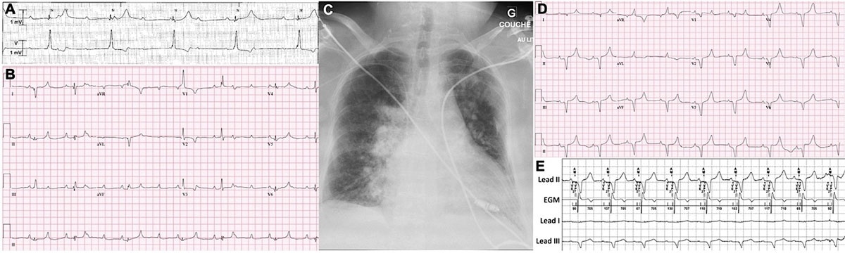
Figure 5
Leadless pacemaker in Eisenmenger syndrome. Atrioventricular synchronous pacing with Micra AV leadless pacemaker (Medtronic Inc) in an adult with Eisenmenger syndrome and complete atrioventricular (AV) block. (A) Rhythm strip (leads II and V) on presentation shows complete AV block with a junctional escape rhythm at 37 beats/min. (B) 12-lead electrocardiogram (ECG) after initiation of an isoproterenol infusion shows high-grade AV block with a narrow QRS (106 ms) rate of approximately 45 bpm. (C) Posteroanterior chest radiograph following implantation of the leadless pacemaker. The leadless pacemaker is positioned in the right ventricular apex. (D) 12-lead ECG shows left bundle branch block morphology with leadless ventricular pacing and 2:1 undersensing of atrial activity that prompted reprograming of the A4 threshold. (E) Intrinsic electrocardiographic tracings (lead II, intracardiac electrogram [EGM], and leads I and III) obtained upon device interrogation at the 3-month follow-up visit. As seen on the marker channel, the ventricular end period and atrial mechanical (AM) period are appropriately sensed following the P wave. The pacemaker spike (VP) follows after the programmed 20 ms AM-VP delay, with appropriate ventricular capture. Reprinted with permission from Ellsevier.57
Dual-chamber leadless pacing systems that are now entering clinical practice may further broaden therapeutic options for patients with ACHD.59,60 For example, reports are emerging of leadless dual-chamber pacing in patients with Fontan palliation using a lateral tunnel or extracardiac conduit in the setting of AV block.61 Although important concerns include right-to-left shunting following large-bore transbaffle access and the risk of thromboembolic events, promising results have been reported using a strategy of shunt closure after implantation and long-term anticoagulation.61 Nevertheless, additional safety data with long-term follow-up are required before this approach can be expanded beyond carefully selected cases.
Conduction System Pacing
Conduction system pacing (CSP), encompassing His bundle pacing and left bundle branch area pacing (LBBAP), has rapidly evolved as a physiologic alternative to conventional ventricular pacing and cardiac resynchronization therapy (CRT). While most evidence arises from acquired heart disease, experience in congenital populations is expanding. Early reports demonstrate that CSP is technically feasible and can achieve narrower QRS durations, improved systemic ventricular synchrony, and stable pacing thresholds in patients with various forms of repaired or unrepaired CHD. Observational series in cohorts with congenitally corrected transposition of the great arteries, AV septal defects, and rTOF have confirmed successful engagement of the conduction system despite marked anatomic variability.17,62,63 Imaging and mapping techniques such as CT-based conduction modeling, electroanatomic mapping, and ICE are increasingly used to identify displaced AV conduction tissue and guide lesion-specific implantation. Among the CSP modalities, LBBAP has gained favor for its broader target zone, lower capture thresholds, and superior sensing stability compared to His bundle pacing, though distal septal injury and lead extraction remain concerns. Despite encouraging procedural success and early functional benefits, data remain limited by small sample sizes, short follow-up, and the absence of randomized trials in CHD. Continued refinement of imaging-guided implantation, lesion-tailored approaches, and long-term registry data will be essential to define the role of CSP as a standard pacing paradigm in CHD (Figure 6).
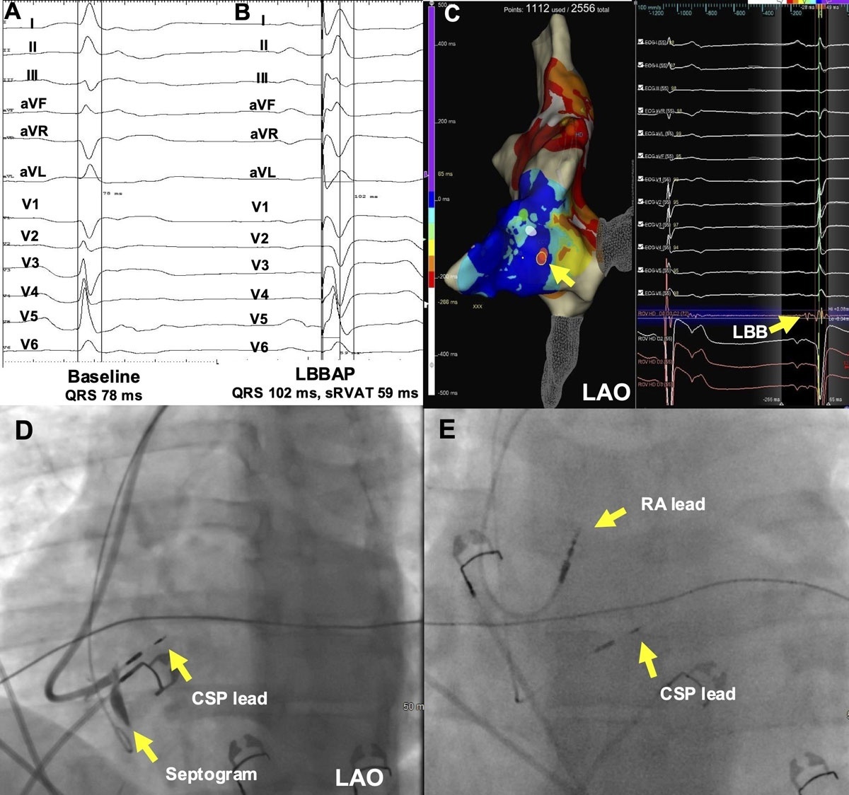
Figure 6
Conduction system pacing (CSP) in congenitally corrected transposition of the great arteries (ccTGA), A 43-year-old patient with ccTGA and intermittent complete heart block underwent electroanatomic mapping-guided pacemaker implantation with a CSP lead. (A) Baseline electrocardiogram (ECG) with a QRS duration of 78 ms. (B) ECG following left bundle branch area pacing (LBBAP) with a QRS duration of 102 ms and systemic right ventricular activation time (sRVAT) of 59 ms. (C) Electroanatomic map showing location and signal of LBB in a left anterior oblique (LAO) orientation. (D) Fluoroscopy image of septogram showing CSP lead 13 mm into the septum. (E) Fluoroscopy image showing final right atrium (RA) and CSP lead position. Reprinted with permission from Elsevier.
Cardiac Resynchronization Therapy
CRT retains an important role for patients with systemic ventricular dysfunction and ventricular dyssynchrony. However, lead placement is often complicated by venous anomalies, surgical baffles, or extensive scar tissue. Procedural adaptations, such as surgical epicardial leads, transbaffle puncture, and endocardial LV pacing, have been employed with variable success.16 In systemic right ventricle and complex anatomy, hybrid pacing strategies combining CSP with coronary sinus or epicardial leads have shown potential for enhanced resynchronization.64 For example, His bundle pacing or LBBAP combined with systemic ventricular epicardial leads has shown promise in achieving effective resynchronization in patients with congenitally corrected transposition of the great arteries and a suboptimal response to CSP.64
Cardiac resynchronization of the subpulmonary right ventricle, such as in patients with rTOF or Ebstein anomaly, has generated much interest. In selected patients, acute hemodynamic benefits have been demonstrated with pacing of the right ventricular basal free wall.65,66 In a small case series of permanent right ventricular basal free wall pacing in patients with rTOF or Ebstein anomaly, right bundle branch block, and moderate-to-severe right ventricular dysfunction, improvements were reported in New York Heart Association functional class, QRS duration, and right ventricular function.67 Larger studies are needed to better identify patients most likely to benefit from this strategy and to evaluate potential downstream effects on SCD risk.
The Wireless Stimulation Endocardially for Cardiac Resynchronization Therapy (WISE-CRT) system, which consists of a leadless sensor in the systemic ventricle and a subcutaneous generator, represents a theoretically attractive strategy for CRT in patients with Fontan palliation because the leadless sensor is designed for the systemic ventricle, and the “co-implant” requirement could potentially be fulfilled by an existing epicardial pacing system.68 The future may see personalized selection between CRT, CSP, and hybrid strategies based on lesion-specific anatomy and location of the conduction system.
Innovations in Risk Prediction and SCD Prevention
Limitations of Conventional Risk Stratification
Conventional risk stratification models for SCD in CHD face inherent methodological and conceptual limitations. Lesion-specific scores, such as those developed for rTOF or the systemic right ventricle, capture relevant anatomic substrates but consider a limited number of potential predictors and are often derived from retrospective cohorts that may reflect historical surgical techniques, center-specific practices, or selectively high-risk populations.48 Their variables may rely on invasive or center-dependent measurements, and calibration deteriorates when applied to contemporary or external cohorts. In contrast, models encompassing heterogeneous CHD populations improve statistical power but sacrifice lesion-level granularity, implicitly assuming shared mechanisms across fundamentally distinct anatomies and thereby diluting clinically meaningful predictors. Both model types typically depend on static parameters and dichotomous cutoffs, overlook time-dependent risk evolution, and rarely incorporate competing nonarrhythmic causes of death that influence net survival benefit from interventions such as ICD implantation (Table 1). As a result, risk estimates remain imprecise for many patients occupying intermediate-risk zones, reinforcing the need for dynamic, multimodal, and lesion-aware frameworks that integrate imaging, electrophysiologic, and biomarker data to support individualized prediction and decision-making.
Table 1
Summary of key sudden cardiac death risk stratification tools by lesion type. Reprinted with permission from Wolters Kluwer Health, Inc. CHD: congenital heart disease; ASD: atrial septal defect; PREVENTION-ACHD: Prospective study on implantable cardioverter-defibrillator therapy and sudden cardiac death in adults with congenital heart disease; TOF: tetralogy of Fallot; VT: ventricular tachycardia; LV: left ventricle; CMR: cardiac magnetic resonance; RVESVi: indexed right ventricular end-systolic volume; ICD: implantable cardioverter-defibrillator; INDICATOR: International Multicenter Tetralogy of Fallot Registry; SCD: sudden cardiac death; TGA: transposition of the great arteries; ccTGA: congenitally corrected TGA; sRV: systemic right ventricle; LVOT: left ventricular outflow tract; MARE: major adverse ventricular arrhythmias and related events; EF: ejection fraction
| LESION TYPE | KEY PREDICTORS | TOOLS/SCORES | LIMITATIONS |
|---|---|---|---|
Mixed cohorts with CHD
|
|
| Models were not devised to tease out predictors unique to specific defects |
| Repaired TOF |
|
|
|
| TGA with Mustard or Senning baffle |
|
| Model lacks competing-risk analyses and integration of advanced imaging and neurohormonal markers |
| Congenitally corrected TGA (ccTGA) |
| MARE risk prediction model | Model is less robust in ccTGA compared to TGA post atrial switch, which may reflect differences in arrhythmic substrates and triggers among the two groups |
| Single ventricle Fontan |
| None | Identification of risk factors have been limited by small sample sizes and low event rates |
Substrate-guided Prevention Framework
A substrate-guided prevention framework emphasizes identifying and modifying the underlying anatomic and electrophysiologic substrates that predispose to ventricular arrhythmias rather than relying solely on probabilistic models of event occurrence. In CHD, advances in CMR and high-density electroanatomic mapping have clarified that regions of fibrosis, surgical scarring, and slow conduction form discrete arrhythmogenic isthmuses that can be targeted for intervention. Late gadolinium enhancement on CMR and 3D reconstruction can delineate these substrates, enabling preemptive or intraoperative ablation strategies in lesions such as rTOF.32 The CATAPULT-TOF study exemplifies this approach by testing whether prophylactic ablation of SCAIs during transcatheter pulmonary valve replacement reduces post-procedural VT.35 This integration of arrhythmia prevention into structural interventions illustrates the paradigm shift toward targeted, mechanism-based SCD prevention.
Genetics
Genetic insights are increasingly relevant to arrhythmic risk in ACHD. While primary arrhythmia syndromes such as Brugada or long QT syndrome are not CHD per se, variants in cardiac transcription factors that cause structural malformations, notably NKX2-5, TBX5, and GATA4, are associated with progressive conduction disease, AV block, and malignant ventricular arrhythmias.69,70 Recognition of these associations has clinical implications for surveillance, device planning, and family counseling. As genomic sequencing becomes more widely adopted, integration of genetic risk markers into predictive models may refine arrhythmic risk assessment in ACHD.
Artificial Intelligence
Artificial intelligence, particularly deep learning applied to ECGs and imaging, offers powerful tools for individualized prediction of arrhythmic outcomes.71 In rTOF, convolutional neural networks trained on 12-lead ECGs have demonstrated superior predictive accuracy for VT, ICD shocks, and death compared with conventional risk factors.18 Beyond ECGs, AI applied to multimodal data, including imaging, hemodynamics, and electronic health records, can generate continuous risk scores and dynamically update predictions over time. These approaches have the potential to shift risk stratification from binary thresholds toward personalized, probabilistic models of arrhythmic risk.
Digital Twins
Digital twin technology—ie, patient-specific computational models calibrated to imaging, electrophysiology, and hemodynamic data—represents a frontier innovation. Personalized digital twin models of rTOF have been shown to identify VT-critical isthmuses with high concordance to invasive mapping.72 In silico testing of ablation strategies within the twin environment allows virtual optimization of lesion sets before invasive procedures, potentially reducing procedural time and risk. Similarly, digital twins may help determine whether ICD implantation is necessary by simulating arrhythmic vulnerability under varying physiologic conditions.73 Early feasibility studies support the accuracy of this approach, though large-scale validation is pending.
Integrated Prevention Strategy
Moving forward, prevention of SCD in ACHD will likely rely on a multimodal strategy. Substrate definition via imaging and mapping, genetic risk markers, AI-driven predictive analytics, and digital twin simulations can be combined to generate individualized profiles of arrhythmic vulnerability. Such precision medicine approaches may allow clinicians to better target ICDs, ablation, and pacing therapies to those at highest risk while sparing low-risk patients from unnecessary interventions.4 Ultimately, the integration of these technologies into prospective multicenter registries and randomized trials is essential to refine risk stratification, reduce arrhythmic mortality, and guide evidence-based clinical practice.
Conclusion
Arrhythmia management in ACHD is entering an era defined by precision, mechanism-based intervention, and integration across disciplines. Advances in mapping, imaging, and device technology are reshaping both diagnosis and therapy, enabling clinicians to move beyond empirical treatment toward lesion-specific, physiology-guided strategies. The convergence of substrate-guided ablation, intraoperative conduction mapping, and physiologic pacing illustrates how mechanistic insights can directly translate into preventive care. At the same time, emerging predictive paradigms, spanning genetics, AI, and digital twin modeling, are contributing to risk assessment by capturing dynamic, individualized patterns of arrhythmic vulnerability. Together, these innovations signal a shift from reactive to proactive management: from treating arrhythmias after they occur to preventing them by modifying their anatomic and electrophysiologic substrates. Continued multidisciplinary collaboration and prospective validation will be essential to realize the promise of personalized, substrate-based arrhythmia prevention and durable rhythm control in the growing ACHD population.
Key Points
Arrhythmogenic substrates in adult congenital heart disease (ACHD) are highly variable and lesion-specific, reflecting congenital anatomy, surgical scarring, hemodynamic sequelae, and remodeling effects.
Detailed mapping and catheter ablation enable mechanistic therapy for a range of tachyarrhythmias, with high-density electroanatomic mapping, image integration, and advances in catheter navigation and ablation modalities allowing precise, tissue-selective substrate modification.
Preventive strategies are emerging, including intraoperative conduction mapping to avert atrioventricular block and prophylactic ventricular isthmus ablation in the peri-pulmonary valve replacement setting to reduce the risk of ventricular tachycardia/sudden cardiac death.
Device therapy is becoming physiologic and anatomy-adapted, encompassing conduction system pacing (CSP), leadless and subcutaneous systems, and hybrid cardiac resynchronization therapy/CSP.
Risk prediction is evolving toward precision modeling, integrating imaging, genetics, artificial intelligence, and digital twins to enable individualized, mechanism-based arrhythmia prevention.