
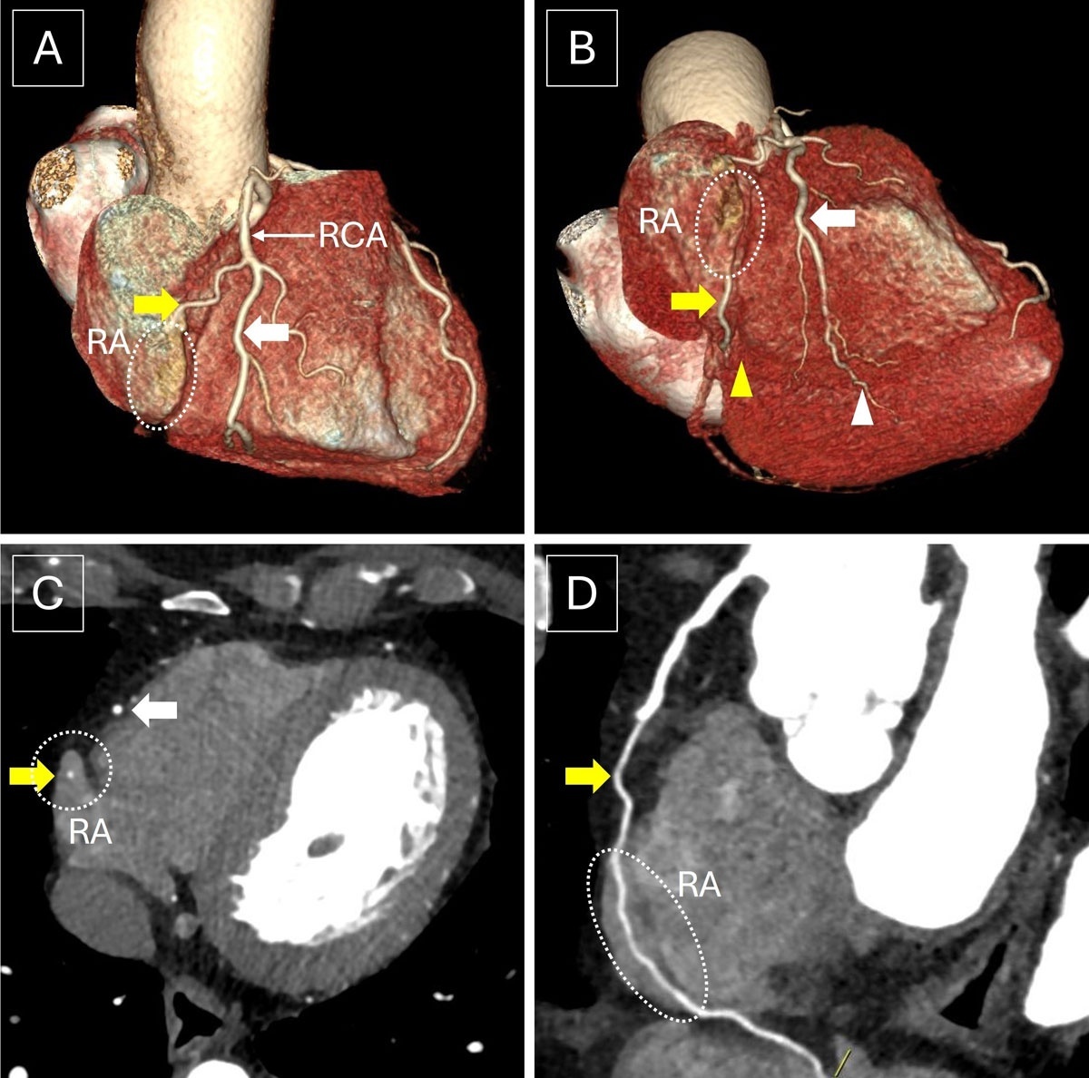
Figure 1
(A, B) Volume-rendered images, (C) axial image, and (D) curved multiplanar reconstruction of computed tomography angiography show the RCA splitting into two trunks. The anterior good-sized trunk (white thick arrow) is seen coursing along the acute margin of the right heart and is seen reaching the distal aspect of the posterior interventricular groove and continuing as the distal posterior descending artery (white arrowhead). The posterior trunk (yellow thick arrow) is seen coursing along the right atrioventricular groove, subsequently entering the RA and having an intra-atrial course (dotted circle) for a length of 3 cm. The posterior trunk is then seen emerging out of the right atrium near the crux of the heart and gives rise to a proximal posterior descending artery along the proximal posterior interventricular groove. RCA: right coronary artery; RA: right atrium
Video 1
Volume rendering of the coronary computed tomography angiography dataset; see also at https://youtube.com/shorts/i2I2_4HVhDY.