
A 79-year-old male presented to the cardiology outpatient clinic with complaints of atypical chest pain. His baseline echocardiogram and blood investigations were within normal limits. Transthoracic echocardiography revealed normal biventricular functions.
Subsequently, the patient underwent echocardiogram-gated coronary computed tomography angiography (CTA), which ruled out any obstructive coronary artery disease. Incidentally, an unusual coronary artery variant anatomy was noted where the proximal right coronary artery (RCA) was seen splitting into two trunks. The posterior trunk coursed within the right AV groove and was seen entering the right atrium (RA) and having an intra-atrial course for a length of 3 cm (Figure 1). The posterior trunk then emerged out of the RA near the crux of the heart and gave rise to the proximal posterior descending artery (PDA) along the proximal part of the posterior interventricular groove. The anterior good-sized trunk deviated from the right atrioventricular (AV) groove and coursed along the acute margin of the right heart. The anterior trunk also reached the distal aspect of the posterior interventricular groove giving off a separate distal PDA (Figure 1; Video 1).
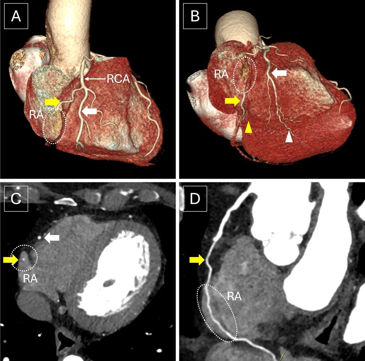
Figure 1
(A, B) Volume-rendered images, (C) axial image, and (D) curved multiplanar reconstruction of computed tomography angiography show the RCA splitting into two trunks. The anterior good-sized trunk (white thick arrow) is seen coursing along the acute margin of the right heart and is seen reaching the distal aspect of the posterior interventricular groove and continuing as the distal posterior descending artery (white arrowhead). The posterior trunk (yellow thick arrow) is seen coursing along the right atrioventricular groove, subsequently entering the RA and having an intra-atrial course (dotted circle) for a length of 3 cm. The posterior trunk is then seen emerging out of the right atrium near the crux of the heart and gives rise to a proximal posterior descending artery along the proximal posterior interventricular groove. RCA: right coronary artery; RA: right atrium
Video 1
Volume rendering of the coronary computed tomography angiography dataset; see also at https://youtube.com/shorts/i2I2_4HVhDY.
A split RCA is characterized by the presence of two variable-sized PDAs arising from anterior and posterior divisions of the RCA. The posterior division usually courses orthotopically along the expected course of the RCA in the right AV groove, reaching the crux of the heart and continues as the proximal PDA that supplies the proximal part of the posterior interventricular septum. The anterior division deviates from the right AV groove and traverses along the acute margin of the heart, ultimately continuing as the distal PDA that supplies the distal part of the posterior interventricular septum and the inferior left ventricular myocardium.1,2
With an estimated prevalence of 0.36%,3 an intra-atrial course of the RCA is a rare anomaly wherein the RCA courses within the lumen of the right atrium. This variant anatomical course predisposes the intra-atrial RCA to risk of inadvertent injury during a vast number of interventional and surgical procedures, such as right heart catheterization, pacemaker insertion, coronary artery bypass surgery, and radiofrequency ablation. A split RCA in conjunction with an intra-atrial course has not been previously reported in literature and is exceedingly rare. Since the “other RCA” is seen coursing along the right heart border, the intra-atrial segment may remain undetected if cross-sectional imaging is not available. The present case thus highlights the importance of coronary CTA in detecting this unusual variant, which would be missed even on invasive coronary angiography.
Competing Interests
The authors have no competing interests to declare.