
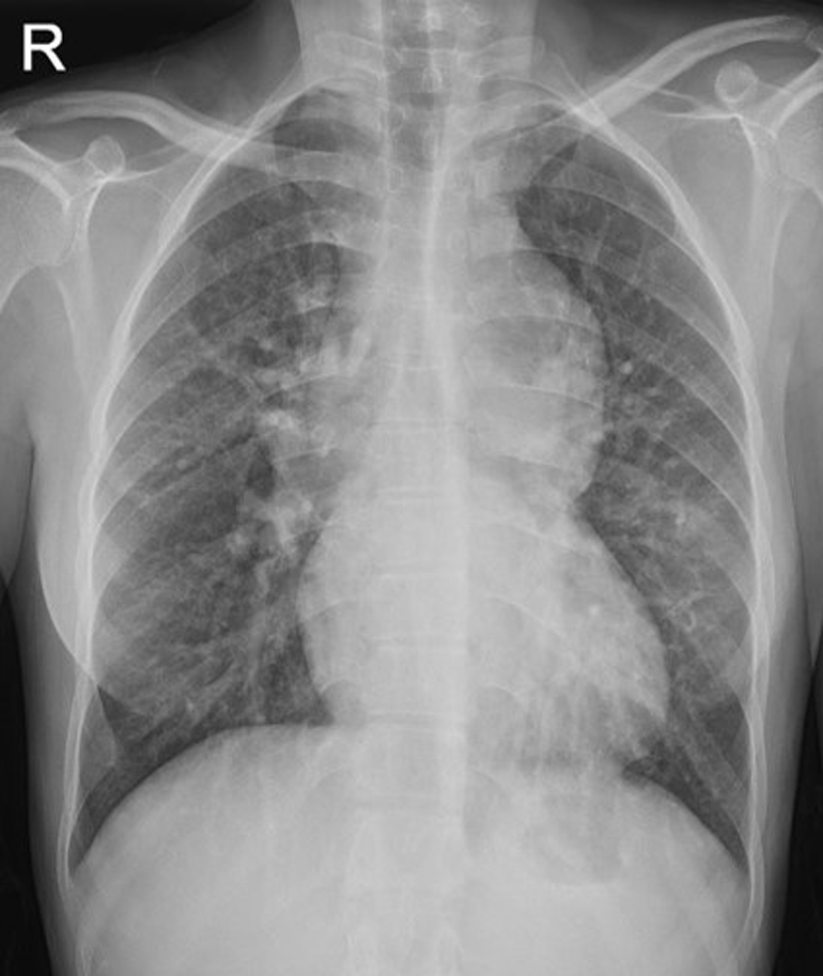
Figure 1
Frontal chest radiograph demonstrates marked mediastinal enlargement, consistent with the classical “snowman sign,” and pulmonary plethora.
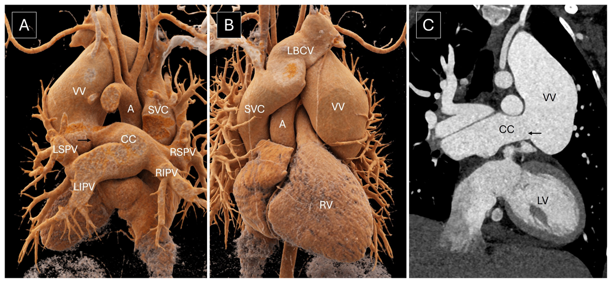
Figure 2
(A, B) Volume rendered images and (C) oblique coronal image show all four pulmonary veins joining to form a common chamber (CC), which drains into the left brachiocephalic vein (LBCV) via an ascending vertical vein (VV). A thin eccentric membranous web (black arrow) is noted at the junction of the common chamber and ascending VV with aneurysmal dilatation of the vertical vein distal to it. A: aorta; LV: left ventricle; RV: right ventricle; SVC: superior vena cava; RSPV: right superior pulmonary vein; LSPV: left superior pulmonary vein; RIPV: right inferior pulmonary vein; LIPV: left inferior pulmonary vein