
A 27-year-old female presented with gradually progressive dyspnea on exertion. Frontal chest radiograph showed marked mediastinal enlargement, consistent with the classical “snowman sign,” and pulmonary plethora (Figure 1). Transthoracic echocardiography demonstrated a supracardiac total anomalous pulmonary venous connection (TAPVC) with a large ostium secundum atrial septal defect.
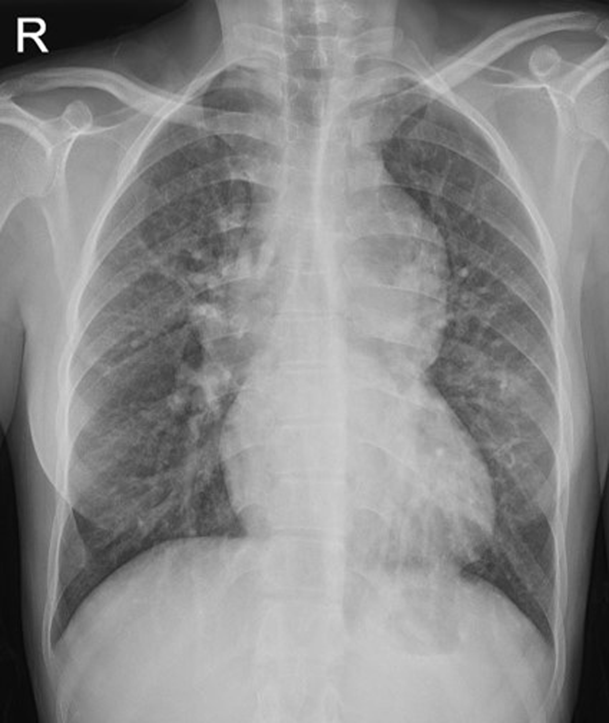
Figure 1
Frontal chest radiograph demonstrates marked mediastinal enlargement, consistent with the classical “snowman sign,” and pulmonary plethora.
The patient subsequently underwent a computed tomography (CT) angiography for further evaluation of the intracardiac anatomy and to rule out any other associated cardiovascular anomalies. CT angiography demonstrated the union of all four pulmonary veins forming a common chamber posterosuperior to the left atrium that ultimately drained into the left brachiocephalic vein via an ascending vertical vein (VV). A thin eccentric membranous web was seen at the junction of the common chamber and ascending VV, with aneurysmal dilatation of the vertical vein distal to it (Figure 2). A large ostium secundum atrial septal defect with dilatation of the right-sided heart chambers was also noted.
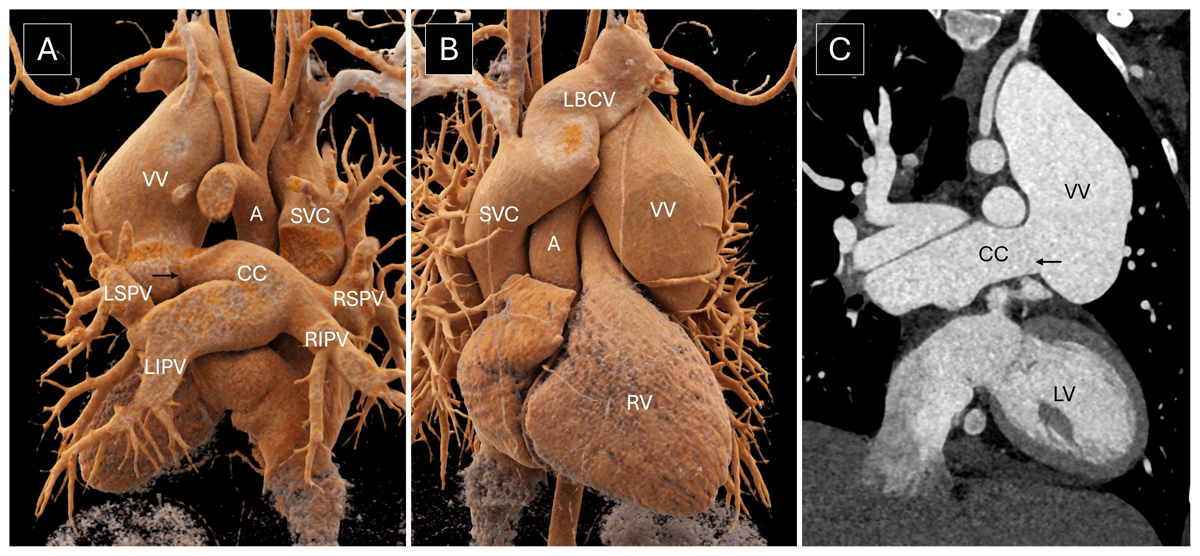
Figure 2
(A, B) Volume rendered images and (C) oblique coronal image show all four pulmonary veins joining to form a common chamber (CC), which drains into the left brachiocephalic vein (LBCV) via an ascending vertical vein (VV). A thin eccentric membranous web (black arrow) is noted at the junction of the common chamber and ascending VV with aneurysmal dilatation of the vertical vein distal to it. A: aorta; LV: left ventricle; RV: right ventricle; SVC: superior vena cava; RSPV: right superior pulmonary vein; LSPV: left superior pulmonary vein; RIPV: right inferior pulmonary vein; LIPV: left inferior pulmonary vein
Aneurysmal dilatation of the ascending VV is exceedingly rare in supracardiac TAPVC, with few cases reported previously. It has been attributed to post-stenotic dilatation distal to the site of extrinsic compression of the VV between the left pulmonary artery and left bronchus1 or distal to a thin membranous web within the VV.2 In the present case, no extrinsic compression over the VV was seen; rather, there was a thin eccentric membranous web at the junction of the common chamber and ascending VV causing focal luminal stenosis. The development of the VV aneurysm in the present case could possibly be attributed to the presence of turbulent flow within the VV, distal to the site of stenosis caused by the membranous web.
Competing Interests
The authors have no competing interests to declare.