
Figure 1
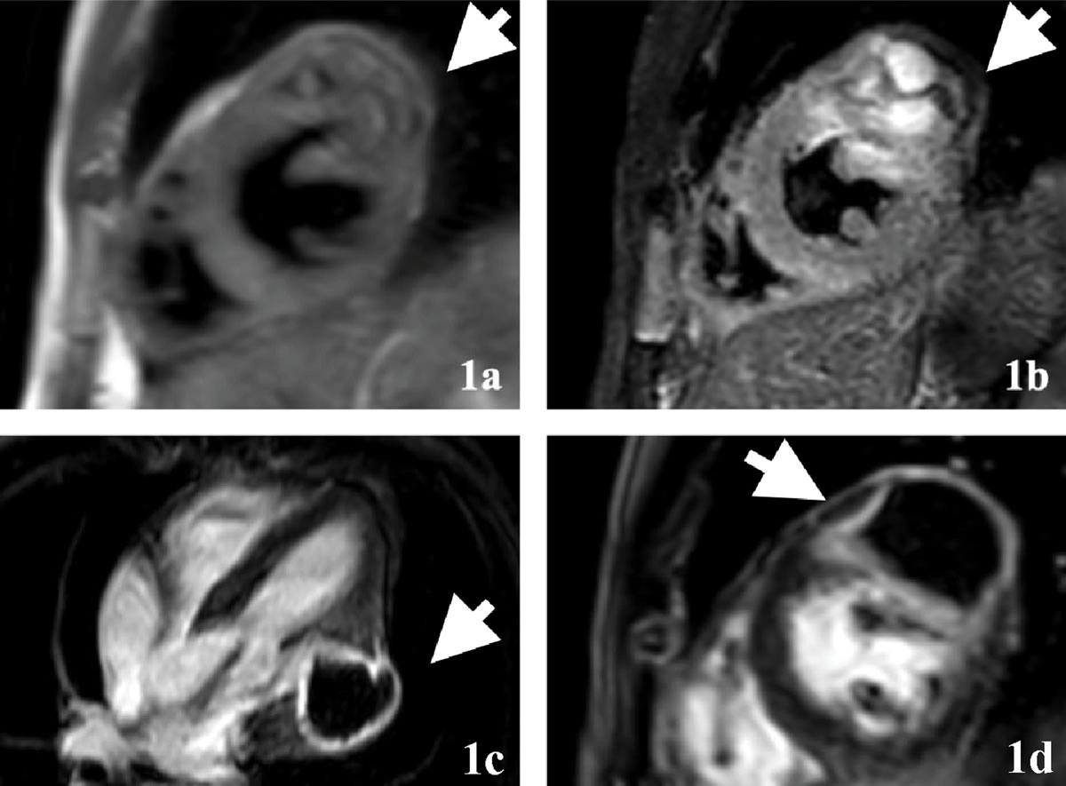
Multicystic lesion in the lateral left ventricular wall: morphology and late gadolinium enhancement findings. (1a) Short-axis double inversion recovery dark blood image at basal to mid-cavity level; (1b) short-axis T2-weighted short-tau inversion recovery image at the basal to mid-cavity level. A multicystic structure is noted with thick septations and fluid-filled cystic structures in the basal-mid lateral/anterolateral segment of the left ventricle bulging outwards towards the pericardial margin as well as mildly towards the cavity. Mild cyst wall enhancement noted on the late gadolinium enhancement (LGE) imaging; (1c) 4-chamber phase-sensitive inversion recovery (PSIR) LGE and (1d) short-axis PSIR LGE.
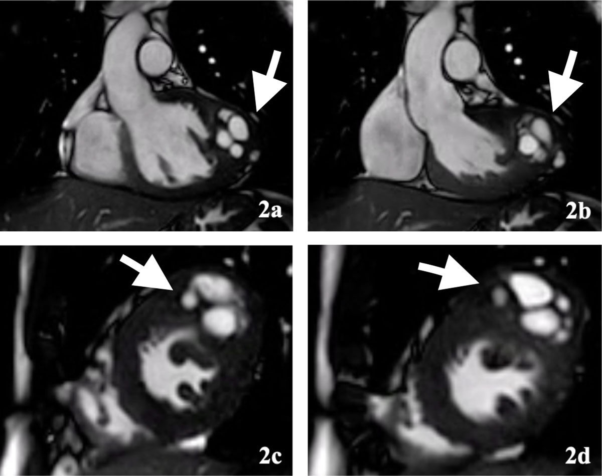
Figure 2
Cine cardiac magnetic resonance imaging depicts dynamic behavior of the multicystic lesion. (2a) Diastolic and (2b) systolic images from cine steady-state free precession-based left ventricular outflow tract; (2c) short-axis cine images at mid-cavity level and (2d) at basal to mid-cavity level, again confirming the multicystic structure in the basal to mid lateral/anterolateral wall of the left ventricle myocardium.
Video 1
Annular plane; see also at https://vimeo.com/1140625664.
Video 2
Apex 1; see also at https://vimeo.com/1140625673.
Video 3
Apex 2; see also at https://vimeo.com/1140625689.
Video 4
Basal 1; see also at https://vimeo.com/1140625693.
Video 5
Basal 2; see also at https://vimeo.com/1140625707.
Video 6
Basal 3; see also at https://vimeo.com/1140625720.
Video 7
Basal most; see also at https://vimeo.com/1140625723.
Video 8
Mid 1; see also at https://vimeo.com/1140625729.
Video 9
Mid 2; see also at https://vimeo.com/1140625737.
Video 10
Mid 3; see also at https://vimeo.com/1140625740.