
A 19-year-old nonsmoking male from rural Pakistan presented with a 1-month history of precordial chest pain, nonproductive cough, and indigestion. Laboratory evaluation was notable for mild eosinophilia (8.2%; reference range: 1–6%) with otherwise unremarkable findings. Transthoracic echocardiography and contrast-enhanced cardiothoracic computed tomography (CT) performed at the referring center demonstrated a cystic mass in the lateral wall of the left ventricle (LV); however, these images were unavailable due to resource limitations. Cardiac magnetic resonance imaging (MRI) showed a multilobulated, thick-walled cystic lesion centered in the basal to mid-lateral LV myocardium, with internal septations, hyperintense T2 signal, and mild peripheral enhancement on late gadolinium enhancement imaging. Findings were consistent with a cardiac hydatid cyst (Figures 1, 2, Videos 1, 2, 3, 4, 5, 6, 7, 8, 9, 10). The patient did not return for further diagnostic testing or treatment.
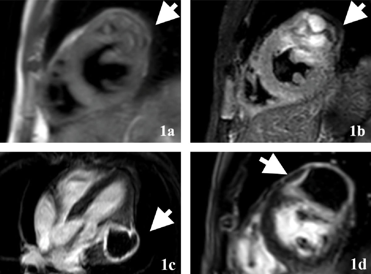
Figure 1
Multicystic lesion in the lateral left ventricular wall: morphology and late gadolinium enhancement findings. (1a) Short-axis double inversion recovery dark blood image at basal to mid-cavity level; (1b) short-axis T2-weighted short-tau inversion recovery image at the basal to mid-cavity level. A multicystic structure is noted with thick septations and fluid-filled cystic structures in the basal-mid lateral/anterolateral segment of the left ventricle bulging outwards towards the pericardial margin as well as mildly towards the cavity. Mild cyst wall enhancement noted on the late gadolinium enhancement (LGE) imaging; (1c) 4-chamber phase-sensitive inversion recovery (PSIR) LGE and (1d) short-axis PSIR LGE.
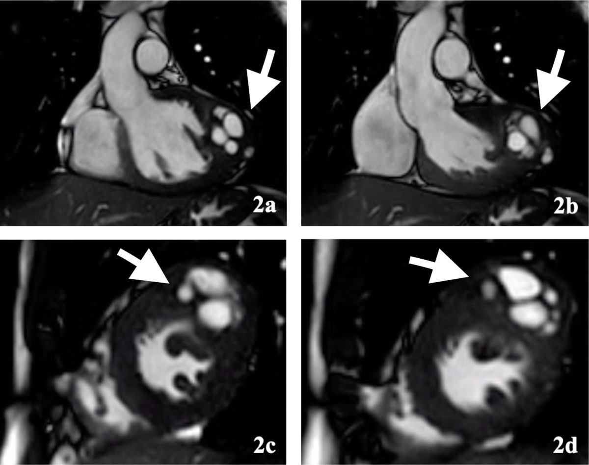
Figure 2
Cine cardiac magnetic resonance imaging depicts dynamic behavior of the multicystic lesion. (2a) Diastolic and (2b) systolic images from cine steady-state free precession-based left ventricular outflow tract; (2c) short-axis cine images at mid-cavity level and (2d) at basal to mid-cavity level, again confirming the multicystic structure in the basal to mid lateral/anterolateral wall of the left ventricle myocardium.
Video 1
Annular plane; see also at https://vimeo.com/1140625664.
Video 2
Apex 1; see also at https://vimeo.com/1140625673.
Video 3
Apex 2; see also at https://vimeo.com/1140625689.
Video 4
Basal 1; see also at https://vimeo.com/1140625693.
Video 5
Basal 2; see also at https://vimeo.com/1140625707.
Video 6
Basal 3; see also at https://vimeo.com/1140625720.
Video 7
Basal most; see also at https://vimeo.com/1140625723.
Video 8
Mid 1; see also at https://vimeo.com/1140625729.
Video 9
Mid 2; see also at https://vimeo.com/1140625737.
Video 10
Mid 3; see also at https://vimeo.com/1140625740.
Hydatid disease most commonly affects the liver (68.8–80%) and lungs (10–22.4%), with cardiac involvement seen in only 0.02% to 1.1% of cases.1 Cardiopericardial hydatid cysts are often asymptomatic but may present with nonspecific symptoms such as chest pain, exertional dyspnea, or palpitations, particularly in larger or septally located lesions.2 Echocardiography is the initial and preferred modality for diagnosing cardiac hydatid cysts, while CT and MRI offer superior delineation of cyst morphology and anatomical relationships, aiding in preoperative assessment.3,4
Competing Interests
The authors have no competing interests to declare.