
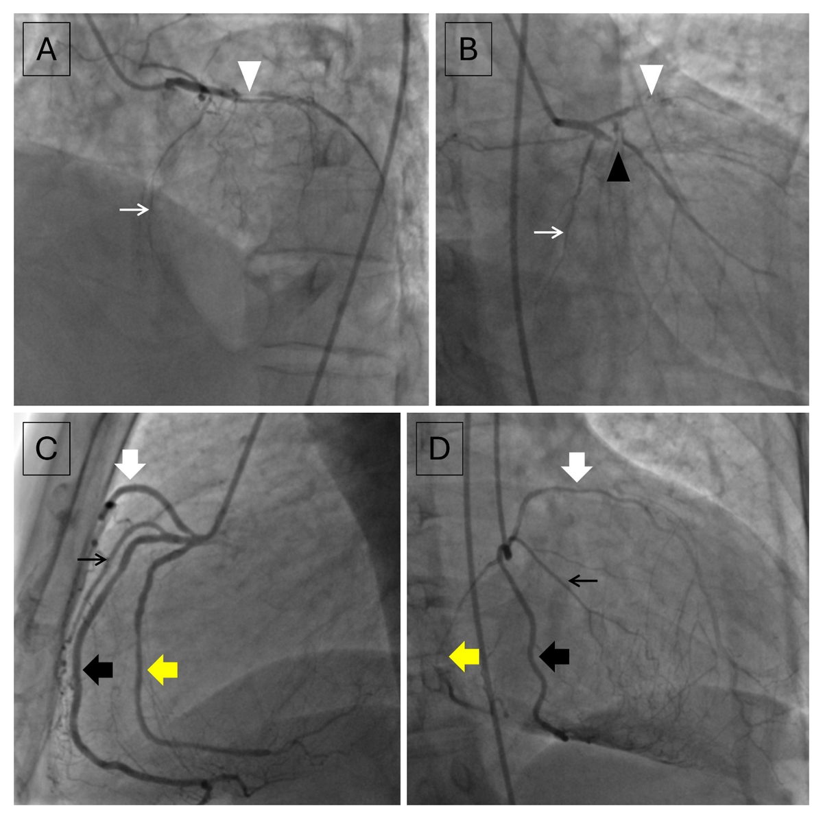
Figure 1
(A, B) Contrast injections in the left main coronary artery show severe diffuse disease in a short left anterior descending artery (white arrowhead) and proximal ramus intermedius (black arrowhead). No significant stenosis was noted in the left circumflex artery (thin white arrow). (C, D) Contrast injections in the right coronary artery show the right coronary artery trifurcating into a longer left anterior descending artery (thick white arrow) and two branches of equal size (anterior branch indicated by thick black arrow and posterior branch indicated by thick yellow arrow) with the anterior branch giving off the acute marginal branch (thin black arrow).
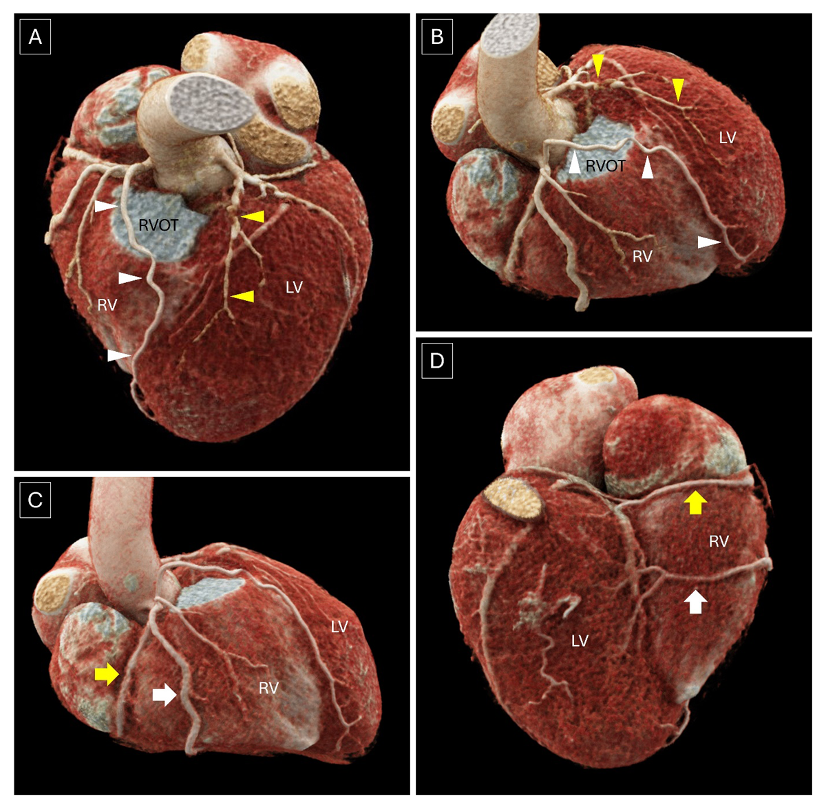
Figure 2
(A, B) Volume rendered images confirm the presence of type IV dual left anterior descending artery with a severely diseased shorter left anterior descending artery (yellow arrowheads) arising from the left main coronary artery and a longer left anterior descending artery (white arrowheads) arising from the right coronary artery having a pre-pulmonic course to enter the anterior interventricular groove, reaching to cardiac apex. (C, D) Volume-rendered images demonstrate a split right coronary artery where the right coronary artery divides into an anterior (white thick arrow) and posterior (yellow thick arrow) branch, both of which reach the posterior interventricular groove to continue as distal and proximal posterior descending artery, respectively. LV: left ventricle; RV: right ventricle; RVOT: right ventricular outflow tract