
A 50-year-old male presented in the outpatient clinic with a history of chronic coronary syndrome with class III angina on exertion. Electrocardiogram (ECG) showed ST elevation with positive cardiac biomarkers. He was taken for primary percutaneous coronary intervention (PCI), which revealed an unusual coronary arterial anatomy. Severe diffuse disease was seen in the shorter left anterior descending artery (LAD) and proximal ramus intermedius. Right coronary artery (RCA) angiogram revealed a longer LAD arising from the proximal RCA coursing towards the anterior interventricular groove reaching to the cardiac apex. The RCA appeared to split into two branches of equal size, both reaching the posterior interventricular groove. No stenosis was seen in the RCA branches (Figure 1). In light of the disease-free longer LAD supplying the left ventricular myocardium, no active intervention was done at the time.
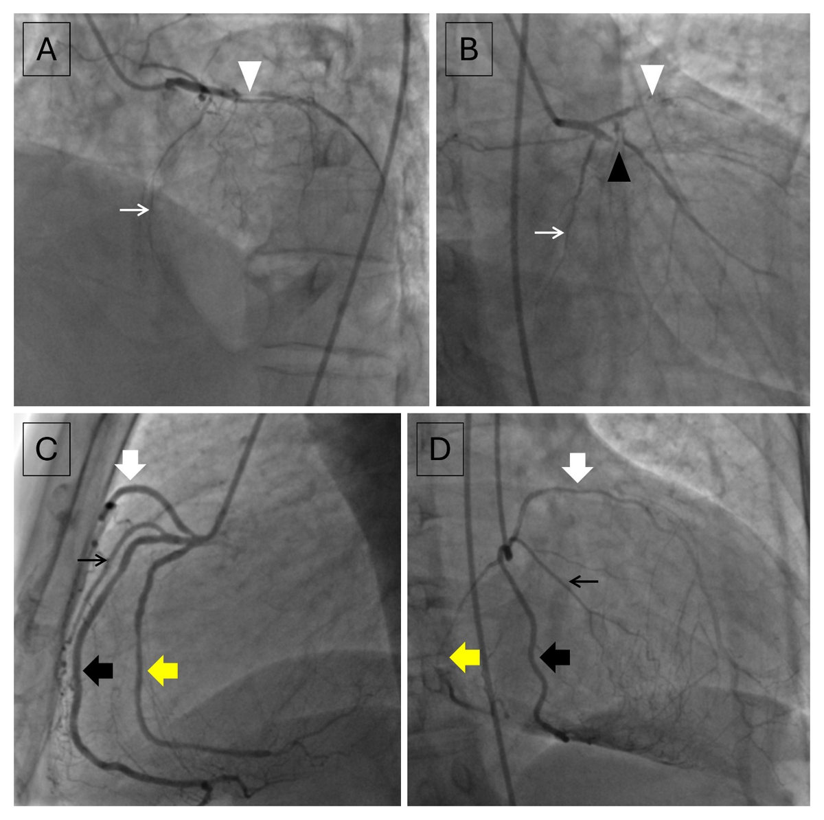
Figure 1
(A, B) Contrast injections in the left main coronary artery show severe diffuse disease in a short left anterior descending artery (white arrowhead) and proximal ramus intermedius (black arrowhead). No significant stenosis was noted in the left circumflex artery (thin white arrow). (C, D) Contrast injections in the right coronary artery show the right coronary artery trifurcating into a longer left anterior descending artery (thick white arrow) and two branches of equal size (anterior branch indicated by thick black arrow and posterior branch indicated by thick yellow arrow) with the anterior branch giving off the acute marginal branch (thin black arrow).
Subsequently, an ECG-gated coronary computed tomography angiography (CCTA) was performed for delineation of the coronary anatomy. CCTA showed the presence of type IV dual LAD, with the severely diseased shorter LAD arising from the left main coronary artery and a longer LAD arising from the right coronary artery having pre-pulmonic course to enter the anterior interventricular groove, reaching to the cardiac apex.1 The RCA was seen trifurcating shortly after its origin into the longer LAD and two other branches (anterior and posterior) of almost equal calibers. The posterior branch was seen coursing orthotopically within the right atrioventricular (AV) groove, reaching until the crux of the heart and terminating as the proximal posterior descending artery (PDA). The anterior branch exited the right AV groove and was seen coursing along the acute margin of the heart, giving off the acute marginal branch. The anterior branch subsequently terminated in the distal half of the posterior interventricular groove to terminate as the distal PDA (Figure 2).
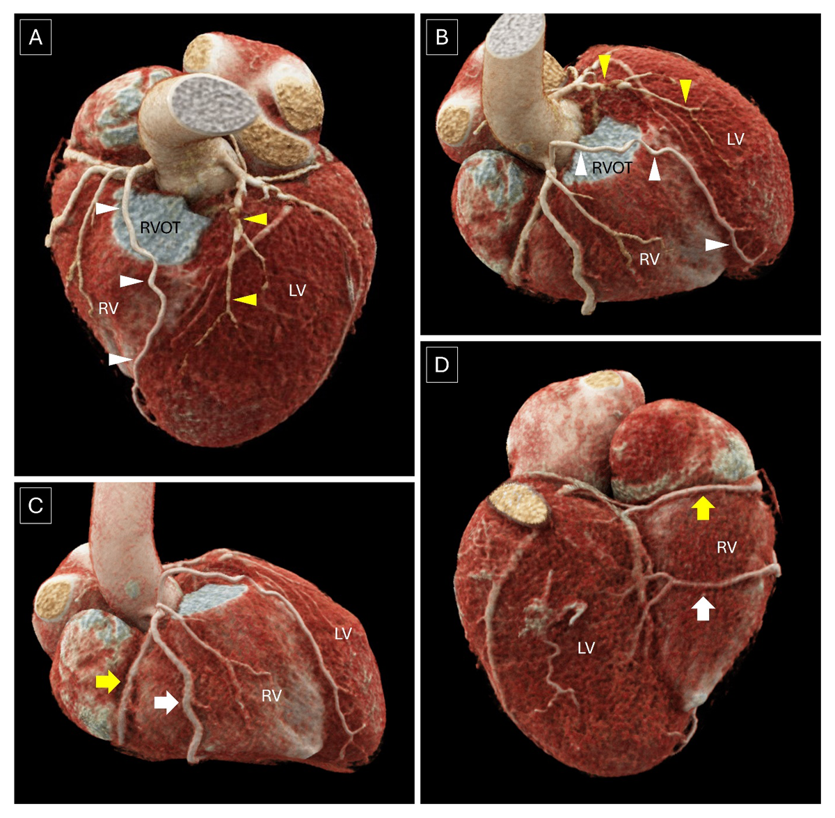
Figure 2
(A, B) Volume rendered images confirm the presence of type IV dual left anterior descending artery with a severely diseased shorter left anterior descending artery (yellow arrowheads) arising from the left main coronary artery and a longer left anterior descending artery (white arrowheads) arising from the right coronary artery having a pre-pulmonic course to enter the anterior interventricular groove, reaching to cardiac apex. (C, D) Volume-rendered images demonstrate a split right coronary artery where the right coronary artery divides into an anterior (white thick arrow) and posterior (yellow thick arrow) branch, both of which reach the posterior interventricular groove to continue as distal and proximal posterior descending artery, respectively. LV: left ventricle; RV: right ventricle; RVOT: right ventricular outflow tract
Split RCA is a rare anatomical variant wherein the RCA divides into anterior and posterior divisions subsequently terminating as two PDA.2, 3 Coexistence of dual LAD with split RCA is rare, with no similar cases reported in literature to date. This case underscores the importance of CCTA in accurately delineating rare coronary artery anatomical variants to avoid both a misdiagnosis and the risk of potential catastrophic outcomes during interventional or surgical procedures.
Competing Interests
The authors have no competing interests to declare.
Author Contributions
Damandeep Singh and Niraj Nirmal Pandey contributed equally to the manuscript and share the first authorship.