
Table 1
Categories of intraoperative data collected in the OR. This table summarizes the six primary categories of intraoperative data commonly collected in modern ORs. Each data type is described with representative examples and its corresponding clinical purpose. These categories encompass physiological monitoring, imaging modalities, surgical video feeds, instrument tracking systems, robotic and kinematic metrics, and environmental/workflow analytics. Together, they form the foundation of intraoperative data analytics and enable real-time surgical decision-making, workflow optimization, and post-procedural evaluation. OR: operating room; SpO2: oxygen saturation; BP: blood pressure; EtCO2: end-tidal carbon dioxide; EEG: electroencephalography; BIS: bispectral index; SSEP: somatosensory evoked potential; MEP: motor evoked potential; ECMO: extracorporeal membrane oxygenation; CT: computed tomography, MRI: magnetic resonance imaging; TCD: transcranial Doppler; AI: artificial intelligence; AR: augmented reality; RFID: radiofrequency identification
| DATA TYPE | DESCRIPTION | EXAMPLES | PURPOSE/USE |
|---|---|---|---|
| Physiological data | Continuous monitoring of patient vital signs and neurological signals during surgery | SpO2, BP, HR, EtCO2, EEG, BIS, SSEP, MEP, ECMO settings | Guides anesthesia and intraoperative decisions; early detection of instability or complications |
| Imaging data | Real-time or intraoperative imaging to guide surgical decisions | Ultrasound, fluoroscopy, Intra-op CT, MRI fusion, TCD, angiography | Enhances accuracy and anatomic guidance; confirms procedural outcomes |
| Surgical video feeds | Visual recordings of procedures through endoscopes, AR headsets, or external cameras | Da Vinci endoscopic video, Apple Vision Pro, CinVivo, microscope feeds, overhead and external cameras | Skill assessment, safety monitoring, AI-based workflow recognition |
| Instrument tracking | Monitoring tool use, movement, and localization using sensor or vision systems | RFID, barcodes, cautery logs, optical/electromagnetic tracking | Optimizes efficiency, ensures instrument accountability, tracks workflow |
| Robotic/kinematic data | High-resolution data from robotic systems that track motion metrics of surgical tools | Da Vinci kinematics, Hugo robotic arm tracking, wrist angles, grip pressure | Assesses technical skill, supports training, enables AI-guided feedback |
| Environmental/workflow data | Metrics that track ambient conditions and room activity during surgery | Temperature, humidity, door openings, team movements, timestamped workflow data | Improves safety, reduces delays, optimizes OR utilization and communication |
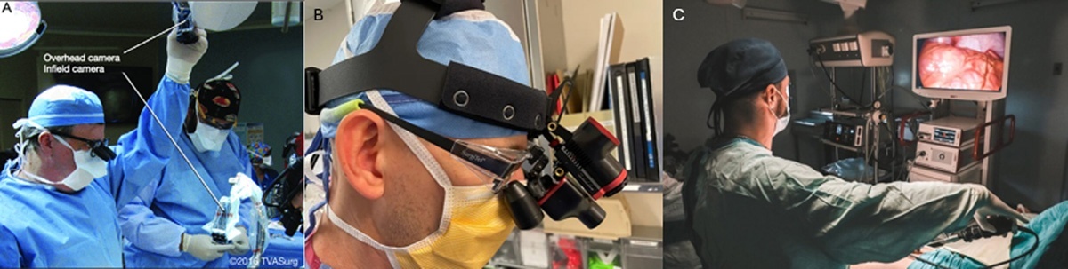
Figure 1
This figure illustrates three commonly used methods for intraoperative video capture in the operating room. (A) Overhead surgical light-mounted cameras provide a wide-angle view of the operative field without obstructing workflow. (B) Head-mounted cameras offer a first-person perspective from the surgeon’s point of view, useful for documentation and education. (C) Laparoscopic or endoscopic camera systems capture high-resolution, magnified views of internal anatomy during minimally invasive procedures. Together, these modalities enable surgical video recording for real-time guidance, postoperative review, technical skill assessment, and machine learning-based analytics.
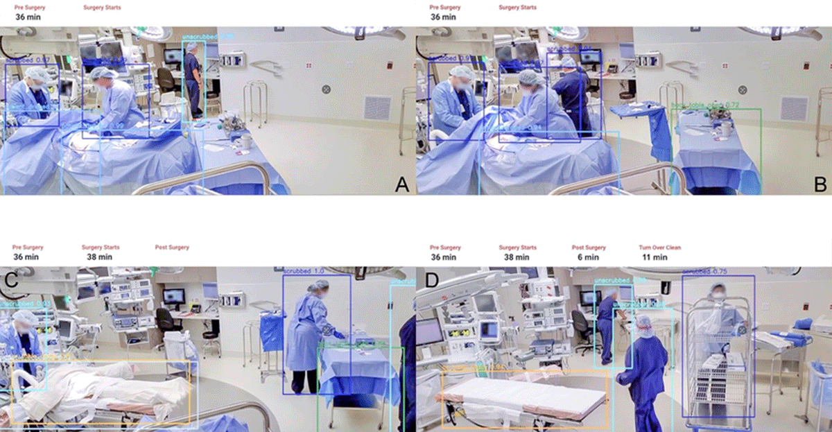
Figure 2
This figure demonstrates the application of surgical video analytics in segmenting and annotating phases of a surgical case using computer vision and time-stamped metadata. (A) Preoperative phase: The anesthesia provider and circulator nurse prepare the patient and confirm presurgical protocols. (B) Setup phase: The surgeon scrubs in while sterile instruments are arranged and surgical timeout is performed. (C) Postoperative phase: The procedure is completed, the patient is undraped, and the anesthesia team manages emergence and airway stabilization. (D) Turnover phase: After the patient exits the operating room, staff begin cleaning and preparing the environment for the next case. These distinct stages can be identified and analyzed through AI-powered video tools, such as Apella, to improve OR efficiency, team coordination, and safety compliance. Reprinted with permission from Apella. AI: artificial intelligence; OR: operating room
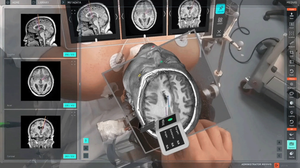
Figure 3
This figure demonstrates the use of an augmented reality surgical navigation platform (Medivis Spine Navigation Platform) that overlays segmented 3-dimensional anatomical structures onto the patient in real time. The system shown utilizes a preoperative computed tomography scan to create a volumetric model of the brain, which is then fused with the patient’s physical anatomy using augmented reality. Multiple orthogonal imaging planes (sagittal, axial, and coronal) are displayed in parallel to guide precise targeting. The holographic overlay allows the surgeon to visualize deep structures without additional incisions or continuous fluoroscopy, enhancing spatial awareness and procedural accuracy. This fusion-based approach is particularly valuable in neurosurgery and other precision-demanding fields, where millimetric accuracy is essential. Reprinted with permission from Medivis SurgicalAR.
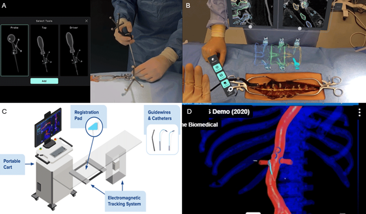
Figure 4
This figure showcases advanced intraoperative navigation technologies that enable real-time tracking and visualization of surgical instruments. (A) Medivis Spine Navigation Platform with a custom fiduciary marker attached to surgical instruments, allowing precise 3-dimensional tracking through augmented reality. (B) Combined augmented reality and image fusion platform used during spinal surgery, integrating tool tracking with preoperative imaging for enhanced navigation. (C) Setup of Centerline Biomedical’s electromagnetic tracking system, which enables intraoperative localization of endovascular catheters and wires without continuous fluoroscopy. (D) Live endovascular navigation using the electromagnetic system, allowing radiation-free tracking of instruments in complex vascular procedures. These technologies represent a shift toward radiation-free, high-precision navigation that enhances safety, accuracy, and intraoperative efficiency. Adapted from Medivis Spine Navigation platform (A-B) and Centerline Biomedical platform (C-D).