
Introduction
Intraoperative data analytics is the real-time collection, processing, and interpretation of data generated during surgical procedures.1 This includes vital signs, medication delivery, surgical instrument usage, imaging studies, video feeds, electronic health records (EHRs), and operating room (OR) workflow metrics. While a single surgery can produce vast quantities of data, these datasets are often dense, heterogeneous, and difficult to interpret.2 By leveraging advanced technologies—such as machine learning (ML), computer vision, spatial computing, and sensor-based monitoring—clinicians and researchers can more efficiently extract meaningful insights through data mining, transformation, and analysis. The field of intraoperative data analytics has rapidly expanded in response to evolving surgical techniques and the demand for improved patient outcomes. Real-time data analytics support personalized treatment, more accurate predictive modeling, and increased OR workflow efficiency.3 Most importantly, these tools contribute directly to patient safety, recognition of patterns in surgical complications, prolonged operating time, or adverse drug events that facilitate early intervention and reduce unfavorable outcomes. Furthermore, healthcare systems can integrate preventative measures and improve protocols. As surgery becomes more data-driven, intraoperative analytics are positioned at the forefront of surgical innovation with the ability to transform information into measurable, reproducible knowledge and better results.
Categories of Intraoperative Data
Operating rooms are gold mines of data: machines constantly collect information on a patient’s physiological signals, hybrid ORs capture imaging scans in the middle of procedures, ORs are fitted with surgical video cameras, and medical instruments and personnel are continuously tracked to ensure their locations. Each method of data collection offers unique insights into surgical performance and can give a comprehensive view of an intraoperative environment (Table 1).
Table 1
Categories of intraoperative data collected in the OR. This table summarizes the six primary categories of intraoperative data commonly collected in modern ORs. Each data type is described with representative examples and its corresponding clinical purpose. These categories encompass physiological monitoring, imaging modalities, surgical video feeds, instrument tracking systems, robotic and kinematic metrics, and environmental/workflow analytics. Together, they form the foundation of intraoperative data analytics and enable real-time surgical decision-making, workflow optimization, and post-procedural evaluation. OR: operating room; SpO2: oxygen saturation; BP: blood pressure; EtCO2: end-tidal carbon dioxide; EEG: electroencephalography; BIS: bispectral index; SSEP: somatosensory evoked potential; MEP: motor evoked potential; ECMO: extracorporeal membrane oxygenation; CT: computed tomography, MRI: magnetic resonance imaging; TCD: transcranial Doppler; AI: artificial intelligence; AR: augmented reality; RFID: radiofrequency identification
| DATA TYPE | DESCRIPTION | EXAMPLES | PURPOSE/USE |
|---|---|---|---|
| Physiological data | Continuous monitoring of patient vital signs and neurological signals during surgery | SpO2, BP, HR, EtCO2, EEG, BIS, SSEP, MEP, ECMO settings | Guides anesthesia and intraoperative decisions; early detection of instability or complications |
| Imaging data | Real-time or intraoperative imaging to guide surgical decisions | Ultrasound, fluoroscopy, Intra-op CT, MRI fusion, TCD, angiography | Enhances accuracy and anatomic guidance; confirms procedural outcomes |
| Surgical video feeds | Visual recordings of procedures through endoscopes, AR headsets, or external cameras | Da Vinci endoscopic video, Apple Vision Pro, CinVivo, microscope feeds, overhead and external cameras | Skill assessment, safety monitoring, AI-based workflow recognition |
| Instrument tracking | Monitoring tool use, movement, and localization using sensor or vision systems | RFID, barcodes, cautery logs, optical/electromagnetic tracking | Optimizes efficiency, ensures instrument accountability, tracks workflow |
| Robotic/kinematic data | High-resolution data from robotic systems that track motion metrics of surgical tools | Da Vinci kinematics, Hugo robotic arm tracking, wrist angles, grip pressure | Assesses technical skill, supports training, enables AI-guided feedback |
| Environmental/workflow data | Metrics that track ambient conditions and room activity during surgery | Temperature, humidity, door openings, team movements, timestamped workflow data | Improves safety, reduces delays, optimizes OR utilization and communication |
Physiological Data
Vital signs such as oxygen saturation (SpO2), blood pressure, heart rate and rhythm, respiratory rate, core and peripheral body temperature, central venous pressure, capnography ventilator settings, urine output, electroencephalogram, bispectral index for anesthesia depth, electromyography, motor evoked potentials, somatosensory evoked potentials, and extracorporeal membrane oxygenation or cardiac bypass settings, are continuously monitored during surgery.4,5,6,7 These signals provide data about the patient’s cardiopulmonary stability and are used to guide anesthesia or surgical decisions. Furthermore, close monitoring of physiological signals allows for earlier identification of possible complications or correction of imbalances.
Imaging Data
Intraoperative imaging technologies—including real-time ultrasound (vascular, cardiac, intravascular, or general), fluoroscopy, transcranial Doppler (TCD), and contrast angiograms—improve surgical accuracy. These images can be compared with previously performed magnetic resonance imaging or computerized tomography (CT) scans, enhancing diagnosing capabilities and guiding later therapy.8,9,10
Surgical Video Feeds
In open surgery, surgeons may use head-mounted cameras, like the CinVivo Titan 4K System (CinVivo), or immersive augmented reality (AR) headsets, such as the Apple Vision Pro (Apple), to capture and share a first-person view of the surgical field.11 In-light cameras provide a wide-angle view of the OR while minimizing disruption to general workflow.12 Surgical endoscopes like these are used for laparoscopic, endoscopic, and robotic surgery and capture high-resolution magnified views of anatomical structures, allowing for precise dissection and real-time guidance during complex procedures.13 Microscopes (used in ophthalmic or neurosurgical procedures) also provide magnified views of anatomical structures. These feeds are increasingly used for real-time analysis, surgical education, and postoperative review (Figure 1).
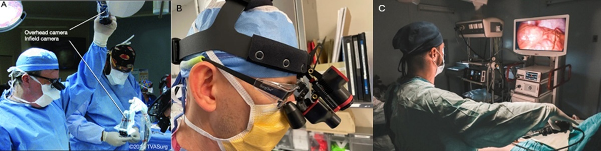
Figure 1
This figure illustrates three commonly used methods for intraoperative video capture in the operating room. (A) Overhead surgical light-mounted cameras provide a wide-angle view of the operative field without obstructing workflow. (B) Head-mounted cameras offer a first-person perspective from the surgeon’s point of view, useful for documentation and education. (C) Laparoscopic or endoscopic camera systems capture high-resolution, magnified views of internal anatomy during minimally invasive procedures. Together, these modalities enable surgical video recording for real-time guidance, postoperative review, technical skill assessment, and machine learning-based analytics.
Surgical video footage provides a rich source of data that can be analyzed to objectively assess technical skill and surgical performance. Computer vision and artificial intelligence (AI)-enabled tracking can extract key motion-based metrics, such as instrument path length, velocity, acceleration, and idle time.14 Beyond skill evaluation, intraoperative video can be segmented to recognize procedural phases and instrument usage timelines.15
Surgical video analytics also have growing utility in safety surveillance and OR efficiency. Systems can detect adverse events such as tool misplacement, bleeding, or skipped protocol steps through pattern recognition and anomaly detection. Environmental analysis using wide-angle or ceiling-mounted cameras can track room traffic, identify distractions or disruptions, and quantify staff movement patterns—all of which affect sterility, room turnover, and surgical flow.16 Furthermore, video datasets provide ground truth for AI model training, supporting the development of autonomous systems that assist with real-time guidance, safety alerts, or predictive analytics. This positions surgical video not just as a documentation tool but as a foundational layer for intelligent, data-driven ORs (Figure 2).
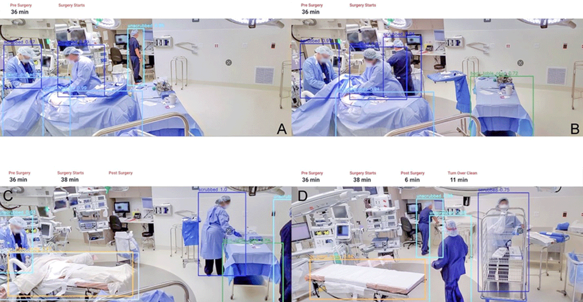
Figure 2
This figure demonstrates the application of surgical video analytics in segmenting and annotating phases of a surgical case using computer vision and time-stamped metadata. (A) Preoperative phase: The anesthesia provider and circulator nurse prepare the patient and confirm presurgical protocols. (B) Setup phase: The surgeon scrubs in while sterile instruments are arranged and surgical timeout is performed. (C) Postoperative phase: The procedure is completed, the patient is undraped, and the anesthesia team manages emergence and airway stabilization. (D) Turnover phase: After the patient exits the operating room, staff begin cleaning and preparing the environment for the next case. These distinct stages can be identified and analyzed through AI-powered video tools, such as Apella, to improve OR efficiency, team coordination, and safety compliance. Reprinted with permission from Apella. AI: artificial intelligence; OR: operating room
Device and Instrument Tracking
Tracking the location, use, and movement of surgical tools and devices helps optimize workflow and ensure surgical safety.17 Technologies such as radiofrequency identification (RFID), as enabled by M&S Biotics, have shown promise and are the focus of ongoing research as a means to provide data on instrument utilization, increase surgical tool retention, and enhance OR efficiency.18,19 Computer vision-based tracking modalities and alternative navigation systems based on optical, fiberoptics, and electromagnetics also allow tool tracking and can be combined with fusion reconstruction of preoperative imaging to track surgical tools during cases.20,21
Robotic and Kinematic Data
Robotic and kinematic data represent a unique and rich category of intraoperative information, particularly in surgeries involving robotic-assisted platforms such as the da Vinci (Intuitive Surgical) or Medtronic Hugo systems. These platforms generate high-frequency data streams that capture instrument joint angles, wrist rotations, grip strength, path length, velocity, and acceleration with subsecond precision.
This kinematic data allows for objective analysis of surgical techniques, including motion economy, tremor suppression, and tool trajectory. It can be used to assess surgeon proficiency, monitor for fatigue, and detect procedural deviations in real time. Additionally, this data is foundational for training AI models that reconstruct surgical workflows, enable autonomous functions, and provide context-aware guidance during procedures.22,23
Environmental and Workflow Data
Environmental and workflow data offer critical context for understanding the dynamics of the OR beyond direct patient care. This category includes information such as ambient temperature, lighting conditions, and humidity, which can influence surgical performance and infection risk. Workflow metrics—such as door openings, staff movements, room traffic, and procedural time stamps—are increasingly captured through ceiling-mounted cameras, RFID systems, or smart OR integration platforms.24,25
These data points help identify inefficiencies, distractions, or breaches in sterility that may compromise patient safety or prolong operative time. Additionally, synchronized audiovisual recordings can track communication patterns and team interactions, offering insights into nontechnical skills such as teamwork and situational awareness. By analyzing environmental and workflow data, hospitals can optimize staffing models, streamline case turnover, and design targeted interventions to reduce delays or errors.
Current State-of-the-art Technologies in Operating Room Data Analytics
Imaging Systems
Advanced imaging modalities now provide detailed real-time visualization that is critical for the planning and execution of surgical procedures. Cone-beam computed tomography, often integrated with mobile C-arm systems, enables intraoperative contrasted 3D imaging without needing to reposition the patient. This is especially useful in endovascular, orthopedic, and cranial procedures, where high-resolution anatomical context can guide precise intervention.26
Augmented fluoroscopy builds upon traditional 2D imaging by fusing preoperative CT or magnetic resonance datasets onto live fluoroscopic views, providing enhanced spatial awareness during device navigation. By using fusion to label the origin or trajectory of artery branches, more precise navigation and treatment (stenting, coiling, ballooning) can be performed with less radiation to the operator and patient. This technique is increasingly used in vascular surgery and neurosurgery, where anatomic complexity and proximity to critical structures demand high precision (Figure 3).21,27,28
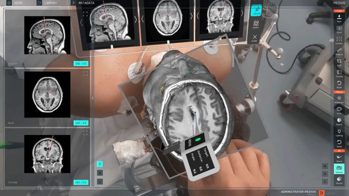
Figure 3
This figure demonstrates the use of an augmented reality surgical navigation platform (Medivis Spine Navigation Platform) that overlays segmented 3-dimensional anatomical structures onto the patient in real time. The system shown utilizes a preoperative computed tomography scan to create a volumetric model of the brain, which is then fused with the patient’s physical anatomy using augmented reality. Multiple orthogonal imaging planes (sagittal, axial, and coronal) are displayed in parallel to guide precise targeting. The holographic overlay allows the surgeon to visualize deep structures without additional incisions or continuous fluoroscopy, enhancing spatial awareness and procedural accuracy. This fusion-based approach is particularly valuable in neurosurgery and other precision-demanding fields, where millimetric accuracy is essential. Reprinted with permission from Medivis SurgicalAR.
Ultrasound, including transesophageal echocardiography and intravascular ultrasound, remains a cornerstone for real-time soft tissue and vascular evaluation. Transcranial Doppler adds further functionality by enabling dynamic assessment of cerebral blood flow, useful in neurosurgical and vascular procedures to detect embolic events or assess perfusion during critical phases where vascular clamps may compromise cerebral blood flow.29,30
Wearables and Sensors
Vital sign monitoring is the cornerstone of intraoperative patient surveillance. In addition to conventional devices, emerging wearable technologies are beginning to augment or replace standard monitors. For example, the BioButton® (BioIntelliSense Inc.) is a compact, chest-worn sensor capable of continuously monitoring respiratory rate, heart rate, skin temperature, and activity level. Though originally developed for post-acute care and remote monitoring, its real-time streaming capabilities are being explored for intraoperative and perioperative use.31
Hemodynamic monitoring during surgery is essential for assessing cardiac output, vascular resistance, and overall perfusion status. Among the advanced tools available, the FloTrac™ system (Edwards Lifesciences) offers a minimally invasive method to continuously measure stroke volume, cardiac output, and dynamic preload indicators such as stroke volume variation using data derived from an arterial line. Unlike traditional pulmonary artery catheters, FloTrac™ does not require central access or external calibration, making it more suitable for a wider range of procedures. It utilizes proprietary algorithms to analyze the arterial pressure waveform in real time, providing clinicians with actionable insights into the patient’s fluid responsiveness and cardiac performance.32
Pressure mapping sensors provide critical data on patient positioning and interface pressures during surgery, helping to prevent positioning-related injuries such as pressure ulcers, nerve compression, or musculoskeletal strain. These thin, flexible sensor arrays are typically placed beneath high-risk anatomical zones—such as the sacrum, heels, or shoulders—where sustained pressure during long procedures can impair perfusion. Systems like ForeSite OR (XSESNSOR) and CONFORMat (Tekscan) use thousands of sensing nodes to create real-time, high-resolution pressure maps, allowing surgical teams to visualize pressure distribution and proactively make adjustments.33,34
Navigation and Robotics
Modern surgical navigation systems increasingly rely on optical and electromagnetic tracking to localize instruments and anatomy in 3D space, enabling greater precision during minimally invasive procedures. In endovascular surgery, the use of electromagnetic tracking with the Intra-Operative Positioning System (IOPS) (Centerline Biomedical) allows real-time navigation of wires and catheters without continuous fluoroscopy. By embedding electromagnetic sensors into endovascular devices and registering them for preoperative CT imaging, IOPS provides a dynamic, radiation-free visualization of instrument movement inside the vasculature (Figure 4).20
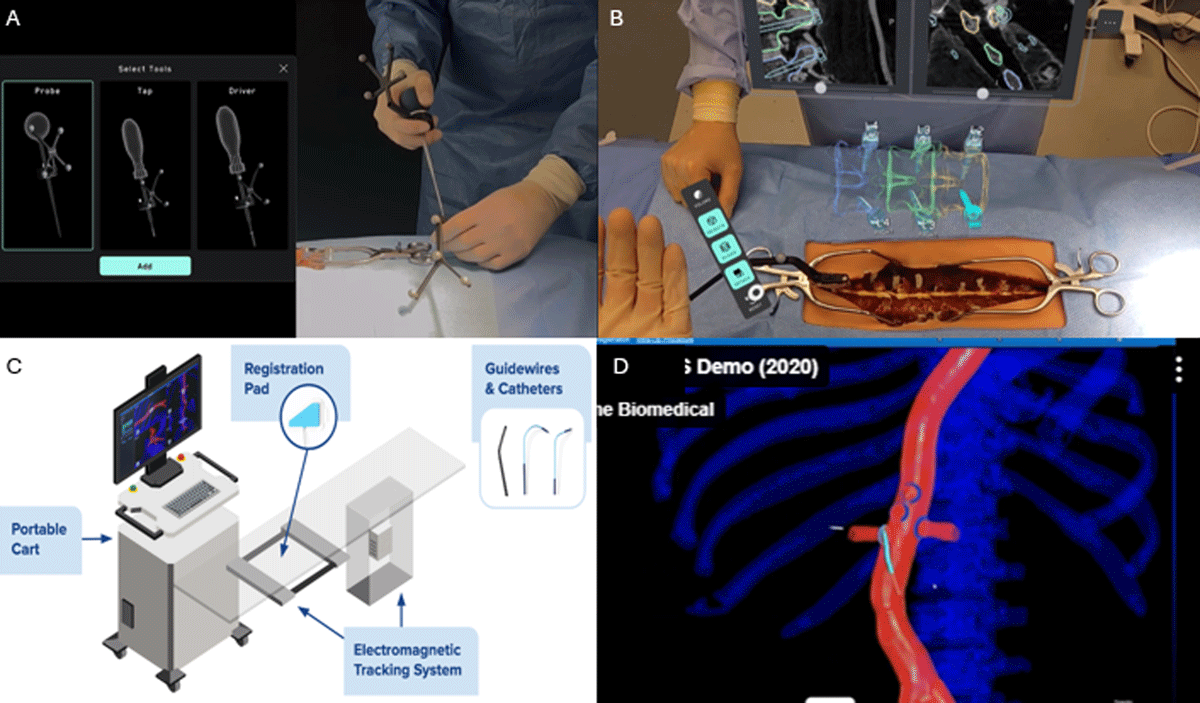
Figure 4
This figure showcases advanced intraoperative navigation technologies that enable real-time tracking and visualization of surgical instruments. (A) Medivis Spine Navigation Platform with a custom fiduciary marker attached to surgical instruments, allowing precise 3-dimensional tracking through augmented reality. (B) Combined augmented reality and image fusion platform used during spinal surgery, integrating tool tracking with preoperative imaging for enhanced navigation. (C) Setup of Centerline Biomedical’s electromagnetic tracking system, which enables intraoperative localization of endovascular catheters and wires without continuous fluoroscopy. (D) Live endovascular navigation using the electromagnetic system, allowing radiation-free tracking of instruments in complex vascular procedures. These technologies represent a shift toward radiation-free, high-precision navigation that enhances safety, accuracy, and intraoperative efficiency. Adapted from Medivis Spine Navigation platform (A-B) and Centerline Biomedical platform (C-D).
Smart tool tracking is revolutionizing surgical workflows by providing real-time visualization of instrument location and movement within the surgical field relative to a reference fusion image from historical CT imaging. For example, the Medivis Spine Navigation Platform, now cleared by the US Food and Drug Administration for spine navigation, integrates optical and infrared tracking with AR and AI. It allows surgeons to see holographic representations of instruments such as drills or catheters in 3D space, align tools to planned trajectories, and interact with imaging data handsfree via gestures and voice controls.21
Robotic surgical platforms like the da Vinci and Medtronic Hugo collect detailed kinematic data—including wrist angles, hand movements, and instrument path lengths—at subsecond resolution. This data is invaluable for training analysis, automation research, and surgical benchmarking. Integration of this kinematic data with AI models allows the reconstruction of procedural steps and error detection in real time.22
Operating Room Integration
Smart OR platforms like DashIQ™ (Stryker) and Loop-X (Brainlab AG) bring together multimodal data streams including video, imaging, device status, and patient vitals into a centralized interface. These systems support synchronized documentation, device control, and real-time collaboration, reducing cognitive load and enhancing situational awareness.35,36
The OR black box (Surgical Safety Technologies) captures synchronized audiovisual, environmental, and patient data throughout a case. When combined with analytics software, this data provides insights into workflow inefficiencies, human factors, and near-miss events, forming a foundation for quality assurance and medicolegal review.37
Artificial Intelligence
Artificial intelligence has begun to transform OR analytics through real-time data interpretation and decision support. One example is Caresyntax, which offers an AI-driven platform that annotates surgical videos.38 Their system applies advanced computer vision to identify and label key events such as incision, stapling, suctioning, and other procedural steps, creating structured timelines that support workflow analysis, surgical documentation, and performance review across various specialties. Another company is Surgical Success, with its SurgAI Video Intelligence Engine that uses AI to detect surgical phases, instruments, and critical events in real time.38,39,40,41
Predictive analytics models, using data from thousands of prior procedures, can forecast complications, blood loss, or prolonged operative time. One notable example of predictive analytics in surgery is MySurgeryRisk, an AI-powered platform developed by University of Florida researchers. Leveraging data from over 74,000 procedures and nearly 58,000 patients spanning 7 years, MySurgeryRisk uses machine learning to forecast complications, prolonged ICU stays, and mortality risk more accurately than standard physician assessment. The system draws on a rich dataset that includes patient demographics, lab values, medication usage, and vital signs recorded during surgery, delivering real-time risk scores to inform intraoperative decision-making.42
Challenges of Data Synchronization
Modern ORs are evolving to support multimodal data acquisition. These environments integrate advanced imaging systems, physiological monitors, robotic surgical tools, and real-time analytics platforms into regular OR workflow. This generates high volumes of data that enhance surgical decision-making, clinician and patient safety, and documentation. Multimodal integration involves capturing and aligning diverse data—physiological signals, imaging data, surgical video feeds, and instrument tracking data—into a cohesive timeline. This is particularly feasible in hybrid ORs, which are designed to support both conventional and image-guided interventions.43
Data synchronization across various sources is crucial for contextual understanding and favorable patient outcomes. Real-time temporal alignment remains a significant challenge because of different sampling rates, data formats, and possible latency between devices. Currently, monitoring systems function independently, and there is yet to be a time-synchronized multimodal monitoring system that efficiently integrates patient vitals during a procedure.44 Thus, OR staff, including surgeons, anesthesiologists, residents, nurses, and scrub technicians, must continuously monitor multiple screens while simultaneously managing patient care, increasing cognitive load and the risk of human error.
However, interoperability standards allow for effective data exchange and reduce human error; in fact, Health Level 7 (HL7 International) and Digital Imaging and Communications in Medicine (DICOM) are widely adopted to support data sharing between clinical systems. They also are utilized in EHR systems such as Epic, where they facilitate communication of critical patient information (such as prior procedures, medications, and clinical history) to support informed intraoperative decision-making. Additionally, some ORs utilize custom protocols tailored to specific surgical workflows or technologies. Seamless interoperability ensures that data from disparate systems can be interpreted together without losing context or fidelity.
Despite the many technological advancements, several challenges remain. For example, temporal alignment of data streams is greatly complicated by varying data resolutions, frame rates, and different units. Physiological monitors may capture vital signals at millisecond intervals, while imaging systems or video feeds may operate at much lower frame rates, making precise synchronization across modalities technically difficult.
Proprietary systems and a lack of standardized application programming interfaces further hinder any integration efforts. Ultimately, the ability to accurately acquire, synchronize, and analyze intraoperative data is foundational to building intelligent surgical systems and fostering a data-driven surgical culture. Therefore, addressing previously mentioned limitations requires engineering solutions and institutional commitment to interoperable designs.
The Potential of Real Time Data Analytics
The integration of data analytics in the OR has moved beyond proof-of-concept to impactful clinical applications that enhance performance, safety, and outcomes. These technologies are being deployed across several domains, from surgical training to personalized procedural planning.
Data analytics has transformed surgical training by enabling objective, quantifiable assessment of technical skills and procedural performance. Traditional methods of evaluation—based largely on subjective observation—have been replaced by metrics derived from motion tracking, instrument kinematics, and video-based analysis.14,15,16 Robotic and laparoscopic platforms routinely capture detailed data such as instrument path length, velocity, precision, and bimanual coordination, all of which allow educators to assess proficiency.13,22 AI-powered video annotation systems can segment procedures into discrete steps, identify errors or inefficiencies, and provide feedback in real time or during postoperative review. These analytics platforms support simulation-based learning by generating personalized performance reports, highlighting areas for improvement, and enabling competence-based progression. As a result, trainees receive more consistent, data-driven evaluations that enhance skill acquisition, reduce variability in training outcomes, and eliminate the need for an expert grader.38,40,41
By integrating physiological signals, imaging, instrument motion, and surgical video, analytics platforms can identify patterns, detect deviations from expected workflows, and offer context-aware guidance during procedures. AI-powered systems are increasingly capable of recognizing anatomical landmarks, suggesting the next procedural step, and even predicting adverse events such as excessive bleeding or prolonged operative time. For instance, predictive algorithms can alert teams to rising complication risk based on live vitals and procedural history, enabling proactive intervention.45 Workflow recognition tools can automatically cue imaging systems or prepare instruments in advance, reducing cognitive load and streamlining the operative sequence.38 This shift from reactive to anticipatory decision-making enhances surgical precision, improves team coordination, and ultimately supports safer, more efficient intraoperative care.
Personalized surgery and digital twin simulations are emerging frontiers. Preoperative imaging and patient-specific models are now used to simulate surgical scenarios, optimize device selection, or rehearse critical steps. Digital twins—dynamic, data-driven representations of individual patients—are being explored for intraoperative guidance and outcome forecasting, offering a personalized roadmap throughout the procedure.46
Collectively, these applications represent a significant leap toward precision, safety, and efficiency in the modern OR, enabling data-driven care that is adaptive to both surgeon skill and patient-specific needs. Ultimately, data analytics is helping to create a more standardized, efficient, and effective pathway for surgical education and professional development.
Challenges and Future Directions
Despite significant progress, the integration of data analytics in the OR faces key challenges. Data overload is a major concern, with high-resolution video, imaging, sensors, and device data generated simultaneously. Processing this information in real time without interrupting workflow requires robust computing infrastructure and efficient algorithm design.
Standardization and regulatory compliance remain significant barriers. Interoperability across devices, surgical platforms, and electronic medical records is limited, impeding data harmonization. Regulatory frameworks for AI-driven tools, particularly those offering real-time decision support, are evolving but currently lag the pace of innovation, which inevitably slows down clinical adoption.47
Privacy, security, and data ownership also present unresolved issues. Continuous data capture in the OR raises questions about consent, surgeon and staff surveillance, and the protection of sensitive patient and procedural data. Clear policies and encryption protocols are critical to maintaining trust.48
Looking ahead, the future of AI-powered intraoperative guidance is promising. Systems that recognize procedural steps, adapt to surgical anatomy, and predict adverse events will become increasingly autonomous and context aware. As these technologies mature, they may evolve from passive support tools to active copilots, enhancing safety, precision, and personalization in real time. Continued collaboration among clinicians, engineers, ethicists, and regulatory bodies will be essential to realize this vision responsibly.
Conclusion
Intraoperative data analytics are reshaping the surgical landscape by transforming the OR into a dynamic, data-rich environment. The integration of real-time physiological monitoring, advanced imaging, surgical video, and smart instrument tracking allows for deeper insights into procedural performance, workflow efficiency, and patient safety. State-of-the-art technologies—including AI-driven video annotation, sensor-enabled tools, and smart OR platforms—are rapidly expanding the capacity for real-time decision support and predictive modeling. These innovations hold the potential to optimize training, reduce variability, and personalize surgical care. However, challenges such as data overload, interoperability, and regulatory gaps must be addressed to fully realize the benefits. Continued interdisciplinary collaboration will be key to ensuring these technologies are implemented ethically, securely, and with clear clinical value, leading to safer surgeries and better outcomes for patients worldwide.
Key Points
Integrating multimodal intraoperative data enables a transition from reactive to anticipatory surgical decision-making.
Advanced monitoring and imaging technologies enhance situational awareness, surgical precision, and patient safety.
Artificial intelligence-driven platforms offer real-time workflow recognition, performance assessment, and predictive risk alerts across diverse surgical specialties.
Smart operating room ecosystems centralize data streams, streamline documentation, and support synchronized team coordination.
Despite rapid innovation, challenges in data overload, interoperability, regulation, and privacy remain critical barriers to clinical integration.
Ongoing development of digital twins and personalized simulations will shape the future of precision data-driven surgery.
Competing Interests
Dr. Lumsden conducts research on behalf of W. L. Gore & Associates; consults for Siemens, Boston Scientific, and W.L. Gore & Associates; and has an ownership interest in Hatch Medical, Egg Medical, and Brijjit. The other authors have no competing interests to declare.