
Introduction
The utilization of robotic-assisted laparoscopic surgery (RALS) has expanded dramatically since the implementation of the da Vinci system in the early 2000s. In 2023, approximately 2,286,000 procedures were performed using the da Vinci surgical system.1 RALS has demonstrated utility in a wide array of surgical specialties—such as gynecology, general surgery, urology, transplant surgery, thoracic surgery, and cardiac surgery—and is often associated with better surgical outcomes and hospital courses.2 Even so, the application of robotic systems in vascular surgery remains relatively nascent, although promising. Petr Stadler, MD, and colleagues have been at the forefront of efforts, publishing results of several hundred RAL vascular surgery procedures.3 Despite these advances, widespread adoption of robotic systems in vascular surgery has been limited by technical challenges and procedural complexity.4
Multiple robotic procedures with core vascular components—such as renal vein transposition, splenic artery aneurysm resection and repair, inferior vena cava (IVC) filter removal, type II endoleak ligation, and robotic aortoiliac reconstruction—are currently being performed. While early outcomes demonstrate safety and technical feasibility, limitations include the lack of haptic feedback, absence of dedicated vascular instruments, and inadequate training pathways within vascular surgery education. With over 13 million robotic procedures performed globally and a growing reliance on robotic systems, vascular surgeons must be prepared to manage intraoperative bleeding complications and embrace robotic-assisted techniques as part of modern vascular practice.
Robotic-assisted vascular surgery presents an opportunity to combine the durability of open repair with the minimally invasive advantages of laparoscopy. To unlock its full potential, the vascular community must invest in specialized training, advocate for the development of dedicated robotic instruments, and define the role of robotic techniques in future clinical practice.
Despite transforming many surgical subspecialties, robotic-assisted laparoscopic surgery has not been widely adopted in vascular surgery. Many specialties have integrated robotic techniques into routine care, often mastering vascular skills in the process. For example, thoracic surgeons perform precise dissections near major vessels, divide the pulmonary vasculature, perform first rib resections, and use intraoperative imaging (Firefly with ICG) for perfusion assessment.5,6 Gynecologists perform hysterectomies with uterine vessel ligation and para-aortic lymph node dissections.7 Transplant surgeons have utilized robotic systems for microvascular anastomoses and control of renal, hepatic, and iliac vessels.8,9,10 General surgeons and surgical oncologists expose retroperitoneal vessels in robotic pancreaticoduodenectomy, adrenalectomy, and median arcuate ligament release.11,12 Urologists routinely perform vascular reconstructions during renal vein transpositions, IVC thrombectomies, and nephrectomies.13 Cardiac surgeons have incorporated robotic mitral valve repair and robotic coronary artery bypass grafting.14,15 These experiences underscore the feasibility of robotic vascular surgery techniques and highlight the importance of cross-disciplinary learning in developing a vascular-specific robotic curriculum.
This review provides a comprehensive clinical perspective on robotic-assisted vascular surgery, including the evolution of surgical robotic platforms, technical capabilities of the da Vinci system, current vascular applications, and the barriers preventing broader adoption among vascular surgeons.
History and Development of Robotic Surgical Platforms
The development of surgical robotics began in the late 20th century, driven by military interest in remote surgical care and a desire for improved precision in minimally invasive procedures. Early prototypes included the Green Telepresence System (SRI International) and ROBODOC (THINK Surgical).16,17 In the 1990s, vascular surgeon Jon Bowersox, MD, led pioneering efforts demonstrating the feasibility of robotic-assisted vascular procedures. Using early robotic platforms, Bowersox and his team successfully performed running sutures on bovine aortas, patch angioplasties, and anastomoses using polytetrafluoroethylene (PTFE) grafts.18
At the same time, two private companies, Computer Motion and Intuitive Surgical, were independently developing their own robotic platforms. Computer Motion introduced the ZEUS system, while Intuitive Surgical developed the da Vinci prototype. Their intense competition culminated in a merger in 2003,19 with the more versatile da Vinci system becoming the dominant platform.20,21 The da Vinci Surgical System received Food and Drug Administration approval in 2000 for general laparoscopic procedures, marking a pivotal milestone in the adoption of robotic surgery.21
Technical Overview of the Da Vinci System
The da Vinci Surgical System is composed of three main components: the surgeon console, the patient-side cart, and the vision cart (Figure 1). The surgeon console serves as the control center, where the surgeon sits in an ergonomic position, looking through a stereoscopic viewer that provides high-definition, magnified 3-dimensional (3D) visualization of the surgical field. Hand and foot controls allow the surgeon to manipulate robotic instruments translating natural wrist and finger movements into micro-motions at the instrument tips. The patient cart houses the robotic arms, typically four, which are docked to the patient and hold either surgical instruments or the endoscopic camera. The vision cart integrates imaging and processing functions, supporting advanced technologies such as Firefly® near-infrared fluorescence imaging and providing communication between the system’s components.22,23,24
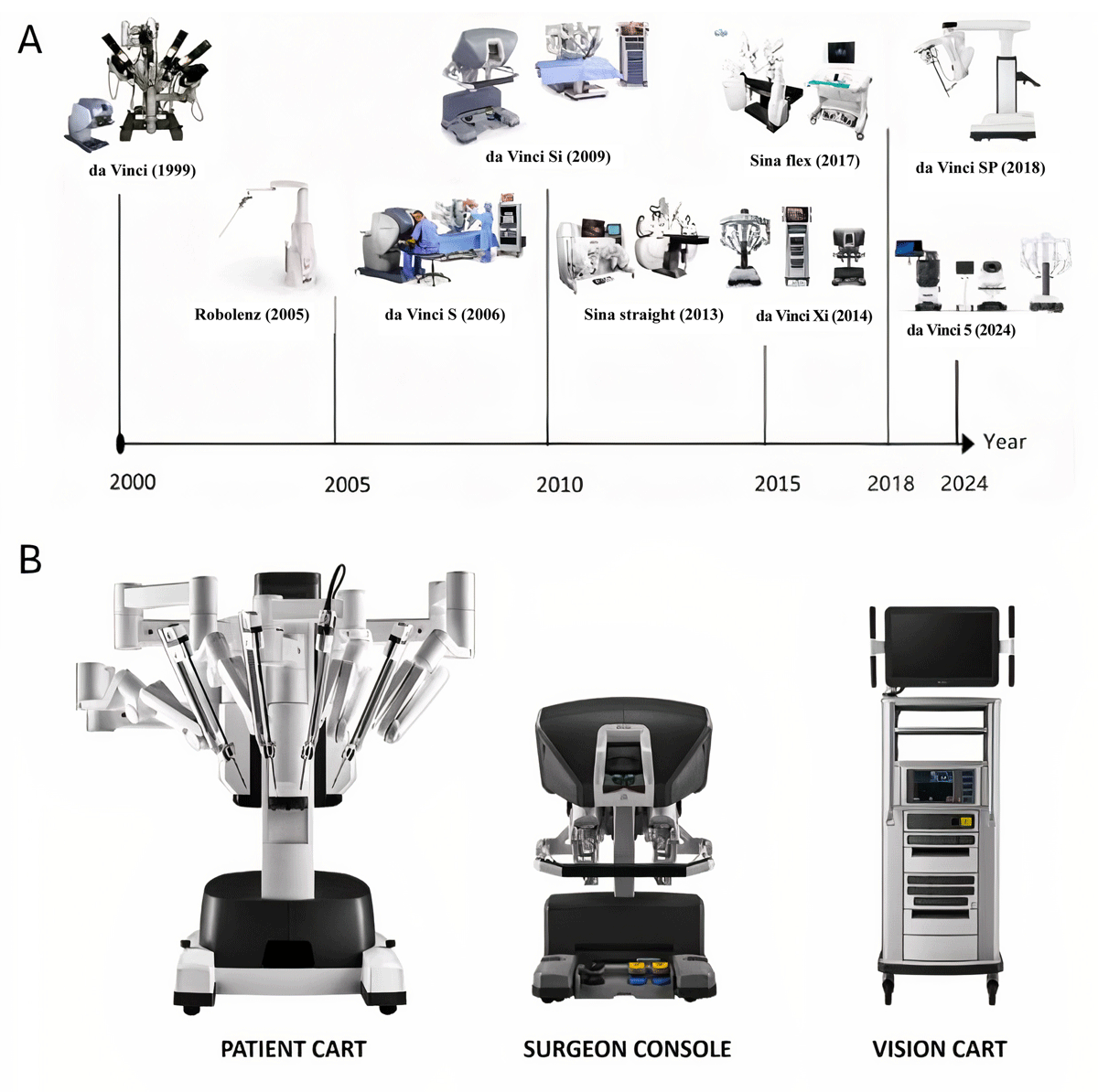
Figure 1
Robotic platform evolution and system components. (A) Timeline illustrating the evolution of robotic surgical platforms from 1999 to 2024. The da Vinci system, originally released in 1999, has undergone several major generational upgrades, including the da Vinci S (2006), Si (2009), Xi (2014), SP (2018), and the most recent da Vinci 5 (2024). The figure also includes other systems such as Robolenz (2005) and the Sina platforms (2013, 2017) to highlight concurrent innovations in robotic surgery. (B) Overview of the core components of the da Vinci Surgical System, which consists of three primary units: the Patient Cart, which holds the robotic arms that execute the surgical tasks; the Surgeon Console, where the surgeon sits and controls the instruments using hand and foot controls while viewing the operative field in 3D; and the Vision Cart, which houses the optical and processing equipment and facilitates communication between the system components.
The system provides high-definition, 3D magnified vision through a stereo endoscopic camera, allowing surgeons to perceive depth and fine anatomical detail. The wristed instruments—known as EndoWrist® technology—mimic the full range of motion of the human hand with seven degrees of freedom (DOF). Motion scaling allows the surgeon to adjust the ratio of hand movement to instrument movement. A 5 mm hand movement may translate to a 1 mm movement inside the patient, allowing extremely precise suturing and dissection in tight anatomical spaces. For vascular surgery, this allows more precise arteriotomies and anastomoses and fewer unintentional intimal injuries. Additionally, the system filters out natural physiologic tremor, resulting in more accurate needle placement and minimizing trauma to fragile vascular structures, leading to better anastomotic integrity and lower risk of postoperative bleeding.25
The da Vinci Surgical System has undergone significant evolution across its generations. The Si system, widely used throughout the early 2010s, offered improved 3D visualization and dual-console capabilities but was limited by bulkier robotic arms and the need for redocking during multi-quadrant procedures.26,27 The introduction of the Xi system in 2014 addressed many of these limitations by incorporating a thinner, more mobile overhead boom, enhanced arm reach and flexibility, and the ability to easily switch the endoscope between ports. These features facilitated faster docking and more efficient workflows, particularly in complex abdominal operations.25,26,27 Most recently, the fifth-generation da Vinci system, unveiled in 2024, introduced haptic feedback (Figure 1).28
An important adjunct to the da Vinci robotic platform is the integration of advanced imaging technologies, most notably FireFly near-infrared (NIR) fluorescence imaging. FireFly allows real-time visualization of perfusion through the use of intravenous indocyanine green dye, which fluoresces under NIR light.29,30,31
Current Clinical Applications in Vascular Surgery
The following section describes vascular procedures currently performed using the da Vinci systems. In terms of procedural volumes, some of these are performed by nonvascular specialists who have mastered essential vascular surgical skills with the use of the robot.
Aortic and Aortoiliac Reconstruction
Robotic-assisted repair of an abdominal aortic aneurysm is a complex surgical procedure, requiring an operator with sufficient experience in both open surgery and robotic technique. It is commonly done using 4 mm to 8 mm ports and 3 mm to 12 mm ports (Figure 2A). Possible robotic interventions include aorto-aortic and aorta-iliac anastomosis, as in the cases of abdominal aorta and iliac artery involvement. During an aortic case, the robot may be used to perform vascular anastomoses, remove aortic thrombus from the aneurysmal sac, ligate the lumbar arteries that bleed back when the sac is opened, and close off the sac from the aortic aneurysm after reconstruction (Figure 2B-E).
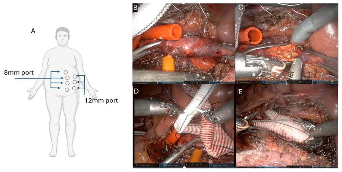
Figure 2
Robotic aortobifemoral bypass: port placement and key operative steps. (A) Schematic of port placement for robotic aortic exposure. Four 8 mm robotic instrument ports and three 12 mm assistant ports are positioned across the left abdomen to allow access for robotic instruments and laparoscopic assistance during aortic dissection and bypass construction. (B) Intraoperative image demonstrating placement of proximal and distal aortic clamps using laparoscopic clamps to control the infrarenal aorta. (C) Robotic removal of intraluminal thrombus from the diseased segment of the aorta following arteriotomy, facilitating a clean landing zone for proximal anastomosis. (D) Robotic tunneling of the bifurcated Dacron graft limbs to the bilateral groins, using a combination of blunt dissection and laparoscopic assistance to ensure proper positioning without kinking. (E) Final view of the completed aortobifemoral bypass with a proximal end-to-side anastomosis to the native aorta and bilateral limb tunneling to femoral targets, demonstrating hemostasis and appropriate graft orientation.
Performing aortic reconstruction requires the ability to control high-pressure arteries, which are often heavily calcified. Choosing the right place for clamping heavily relies on preoperative imaging, although some visual clues are helpful, such as the color of the vessel wall or how it reacts to movement and palpation with the instruments. Clamping can be done either by inserting a laparoscopic clamp through an assist port or by inserting a DeBakey clamp through a small incision (Figure 2B). Balloon occlusion of the iliac arteries can be performed as well. No specialty-focused vascular robotic instruments, such as dedicated aortic robotic clamps, are currently available.
Identification and control of lumbar arteries before opening the aneurysm sac is another key element in the safety of these operations, as uncontrolled bleeding from these can cause major issues. Preoperative imaging and image fusion (overlaying preoperative imaging onto the live surgical view) could play a major role in this topic.4,32,33,34,35 Most of these studies come from a few centers and a relatively small number of cases.
Iliofemoral and Aortofemoral Bypass
In the era of endovascular therapy, the number of iliofemoral reconstructions is constantly decreasing. Robotically assisted iliofemoral reconstructions are done mainly for the purpose of training and practicing robotic vascular procedures. For a minimally invasive strategy, the most common types of robotically assisted vascular reconstruction include iliofemoral reconstruction for occluded external iliac artery occlusion and patent common iliac artery, aortofemoral bypass for unilateral occluded iliac artery, and aortobifemoral bypass for bilateral occluded iliac arteries.4,36
Iliac Artery Aneurysm
Isolated aneurysm of the iliac artery is a relatively rare condition. In robotic surgery, four robotic arms are used, in this case using the fourth arm as a retractor in the pelvic region. The position of the patient is similar to robotic-assisted aortofemoral surgery using the standard six trocars.4,36
Thoracofemoral Bypass
Thoracofemoral bypass has better patency rates than axillofemoral bypass but requires a patient who can tolerate thoracic exposure and clamping of the descending aorta. By using robotic assistance, the time taken for clamping and the anastomosis can be shortened. However, this procedure is rarely done due to the narrow group of ideal patients and advances in endovascular therapy.37
Treatment of Type II Endoleak
Guidelines recommend reintervention in the presence of sac enlargement during follow-up.23 Most treatment options consist of endovascular techniques, but when these fail, open reconstruction may be required. For these patients, a less invasive treatment can be provided by robot-assisted techniques. In 2009, Lin et al. presented a case with successful robot-assisted ligation of the inferior mesenteric artery (IMA), which was the source of a type II endoleak, causing sac enlargement in an 84-year-old male. The total operation time was 249 min, of which console time (when the surgeon is seated at the robotic console) was 180 min. The estimated blood loss was 50 mL. He discharged on postoperative day two. The 3-month follow-up computed tomography scan confirmed the occlusion of the IMA and stabilization of the aneurysm sac size.38
In 2019, Morelli et al. shared their experience with their first two patients who underwent total robotic type II endoleak repair and reported equally promising results. The operation consisted of two phases: the ligation of the IMA and then the posterior mobilization of the aneurysm sac to make the selective clipping of lumbar arteries. Preoperative computed tomography angiography imaging was used for the identification of feeding vessels in these cases. After target ligation was complete, verification of the absence of backflow was carried out with a dedicated ultrasound probe, inserted through one of the assistant ports.39
One of the biggest challenges lies in identifying the correct feeding vessels on preoperative imaging and translating this finding to the robotic platform. Another challenge is finding an efficient method to expose both the left- and right-sided lumbar arteries, or the medial sacral artery, which often presents as a cause of endoleaks.
The modified transperitoneal approach involves robotic ports placed intraperitoneal and opening the retroperitoneum on the left side of the aorta to expose from its bifurcation to left renal vein. Described by Stadler et al., it is adequate for exposing the left-sided side branches, and port placement is described in Figure 3A. Going under an often heavily calcified aorta to reach feeding branches on the other side is a difficult and risky maneuver.40 In our experience, exposing the aorta from the right side is unlikely the answer to this dilemma due to the closeness of the inferior vena cava and the need for redocking and repositioning of ports, which would make the operation significantly longer and more complex.
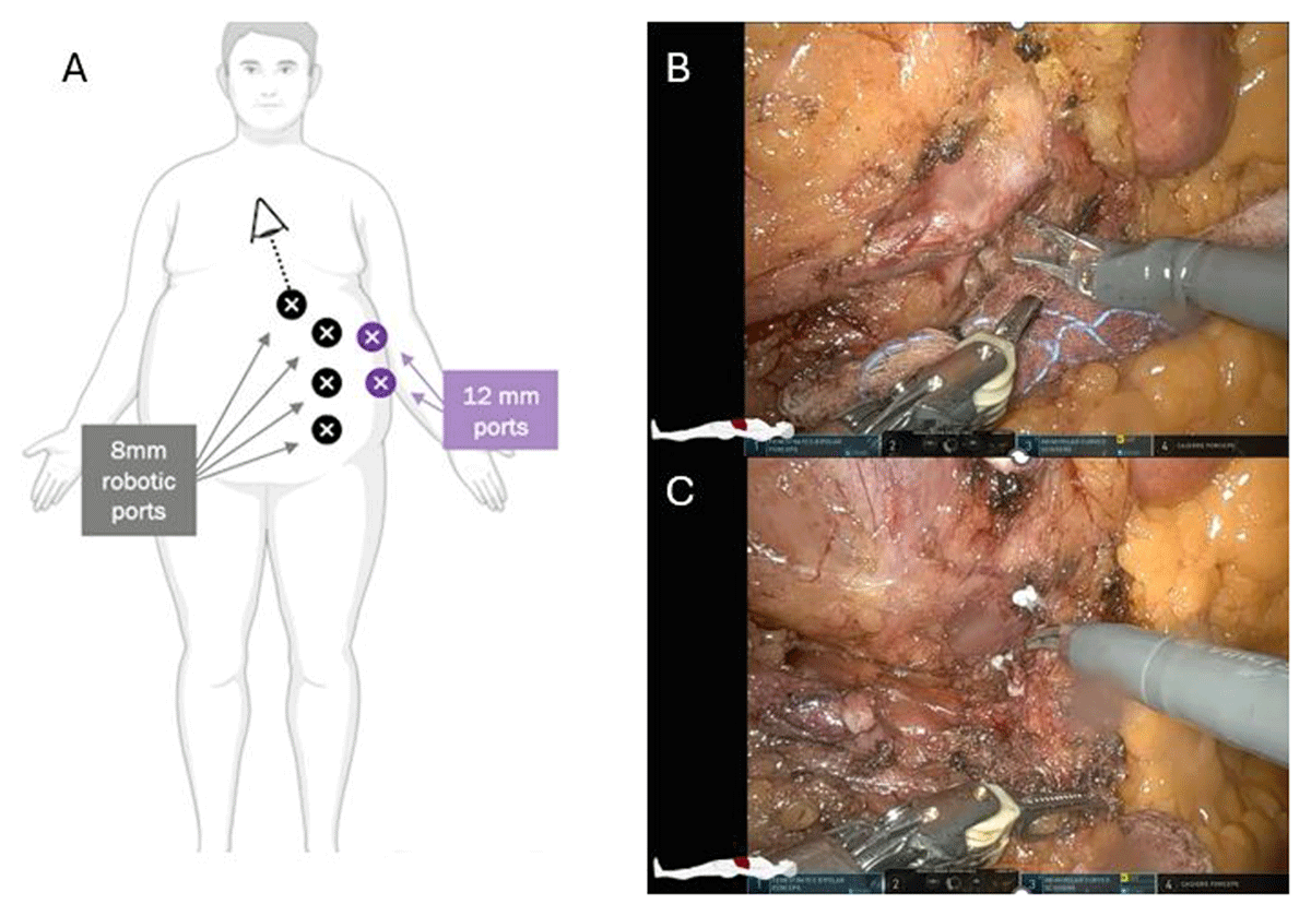
Figure 3
Robotic ligation of type ii endoleak vessels. (A) Port placement strategy for robotic retroperitoneal exposure in the treatment of type II endoleaks originating from the inferior mesenteric artery, left-sided or posterior lumbar arteries, or the median sacral artery. A combination of 8 mm robotic ports (black) and 12 mm assistant ports (purple) are positioned to allow optimal triangulation for left lower quadrant and midline retroperitoneal access. (B) Intraoperative image showing robotic exposure of the inferior mesenteric artery, dissected free from surrounding retroperitoneal tissue in preparation for ligation. (C) Robotic ligation of a left-sided lumbar artery contributing to persistent type II endoleak, with clear visualization of vascular control and surrounding tissue dissection.
Other Vascular Interventions
Splenic Artery Aneurysm
Splenic artery aneurysm is the most common type of visceral aneurysm, with a prevalence of around 0.8% in the general population. Generally, diameters exceeding 30 mm are to be treated, or any size in pregnant women and symptomatic cases. The first treatment of choice if feasible is an endovascular procedure, but open reconstructions also provide viable options depending on aneurysm location, tortuosity, and feeding/draining branches. A 2024 meta-analysis by Jozwik et al. describes 28 patients who underwent robotic-assisted end-to-end anastomosis, ligation, or graft placement for splenic artery aneurysm. They reported a mean operative time of 210 minutes and two conversions to open surgery (7.1%) (Figure 4A-E).41,42
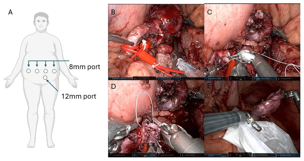
Figure 4
Robotic resection and reconstruction for splenic artery aneurysm. (A) Port placement schematic for robotic splenic artery aneurysm resection. Four 8 mm robotic ports and one 12 mm assistant port are placed across the upper abdomen to allow optimal exposure and instrument access to the splenic artery along the superior border of the pancreas. (B) Intraoperative view showing careful dissection and identification of the splenic artery aneurysm, with vascular control obtained proximally and distally using vessel loops. (C) Following aneurysm resection, the robotic instruments begin end-to-end anastomosis of the splenic artery using fine suture, reestablishing in-line flow. (D) Near completion of the end-to-end splenic artery reconstruction. (E) The resected splenic artery aneurysm specimen being retrieved into an endo-catch bag for removal from the abdominal cavity.
Renal Artery Reconstruction
Robotic-assisted renal artery reconstruction is a rarely performed intervention and has been performed via a six-port modified transperitoneal approach. This robotic-assisted intervention may also be done during an aortofemoral reconstruction or as a renal artery bypass.43
Median Arcuate Ligament Release (MALS)
A few studies have presented small to medium numbers of cases of MALS release with robotic assistance. All reports show favorable outcomes and technical feasibility with minimal conversion rates and short in-hospital stays, providing good long-term results in terms of symptom relief and decrease of peak systolic velocity during ultrasound control (Figure 5A,B).44,45,46
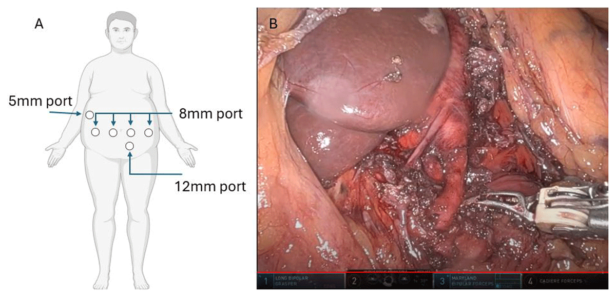
Figure 5
Robotic median arcuate ligament release. (A) Port placement schematic for robotic-assisted median arcuate ligament release. Four 8 mm robotic ports, one 5 mm port in the right upper quadrant for placement of liver retractor, and one 12 mm assistant port to facilitate precise dissection along the diaphragmatic crura and access to the supraceliac aorta and celiac axis. (B) Intraoperative view demonstrating completed dissection with clear exposure of the supraceliac aorta and the origin of the celiac artery following division of the median arcuate ligament and thorough neurolysis of the celiac plexus.
Renal Vein Transposition (Nutcracker)
Several small case series describing robotic left renal and/or ovarian vein transposition and renal auto-transplantation report comparable operative time, low blood loss, and decreased length of stay, with low complication rates and good clinical outcomes in terms of symptom relief (Figure 6A-E).13,47,48
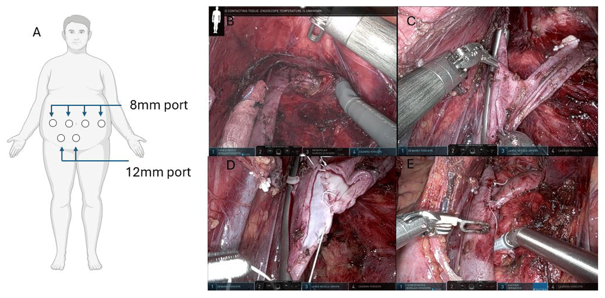
Figure 6
Robotic left renal vein transposition for nutcracker syndrome. (A) Port placement schematic for robotic-assisted left renal vein transposition. Four 8 mm robotic ports and two 12 mm assistant ports are positioned across the mid to lower abdomen to allow optimal access and visualization of the infrarenal IVC and left renal vein. (B) Intraoperative view showing careful dissection and identification of the left renal vein and infrarenal IVC, with vascular control established to facilitate safe mobilization. (C) Mobilization and positioning of the left renal vein for transposition, demonstrating alignment for a tension-free anastomosis to the infrarenal IVC. (D) Robotic construction of the end-to-side anastomosis between the left renal vein and the IVC using fine monofilament suture, ensuring unobstructed venous outflow. (E) Completed left renal vein transposition with hemostatic anastomosis and restored anatomic orientation, confirming resolution of the compression. IVC: inferior vena cava
Inferior Vena Cava Filter Removal
The retrieval rate for IVC filters is only around 25% to 30% in the United States.49 Endovascular approach remains the first choice when an IVC filter is to be removed; however, this can be unsuccessful or considered high risk because of possible extrusion of the filter. Robot-assisted surgery offers an alternative to an open approach, providing a minimally invasive solution. Few case series have been published on robot-assisted IVC filter removal, each of which presents good results, with high success rate, low number of postoperative complications, and short length of stay (Figure 7A-E).49,50
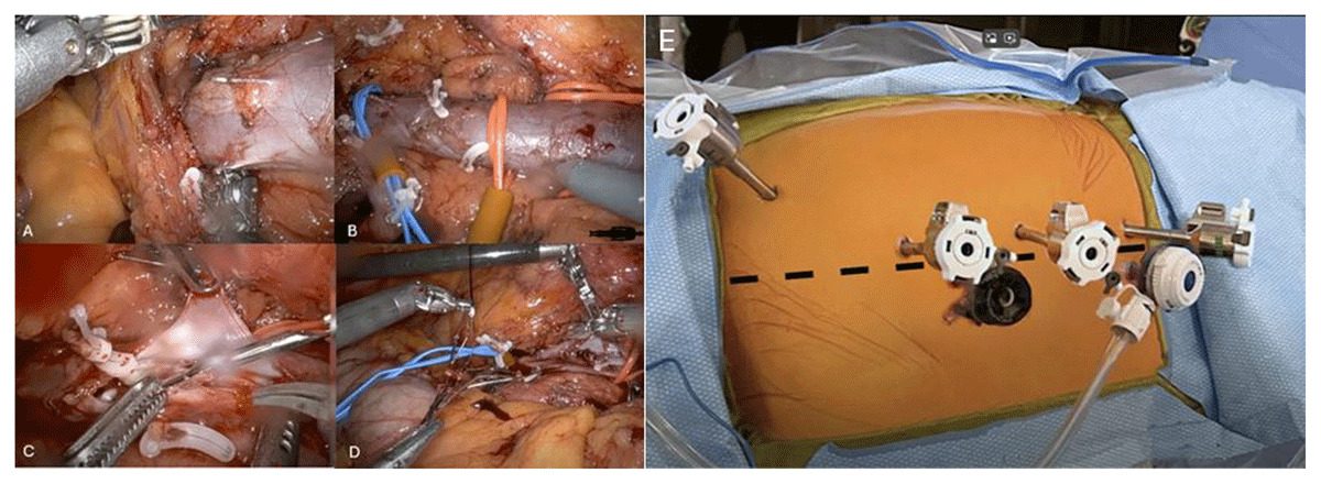
Figure 7
Robotic IVC filter retrieval. (A) Intraoperative view showing robotic exposure of the infrahepatic IVC with identification of IVC filter legs protruding through the caval wall, often associated with surrounding fibrosis or inflammation. (B) Placement of vessel loops and vascular clips for proximal and distal control of the IVC to ensure safe cavotomy. (C) Cavotomy with initial opening of the IVC reveals the embedded filter struts and associated synechiae. (D) Robotic-assisted removal of the IVC filter from within the IVC lumen using atraumatic graspers and dissection techniques to safely extract the device while minimizing damage to the caval wall. (E) Port placement schematic for robotic-assisted IVC filter retrieval. Four 8 mm robotic ports and two 12 mm assistant ports are placed across the lower and lateral abdomen to provide adequate access and triangulation for precise vascular dissection and suturing. IVC: inferior vena cava
Robotic First Rib Resection
A case series of 83 patients undergoing robotic first rib resection was presented in 2018. The robot was used for the dissection of the first rib, disarticulation of the costosternal joint, and division of the scalene muscles. The average operative time was 127 min (± 20 min). Median hospitalization was 4 days, and no surgical or neurovascular complication was reported.51 A systematic review comprising 12 studies of 379 patients with thoracic outlet syndrome (TOS) suggested that robotic technique is an effective method in the treatment of TOS. It offers improved exposure, reduced risk of neurovascular injury, and shorter hospitalization.52
RALS Aortic Mass Biopsy
Robotic-assisted laparoscopic biopsy offers a precise, minimally invasive approach for tissue diagnosis of retroperitoneal and aortic-adjacent masses. After gaining transperitoneal access and mobilizing overlying bowel structures, the mass is carefully dissected from surrounding vasculature, with particular attention to preserving the adventitial plane (outermost layer) of the aorta. Case reports show that robotic biopsy of aortic-associated masses is feasible, safe, and provides adequate tissue for histopathologic analysis.53
Aberrant Right Subclavian Artery Transposition
RAL aberrant right subclavian artery (ARSA) transposition is performed to treat symptomatic vascular compression syndromes such as dysphagia lusoria. Utilizing the da Vinci robotic platform, the ARSA is mobilized from its origin, typically arising distal to the left subclavian artery on the aortic arch, through a right-sided chest wall approach. The esophagus is retracted to expose the retroesophageal segment of the ARSA, which is then carefully ligated and divided. Ligation has been described using a robotic vascular stapler. The artery is subsequently transposed and reanastomosed to the right carotid or subclavian artery through a supraclavicular incision. Case reports show reduced postoperative pain, shorter hospital stays, and quicker recovery compared to traditional open or hybrid approaches.54
Robot-Assisted IVC Thrombectomy
Associated with renal cell carcinoma, this procedure is mainly performed by urologists and may involve dissection and even reconstruction of major vessels. One of the main challenges of this operation is vascular control. Temporary occlusion of the IVC can be done by clamps introduced to the abdomen through an assistance port or simple stab incision, vessel loops applied circumferentially and then creating a modified Rummel tourniquet using a small rubber tube, or the use of a temporary IVC occlusion balloon.55,56
Robot-Assisted Kidney Transplantation
A meta-analysis published in 2022 demonstrated that robot-assisted kidney transplant is safe and feasible. Compared to the open approach, it is associated with lower risk of surgical site infection, less postoperative pain, and shorter length of hospital stay, with no difference in renal function, graft, and patient survival. It can be especially beneficial for obese patients due to the assessed lower risk of surgical site infections.57
Robot-Assisted Lung Transplantation
In 2023, the first robot-assisted lung transplantation was performed successfully. The robot was used for the removal of the recipient’s diseased right lung and, after the donor’s lung was inserted into the chest, the bronchial and the left atrial anastomosis were created with robotic assistance. The patient recovered without any major adverse events and discharged on the 11th postoperative day. Since then, several more robot-assisted lung transplants have been performed.58
Portal Vein Reconstruction in Robot-Assisted Pancreaticoduodenectomy
A patient is considered a candidate for robotic pancreaticoduodenectomy in cases of venous involvement with less than 180° circumferentially with the vein being patent. After resection is complete, reconstruction is required. According to the International Study Group of Pancreatic Surgery (ISGP) classification, types of vein resection can be divided into four categories. Type I resection means a small side wall resection, which can be closed with direct suture. In case of type II resection, patch closure is required. In the case of type III and IV resections, a complete segmental resection is required, which can be reconstructed with direct repair in the former, and only with interposition in the latter.59
Robotic Coronary Artery Bypass Grafting
Using the da Vinci system, the internal thoracic artery can be harvested with exceptional accuracy through small thoracoscopic ports, eliminating the need for full sternotomy. Robotic coronary artery bypass grafting has been reported to have reduced operative blood loss, lower complication rates, and faster postoperative recovery compared with conventional open techniques.60
Robotic Heart Transplantation
In June 2025, Khaliel et al. reported the first-in-human, totally robotic orthotopic heart transplant. The procedure involved peripheral cannulation for cardiopulmonary bypass and a minimally invasive thoracic approach to explant the native heart followed by robotic-assisted anastomosis of atrial cuffs and great vessels. Remarkably, the ischemic time was minimized, the patient was extubated within 24 hours, and expedited mobilization was achieved.61
Advantages and Barriers to Adoption in Vascular Surgery
Compared to traditional laparoscopy, the da Vinci system provides a significantly shorter learning curve due to its intuitive instrument controls, wristed articulation, and immersive 3D visualization. These features allow for precise dissection and suturing in confined spaces, with reduced surgeon fatigue due to the system’s ergonomic design. Additionally, the minimally invasive nature of robotic surgery may lead to shorter hospital stays, less postoperative pain, and faster recovery—benefits particularly valuable for high-risk vascular patients.4
However, barriers to adoption remain significant. The high cost of acquiring and maintaining the robotic platform combined with the need for specialized staff training can limit access to the technology, especially in lower-volume or resource-constrained institutions. Vascular surgery also suffers from a lack of dedicated robotic instruments, such as vessel loops, aortic clamps, or fine needle drivers designed specifically for small-caliber vascular work. Furthermore, earlier generations of robots lack haptic feedback, making delicate tissue handling and suture control more challenging, although the latest models are beginning to address this.21 Finally, the institutional logistics—including operating room scheduling, equipment storage, and interdepartmental coordination—can be complex.
Training and Curricular Pathways for Vascular Robotic Surgeons
One of the major barriers to the adoption of robotic surgery in vascular practice is the lack of formal training within current vascular surgery residencies and fellowships. Unlike general surgery or urology, where robotic training has become standardized and often mandatory, most vascular trainees graduate with little to no exposure to robotic platforms. To address this gap, a tiered training pathway has been proposed, beginning with simulation and then progressing to low-risk procedures, then to moderate, and then high complexity vascular operations such as renal vein transposition, visceral aneurysm reconstruction, and aortic reconstruction.
Simulation platforms—such as those offered by Intuitive Surgical—enable residents and fellows to build console skills in a controlled environment. Additionally, structured proctoring by experienced robotic surgeons is essential for ensuring safety and competency as vascular trainees begin to perform real cases.62,63,64
Future Directions and Technology Innovation
One of the most anticipated developments is the integration of artificial intelligence and machine learning into robotic platforms, enabling real-time surgical guidance, automated recognition of anatomical structures, and enhanced safety through predictive analytics. Augmented reality and image fusion technologies may allow intraoperative overlay of preoperative imaging onto the surgical field, improving orientation and precision during complex vascular dissections.65 The continued development of haptic feedback, now introduced in the fifth-generation da Vinci system, is expected to evolve further, offering refined tactile sensation critical for vascular tissue handling and suture control. Additionally, the field urgently needs dedicated robotic vascular instruments, such as aortic clamps, fine needle drivers, and vessel loops, to safely expand the scope of robotic vascular interventions.
The da Vinci SP (Single Port) surgical system, also developed by Intuitive Surgical, is designed to perform complex procedures through a single 2.5 cm incision. Unlike multi-arm systems, the SP robot features a single cannula that houses a fully wristed, multi-jointed 3D high definition camera and three double-jointed instruments, allowing for precise, 360-degree access in confined anatomical spaces. This allows access to deep or narrow anatomical sites. While its use in vascular surgery remains limited, early cadaveric studies show the ability to perform extensive dissection in the upper and lower extremities and easily incorporate fluoroscopic imaging due to the smaller profile of the SP cart.66
Conclusion
The large incision “delivery system” of open surgery is unappealing to patients, despite the superior durability of procedures like open aortic repair using Dacron grafts. Conversely, endovascular techniques offer minimally invasive access but often lack long-term durability and have higher reintervention rates.67 Robotic surgery offers the durability of an open repair with a minimally invasive approach that better allows patients to tolerate the perioperative phase of care.
While early clinical experience demonstrates promising safety, feasibility, and patient recovery benefits, broader adoption remains constrained by high costs, limited training opportunities, and the lack of robotic platforms and instruments specifically designed for vascular applications. To fully realize the potential of this technology, the vascular surgery community must prioritize structured training programs, surgical curriculum integration, and collaborative research efforts. Partnering with engineers, industry stakeholders, and cross-specialty robotic users can accelerate this field.
Key Points
Robotic surgery offers minimally invasive options for traditionally open vascular procedures.
This manuscript presents standardized port placement schematics for six common robotic vascular surgery procedures.
Adoption of robotic vascular surgery is limited by cost, lack of training, and dedicated tools.
Early outcomes for robotic vascular surgery show safety and feasibility in select procedures.
Future adoption of robotic vascular surgery hinges on curriculum development and technology innovation.
Multidisciplinary collaboration can accelerate progress in robotic vascular surgery.
Competing Interests
Dr. Lumsden conducts research on behalf of W. L. Gore & Associates; consults for Siemens, Boston Scientific, and W.L. Gore & Associates; and has an ownership interest in Hatch Medical, Egg Medical, and Brijjit. The other authors have no competing interests to declare.