
Introduction
Atherosclerosis and autoimmune rheumatologic diseases (ARD) are multifactorial, chronic inflammatory conditions. Patients with ARDs face a higher risk of developing atherosclerotic cardiovascular disease (ASCVD) and often present with the disease at a younger age than the general population.1 The ARD population bears a significant burden of traditional risk factors, including dyslipidemia, hypertension, obesity, smoking, and diabetes. Additionally, they carry unique risk factors such as chronic steroid use, depression, and a sedentary lifestyle due to arthritis. However, even after adjusting for these variables, the risk for ASCVD remains elevated.2 As chronic inflammation is not accounted for in traditional cardiovascular (CV) risk calculating tools, this has led to an alarming risk underestimation in this population.1
Multimodality cardiovascular imaging, including echocardiography, computed tomography, cardiac magnetic resonance imaging (CMR), single photon emission computed tomography (SPECT), and positron emission tomography (PET), provides valuable insights in ARD patients.
In this review, we examine the potential applications of various cardiovascular imaging modalities in diagnosing coronary artery disease (CAD) among patients with ARD (Figure 1). We aim to highlight the current strengths and weaknesses of each imaging technique and explore potential future applications for CAD detection in ARD patients. Findings by these imaging modalities highlight the common inflammatory pathways that link ARDs and CAD, contributing to endothelial damage and atherosclerosis. This knowledge can be instrumental in developing detection and prevention strategies for individuals with rheumatic diseases and the general population.
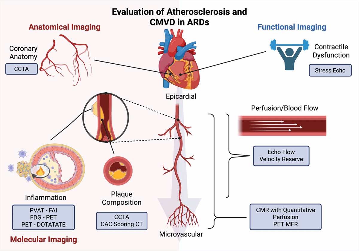
Figure 1
Multimodality cardiovascular imaging for the evaluation of atherosclerosis and CMVD in ARDs. CMVD: coronary microvascular disease; ARD: autoimmune rheumatic disease; CCTA: coronary computed tomography angiography; PVAT: perivascular adipose tissue; FAI: fat attenuation index; PET: positron emission tomography; CAC: coronary artery calcium score; CT: computed tomography; CMR: cardiac magnetic resonance; MFR: myocardial flow reserve. Created in BioRender. Williams K. (2025) https://BioRender.com/zynjp50
The Interrelated Physiology of Coronary Artery Disease in Rheumatologic Diseases
Obstructive CAD is characterized by a mismatch between myocardial oxygen supply and demand, which can occur from the coronary vessel take-off to the microvascular capillary level.3 This can present as a spectrum from asymptomatic stable CAD to acute coronary syndrome.4 Rheumatologic diseases and atherosclerosis share similar inflammatory pathways, which are mediated by immune cells, autoantibodies, acute-phase reactants, and inflammatory cytokines.3
Atherosclerosis begins with endothelial dysfunction, which is followed by a cascade of events leading to the subendothelial accumulation of lipid particles, fibrous elements, and calcification, creating an artery-lumen-narrowing atheroma. Traditionally, these processes were thought to be attributed to age, sex, smoking, hypertension, hyperlipidemia, obesity, diabetes, and physical inactivity as presented by the Framingham study.5 While these are proven independent factors, the role of inflammation has been increasingly highlighted as a driving force behind atherosclerosis. Numerous studies have shown that elevated markers of inflammation, such as high sensitivity C-reactive protein (hs-CRP), are independently associated with adverse cardiovascular events, and it is now well-established that autoimmune disease is an independent risk factor for ASCVD.6 Furthermore, an extensive population-based study found that the presence of any ARD is associated with an increased incidence of ASCVD, and that this risk is particularly high in patients with systemic sclerosis (SSc) or systemic lupus (SLE), as well as in those with multiple ARDs.7
The mechanistic processes behind autoimmune-driven atherosclerosis are complex, with the common link beginning with endothelial dysfunction. Prolonged systemic inflammation, prevalent in ARDs, results in chronic endothelial activation. Proinflammatory cytokines, including tumor necrosis factor-α (TNF-α), interleukin-1 (IL-1), and IL-6, promote the expression of adhesion molecules such as vascular cell adhesion molecule-1 (VCAM-1) and intercellular adhesion molecule-1 (ICAM-1) on endothelial cells, which recruit leukocytes into the vessel wall, perpetuating the cycle of inflammation.8 Numerous other downstream effects of inflammation, both directly and indirectly, promote atherosclerosis.
The results of the CANTOS (Canakinumab Anti-Inflammatory Thrombosis Outcomes Study) trial supported the inflammation-induced CAD hypothesis by demonstrating that monoclonal antibody therapy targeting IL-1, a key chemokine of the innate immunity pathway, led to decreased recurrent ASCVD events and lowered CRP levels in a general population post myocardial infarction (MI).9 Several studies followed this landmark trial, including the low-dose colchicine trial (LoDoCo) and LoDoCo2 trials that found a lower risk of cardiovascular events in patients with chronic CAD taking low-dose colchicine.10,11 Furthermore, the COLCOT (Colchicine cardiovascular outcomes) trial demonstrated that low-dose colchicine lead to a significantly lower risk of ischemic cardiovascular events compared to placebo in the acute phase following MI (within 30 days).12 Collectively, these trials emphasize the central role of inflammation in cardiovascular disease and the therapeutic benefits of targeting this inflammation.
While atherosclerosis in large vessels is prioritized, it is important to discuss how autoimmune disease contributes to coronary microvascular dysfunction (CMVD). CMVD is challenging to diagnose by traditional imaging techniques, may present with functionally limiting symptoms such as angina, and is known to lead to significant adverse cardiovascular events (MACE).13 Numerous ARDs have been linked to CMVD, including SSc, rheumatoid arthritis (RA), SLE, and psoriatic arthritis.14, 15, 16, 17
Dyslipidemia is also highly prevalent in patients with ARDs.2 Furthermore, chronic inflammation functionally diminishes high density lipoprotein (HDL) protective actions, resulting from reactive oxygen species and acute phase proteins displacing apolipoprotein-a1 from HDL surfaces.18 In addition, the oxidation of low density lipoprotein (LDL) particles, a key step in forming atherosclerotic plaque, is increased by systemic inflammation and tissue oxidative stress.19 Lastly, the so-called “lipid paradox” in RA highlights the complex relationship between inflammation, anti-inflammatory therapy, and lipid levels, where lower levels of LDL cholesterol are paradoxically associated with an increased risk of cardiovascular disease.20 Importantly, recent results also indicate that ARD patients are less likely to receive lipid-lowering therapy compared with controls, even in the presence of ASCVD.21 The emerging data highlighting accelerated atherosclerosis in ARDs underscores the need to screen and aggressively modify risk factors in this patient population.
Imaging Modalities for CAD in Rheumatologic Diseases
Advances in noninvasive cardiovascular imaging have shed light on the shared pathophysiology between CAD and ARDs, allowing for more timely diagnosis and better risk stratification in these patients.22,23 Noninvasive CAD analysis can be classified into anatomical or functional evaluations. Due to the underestimation of risk by traditional non-imaging-based ASCVD risk calculators, cardiovascular imaging has emerged as an essential tool in the cardiovascular risk stratification of the ARD population.
Transthoracic Echocardiography
Transthoracic echocardiography is the cornerstone of noninvasive cardiovascular evaluation, providing qualitative and quantitative information on systolic and diastolic cardiac function.
Stress Echocardiography
Stress echocardiography is a widely available modality to detect wall motion abnormalities aiding in the diagnosis and prognosis of CAD. Results indicate that patients with RA experience a two-fold higher incidence of positive exercise stress echocardiogram compared to controls, along with an increased all-cause mortality of 14.9% versus 4.3% for negative stress tests.24 Additionally, a dobutamine stress echocardiogram study indicated that RA patients had a similar incidence of asymptomatic myocardial ischemia without obstructive CAD compared to patients with diabetes mellitus.25
Echocardiographic Coronary Flow Velocity Reserve
The coronary flow velocity reserve (CFVR) of the left anterior descending artery (LAD), derived from Doppler echocardiography, shows high correlation with invasive coronary angiographic coronary flow reserve assessment.26 CFVR is calculated by the ratio of peak diastolic velocity during stress to peak diastolic velocity at rest. A CFVR > 2 is consistent with normal coronary flow, while a ratio < 2 is a surrogate indicator of CMVD or obstructive CAD.27
Numerous studies highlighted impairment of CFVR in ARDs, such as RA, SLE, ankylosing spondylitis, and psoriatic arthritis, indicating premature atherosclerosis and presence of CMVD.15,28, 29, 30, 31, 32 Among others, Hirata et al. found that young SLE patients without cardiovascular risk factors showed a decreased CFVR compared to controls.16 This was corroborated by Sincer et al., who also found lower CFVR in SLE compared to controls, and noted that lower CFVR correlated with total antioxidant levels and inversely correlated with hs-CRP.33 Furthermore, in ARD patients without a history of CAD, treatment with IL-12 inhibitors in psoriatic arthritis and IL-1 inhibitors in RA led to decreased inflammatory markers and improved CFVR.34,35 This underscores the clinical potential of anti-inflammatory therapy response in the ARD population.
Computed Tomography
Cardiac CT is routinely performed to gain insight into cardiac or coronary anatomy. CT with and without contrast allows for noninvasive 3-dimensional visualization of the coronary vasculature relatively quickly and inexpensively.
Non-contrast Chest CT
Coronary artery calcification (CAC) has been recognized as an independent predictor of MACE and overall mortality.36 Using non-contrast gated breath-held chest CT, the extent and distribution of mineralized atherosclerotic plaques can be quantified using standardized methods (Agatston score). A large meta-analysis of 19 studies found that ARD subjects had a higher CAC score than controls and that higher CRP levels were related to higher CAC score, suggesting that chronic inflammation plays a key role in the development of CAC.37 A study by Romero-Diaz et al. also showed that SLE patients were 10 times more likely to have CAC compared to age-sex-matched controls, suggesting premature atherosclerosis in SLE.38 Patients with RA also have been shown to have significantly higher CAC scores associated with IL-6 and TNF-α, independent of the Framingham risk score and diabetes.39 This supports the role of inflammatory pathways in the formation of calcified plaques. In an analysis of the Western Denmark Heart Registry, ARD was associated with any CAC, with an adjusted odds ratio of 1.39, and a severe CAC of > 90th percentile, with an adjusted odds ratio of 1.53.1 Importantly, these authors found that ASCVD events were strongly associated with all CAC scores but even occurred in ARD patients with a CAC score of zero.1
The strength of CAC scoring lies in offering affordable cardiovascular risk assessment for the ARD patient population that is more accurate than clinically calculated risk models. In addition to traditional CAC scoring with ECG-gated, breath-held images, several studies propose that non-dedicated, non-ECG gated chest CT examinations can be used opportunistically to quantify CAC burden in a semiquantitative or quantitative way.40,41 Indeed, a recent study demonstrated in SSc that these non-gated chest CT scans can be used to track progression of atherosclerosis and to identify disease-specific factors associated with atherosclerosis progression.42 As the ARD population often receives a non-contrast CT chest to diagnose/monitor pulmonary manifestations, this offers an opportunity for early diagnosis and primary prevention.
Coronary CT Angiography
The high accuracy and high spatial resolution of CCTA have made it an established and reliable method for assessing the presence, severity, and composition of atherosclerotic plaques.43 The advantage of CCTA beyond obstructive CAD detection lies in its ability to detect nonobstructive CAD, which may be overlooked by traditional stress myocardial perfusion imaging (MPI). Furthermore, CCTA has an advantage over non-contrast CAC scoring CT in detecting non-calcified plaques (NCP) that pose a significant risk for acute coronary syndromes.44
Coronary plaque features have been extensively assessed using CCTA. High-risk plaque characteristics include spotty calcification, thin-cap fibroatheroma, napkin ring sign, positive remodeling, high plaque burden, low attenuation plaque, and NCP.45 It has been shown that ARD patients have a higher incidence of any plaque and a more significant burden of NCP than non-ARD controls. A study involving 150 patients with RA who exhibited no current or prior symptoms of CAD revealed a higher prevalence of NCP, calcified plaque, and mixed plaque compared to subjects without ARD.46 Furthermore, Stojan et al. found that SLE patients had a higher burden of high-risk low attenuation plaque compared to controls.47
These results show that CT imaging has potential for identifying and quantifying high risk CAD in ARD patients. Furthermore, as recently artificial intelligence-enabled CT-based plaque analysis received expanded Medicare coverage, the quantification of plaque burden may become more available for ARD patients and may help in clinical decision-making for preventive strategies.
CCTA alone is limited in its ability to discern significant hemodynamic obstruction of coronary atherosclerotic lesions. The advent of CT fractional flow reserve (FFR) overcomes this deficiency by estimating the hemodynamic significance of coronary lesions.48 To our knowledge, no reports on CT FFR use have been published specifically for the ARD patient population.
Perivascular adipose tissue (PVAT) is the outermost layer surrounding the adventitial layer of arteries. It provides structural support and has been indicated to play a key metabolic role in arterial function, including vascular tone, remodeling, and inflammation. In the context of ARD, women with SLE were noted to have higher volumes and higher density of PVAT of the thoracic aorta.49 Aortic PVAT density in this group correlated with the degree of aortic calcification and serum CRP.50 Antonopoulos et al. developed the fat attenuation index (FAI) to quantify inflammation in the peri-coronary adipose tissue by quantifying fat stranding, an inflammatory phenomenon first described by abdominal CTs.51,52 The FAI score increases with the degree of inflammation observed.
The CRISP-CT (Cardiovascular RISk Prediction using Computed Tomography) trial demonstrated that high FAI provides an early marker of coronary inflammation and is associated with increased cardiac mortality.53 Patients with psoriatic arthritis on biologic therapy showed lower FAI (eg, healthier fat phenotype) than those not on biologic therapy, and the use of biologic therapy in psoriatic arthritis demonstrated a significant reduction in FAI after one year of treatment.54 Future studies are needed to better understand the shared inflammatory pathophysiology between CAD and ARDs and the influence of treatment on disease outcomes.
Magnetic Resonance Imaging
Cardiovascular magnetic resonance (CMR) provides a comprehensive assessment of cardiac function, inflammation, perfusion, and scar. CMR in the context of ARD offers new insights into the effects of chronic inflammation on the heart. CMR can help evaluate scar burden by assessing late gadolinium enhancement (LGE), which also can be used for viability assessment.55 The most commonly observed LGE patterns in patients with ARDs include right ventricular insertion site LGE, subendocardial/transmural LGE, midmyocardial/subepicardial LGE, and diffuse subendocardial fibrosis.56 CMR can also be a helpful tool in the evaluation of acute coronary syndrome, including diagnosing microvascular obstruction, intramyocardial hemorrhage, and MI with nonobstructive coronaries (MINOCA).57
Stress CMR
Evaluating myocardial perfusion with CMR and vasodilator stress is an established and accurate method for measuring function, detecting ischemia, predicting viability, and providing prognosis. Observing wall motion abnormalities and perfusion defects with first-pass myocardial contrast enhancement allows for high-resolution qualitative assessment of CAD.57 Stress CMR has been reported to have a higher sensitivity in detecting significant CAD compared to SPECT MPI.58
A significant advantage of stress CMR is the possibility of quantification of absolute myocardial blood flow (MBF) by tracking gadolinium, thus enabling the detection of severe multivessel disease and CMVD. While CMR quantitative stress perfusion is not widely available currently, it is a promising tool for further understanding ischemic pathophysiology and prognosis.59
Multiple stress CMR studies have demonstrated a higher incidence of perfusion defects and reduced MBF in the ARD population. Kobayashi et al. found that RA disease activity and systemic inflammatory markers significantly correlated with perfusion defects in RA patients without known cardiac disease.60 Sacré et al. demonstrated reduced myocardial perfusion reserve index (MPRI, a semiquantitative CMR measure of stress myocardial perfusion) and increased late gadolinium enhancement in antiphospholipid syndrome patients without known cardiac disease compared to healthy controls.61
Ishimori et al. found that SLE patients without obstructive CAD with anginal pain-like symptoms had a 44% prevalence of perfusion defects and a significantly lower MPRI.62 The pattern of visual perfusion defect was circumferential and subendocardial in all patients with abnormal results, indicating diffuse CMVD. This SLE cohort was followed for 5 years and demonstrated that most patients experienced persistent angina with unchanged or worsened MPRI.63 Interestingly, 75% of patients with improved MPRI were on aspirin, beta-blockers, and a cholesterol-lowering agent.
Dumitru et al. identified that patients with SSc had significantly lower myocardial flow reserve (MFR) than healthy individuals (median 1.9 vs 3). Late gadolinium enhancement was found in 17 out of 83 SSc patients but was absent in healthy controls and was directly correlated with troponin I and N-terminal pro-brain natriuretic peptide levels.64 Collectively, these results highlight the significant burden of CMVD in ARD patients and the potential utility of CMR in this population.
Positron Emission Tomography Imaging
While SPECT MPI is the most frequently performed non-invasive imaging modality for detecting CAD, it has significant limitations in detecting balanced ischemia and CMVD. With robust literature on MBF quantification, PET MPI is considered the noninvasive gold standard for evaluating CMVD. Furthermore, PET implemented with molecular radiotracers is a promising field for understanding the pathophysiology of ARD-related cardiovascular disease.
PET Myocardial Perfusion Imaging
With PET MPI, MFR can be estimated by calculating the stress to rest MBF ratio, which is a validated diagnostic and prognostic marker for epicardial atherosclerotic CAD and CMVD.65 In the context of ARD, multiple publications demonstrated the use of PET MPI for the detection, risk-stratification, and monitoring of CAD and/or CMVD.
The LiiRA study (Lipids, inflammation, and cardiovascular risk in Rheumatoid Arthritis) evaluated the effects of ARD-modifying TNF-inhibition therapy on CAD with PET MPI and serum inflammatory marker testing before and after TNF-inhibition.66 Prior to treatment initiation, the average ASCVD risk was 2.7%, below the primary prevention threshold, but 47% of subjects had evidence of CMVD on PET MPI, with IL-6 levels 44% higher in patients with CMVD compared to those without.67 Interestingly the authors detected no significant change in MFR after 24 weeks of TNF inhibitor treatment.66 Amigues et al. found significantly reduced MFR in approximately a third of RA patients without a history of ASCVD, and MFR was inversely correlated with IL-6 levels.68 Moreover, reduced MFR was documented in SSc patients in comparison to patients with primary Raynaud phenomenon and healthy controls without autoimmune conditions.69
Feher et al. also found that ARD patients had significantly lower global PET MFR than matched controls without ARD.70 Furthermore in this study, ARD patients with an MFR under 1.5 experienced significantly worse event-free survival (combined outcomes of death, MI, or heart failure admission) in contrast to non-ARD patients with an MFR less than 1.5, as well as those with ARD but an MFR greater than 1.5.70 Weber et al. revealed similar findings in a cohort of RA, SLE, and psoriasis patients.71, 72, 73 ARD patients with impaired MFR (< 1.65) experienced higher all-cause mortality and reduced MACE-free survival when compared to those with preserved MFR.71
These studies indicate that ARD patients are at increased risk for the development of CMVD, which serves as a marker of adverse prognosis in these patients. PET MPI is instrumental in elucidating CMVD in ARD patients, helping to predict adverse outcomes and ultimately improving patient management.
PET Molecular Imaging
F-18 fluorodeoxyglucose (FDG) PET is well known for its use in oncology to detect metabolically active cancer cells. This technique has many applications in cardiovascular PET imaging, including viability assessment and evaluation for cardiac sarcoidosis. An additional application of FDG PET has been explored to describe inflammatory metabolic activity in atherogenesis. FDG is absorbed as an energy source by macrophages involved in atherosclerosis formation. PET can detect active vascular inflammation, which correlates with increased cardiovascular risk.74,75 Although FDG PET in ARDs has been explored to assess disease activity at a systemic level and identify areas of inflammation,76 only a handful of FDG PET studies have evaluated metabolically active atherosclerosis in ARDs.
A prospective study revealed that biologic therapy led to a notable decrease in vascular inflammation as indicated by FDG PET uptake in the thoracic aorta among patients with psoriatic arthritis.77 Furthermore, unlike non-responders, those who exhibited a significant anti-inflammatory response on FDG PET imaging could maintain their MFR on PET MPI. These findings align with a study investigating RA patients, which showed that anti-TNF-α therapy also reduced vascular inflammation on FDG PET imaging after 8 weeks of treatment.78 Simultaneous improvement was also documented in joint inflammation and arterial inflammation by FDG PET in RA patients after 12 week of IL-6 targeting tocilizumab therapy.79 In addition, multiple radiotracers are under development for the molecular imaging of atherosclerosis by targeting microcalcifications (F-18 sodium fluoride imaging), somatostatin receptor 2 (68-Gallium DOTATATE), or C-X-C Motif Chemokine Receptor 4 (68-Gallium pentixafor).80 However, these have not been tested in the ARD population.
Conclusion
The evaluation of atherosclerosis in ARDs is a significant area of research that bridges two crucial fields, leading to new insights and potential breakthroughs in both CAD and ARD treatment. Each imaging modality discussed has strengths, limitations, and emerging applications, and the selection of the most optimal imaging technique for the evaluation of CAD has to be informed by local expertise. Promising future innovations include integrating multi-modal cardiovascular imaging and the implementation of artificial intelligence to improve understanding of pathology, diagnosis, treatment, and prognostication of disease-related events. We call for continued research into refining imaging technologies and a better understanding of the unique cardiovascular risks faced by patients with rheumatologic diseases. This ongoing research is crucial in advancing our ability to diagnose and manage CAD in these high-risk populations.
Key Points
Patients with autoimmune rheumatologic diseases face a heightened risk for atherosclerosis due to chronic inflammation not fully explained by traditional risk factors.
Advanced imaging techniques are vital for the timely diagnosis and risk stratification of coronary artery disease in patients with rheumatologic diseases.
Inflammation is a key driver in autoimmune rheumatologic diseases-related atherosclerosis; elevated inflammatory markers are linked to higher cardiovascular event risks, emphasizing the need for targeted anti-inflammatory therapies.
Emerging techniques like absolute myocardial blood flow quantification with PET and cardiac magnetic resonance imaging, fat attenuation index, and PET molecular imaging are enhancing the detection and management of coronary artery disease in autoimmune rheumatologic disease patients.
Competing Interests
The authors have no competing interests to declare.