
Figure 1
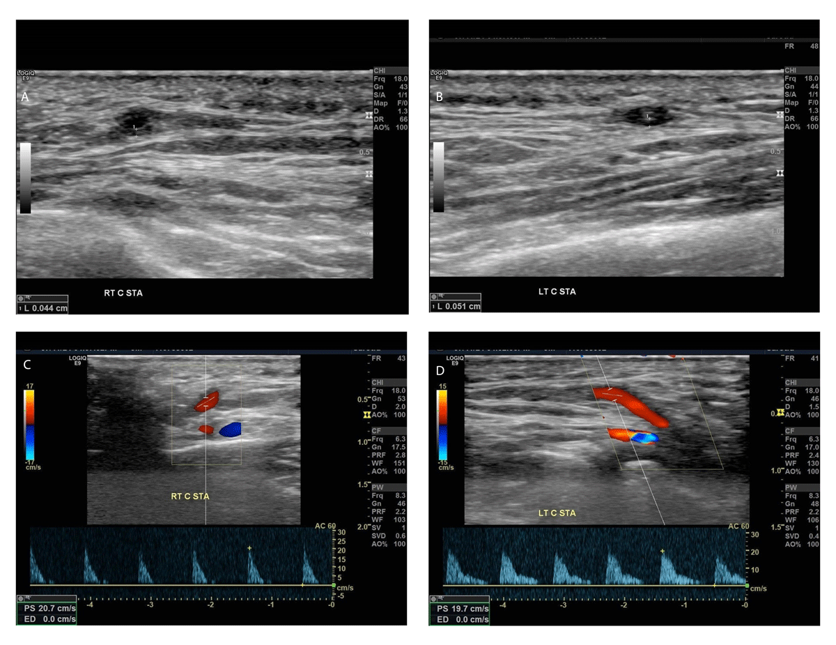
(A) No halo sign is seen in the right superficial temporal artery, (B) whereas a halo sign is present in the left common superficial temporal artery (wall thickness > 0.05 cm). (C, D) Flow velocities are remarkably decreased in both vessels; color Doppler mode increases the visibility of the thickened hypoechoic vessel wall.
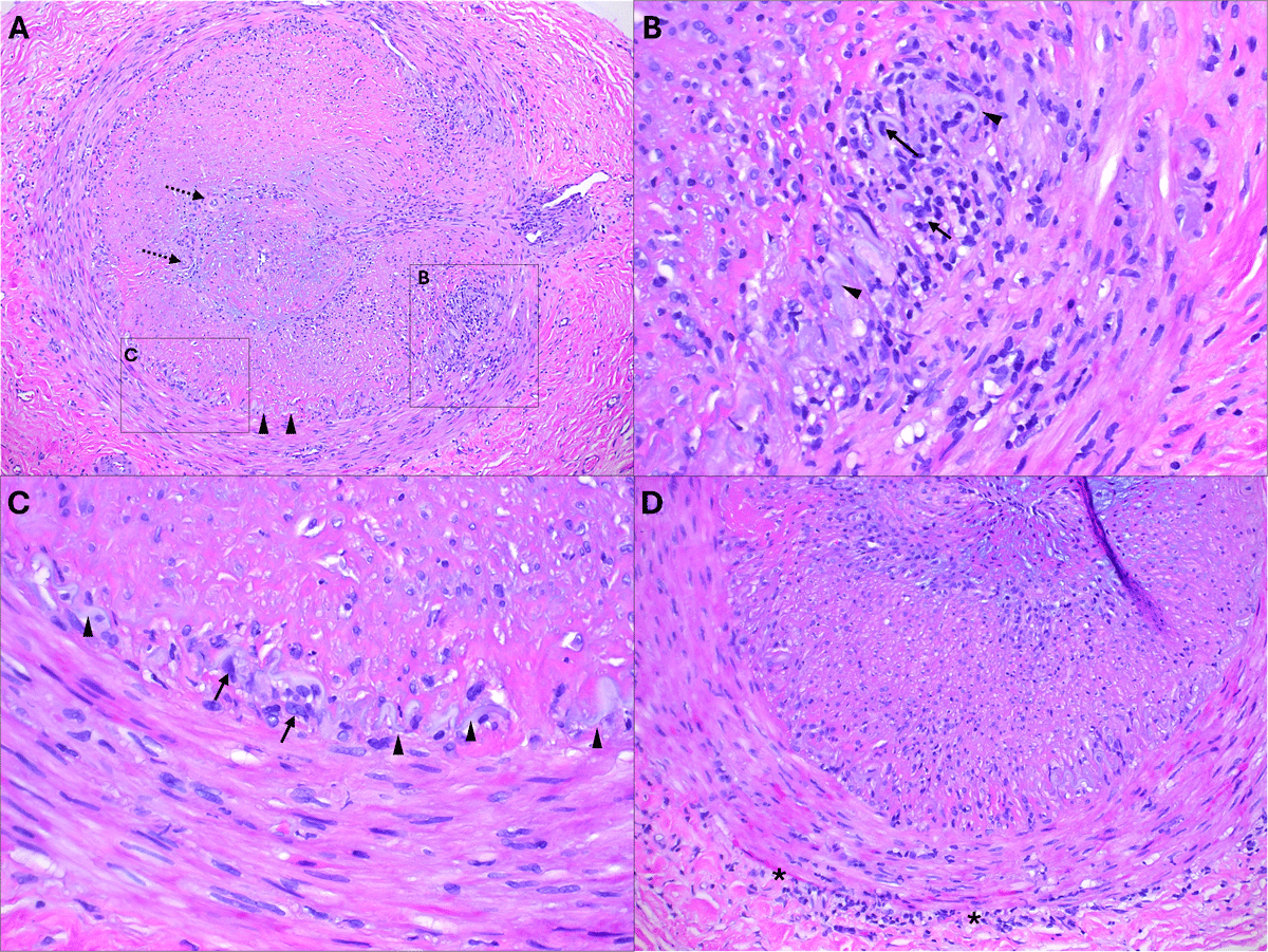
Figure 2
(A) Low-power view of a cross-section of the temporal artery showing complete obstruction by a hyperplastic intima with a mild mixed inflammatory infiltrate and areas of recanalization (dashed arrows). The media also exhibits foci of inflammation (square “B”). The elastic lamina (arrowheads) is focally present and associated with histiocytes (square “C”). (B) Higher magnification image of an area of active inflammation, where the elastic lamina (arrowheads) is obscured by lymphocytes and histiocytes (arrows). (C) Higher magnification image showing an area with less inflammation around the elastic lamina (arrowhead); however, histiocytes remain evident (arrows). (D) Low-power view of a cross-section of the temporal artery displaying a mild lymphocytic infiltrate at the media-adventitia junction (asterisk), indicating healing/treatment effect.
Table 1
American College of Rheumatology classification for giant cell arteritis updated in 2022. ESR: erythrocyte sedimentation rate; CRP: C-reactive protein; TAUS: temporal artery ultrasound; FDG-PET: fluorodeoxyglucose-positron emission tomography
| ABSOLUTE REQUIREMENT | |
| Age ≥ 50 years at time of diagnosis | |
| ADDITIONAL CLINICAL CRITERIA | |
| Morning stiffness in shoulders/neck | +2 |
| Sudden visual loss | +3 |
| Jaw or tongue claudication | +2 |
| New temporal headache | +2 |
| Scalp tenderness | +2 |
| Abnormal examination of the temporal artery | +2 |
| LABORATORY, IMAGING, AND BIOPSY CRITERIA | |
| Maximum ESR ≥ 50 mm/hour or maximum CRP ≥ 10 mg/liter | +3 |
| Positive temporal artery biopsy or halo sign on TAUS | +5 |
| Bilateral axillary involvement | +2 |
| FDG-PET activity throughout aorta | +2 |