
Introduction
Giant cell arteritis (GCA) is the most common form of systemic vasculitis and typically affects medium and large extracranial vessels.1 Its incidence is 15 to 25 cases per 100,000 people over age 50.2 Age is the greatest risk factor, and women are three-times more likely to develop GCA than men.3 This condition primarily affects the arteries of the scalp and head, particularly the temporal, ophthalmic, posterior ciliary, and vertebral arteries.4
The most common symptom of GCA is a new onset headache, often centered around the temples. Other general symptoms include fatigue, loss of appetite, weight loss, flu-like symptoms, and jaw pain while chewing. If GCA affects the blood supply to the eye, it may cause vision disturbances. The most serious complication is permanent blindness, which makes early diagnosis and immediate treatment critically important.
Temporal artery biopsy (TAB) has long been considered the diagnostic gold standard, but its sensitivity is limited (39–80%), and a negative result does not rule out the disease.3,5,6 High-dose prednisolone has been shown to be an effective treatment for GCA.7 To avoid delayed diagnosis and prevent complications such as irreversible blindness, corticosteroid therapy is often started empirically despite potential adverse effects, particularly in the elderly.8,9 Therefore, improving the overall diagnostic is essential in the management of GCA.
While TAB remains widely used, temporal artery ultrasound (TAUS) is emerging as a valuable noninvasive diagnostic tool and is now recognized in the 2022 American College of Rheumatology (ACR) classification criteria.10 TAUS improves diagnostic sensitivity, helps guide biopsies, and—unlike TAB—allows for noninvasive monitoring of treatment response, which is a significant advantage in managing GCA.3,7,10,11 However, the correct sequence of performing TAUS before TAB is often overlooked in clinical practice, potentially affecting diagnostic accuracy.3,7 Here, we present a case that underscores the importance of properly integrating TAUS into the diagnostic pathway for GCA and highlights its potential to enhance patient care.
Case
This patient is a 70-year-old woman with a medical history of hypertension, hyperlipidemia, and Crohn’s disease. She visited a dentist due to bilateral jaw pain that was predominantly affecting her left side. The dentist suspected teeth grinding as the cause and recommended the use of a mouthguard. Two weeks following the initial event, the patient reported intermittent white clouding affecting her vision in the right eye. This symptom subsequently developed in her left eye 1 week later. After another 2 weeks, she sought care from an ophthalmologist, who suspected GCA and prescribed 60 mg of prednisolone daily. The ophthalmologist also referred her for a TAB.
After beginning prednisolone treatment, the patient’s symptoms started to improve. However, before the TAB could be performed, her blood pressure became uncontrollable. This necessitated an emergency department (ED) visit, where the attending physician concluded that the hypertensive episode was a side effect of her medication; he reduced the daily prednisolone dose to 40 mg and started more aggressive antihypertensive therapy. The ED event also led to the cancellation of the initial TAB appointment.
The patient’s outpatient physician then referred her to Houston Methodist Hospital, where a TAB appointment was scheduled, followed by a TAUS appointment a few days later. However, the TAB was again canceled, this time due to unforeseen circumstances that prevented patients and staff from reaching the hospital. The TAUS examination was conducted as scheduled, during which the “halo sign” was detectable in the left common superficial temporal artery, indicating GCA (Figure 1). The halo sign refers to a homogeneous, hypoechoic thickening of the arterial wall, localized to the intima-media complex.10,12
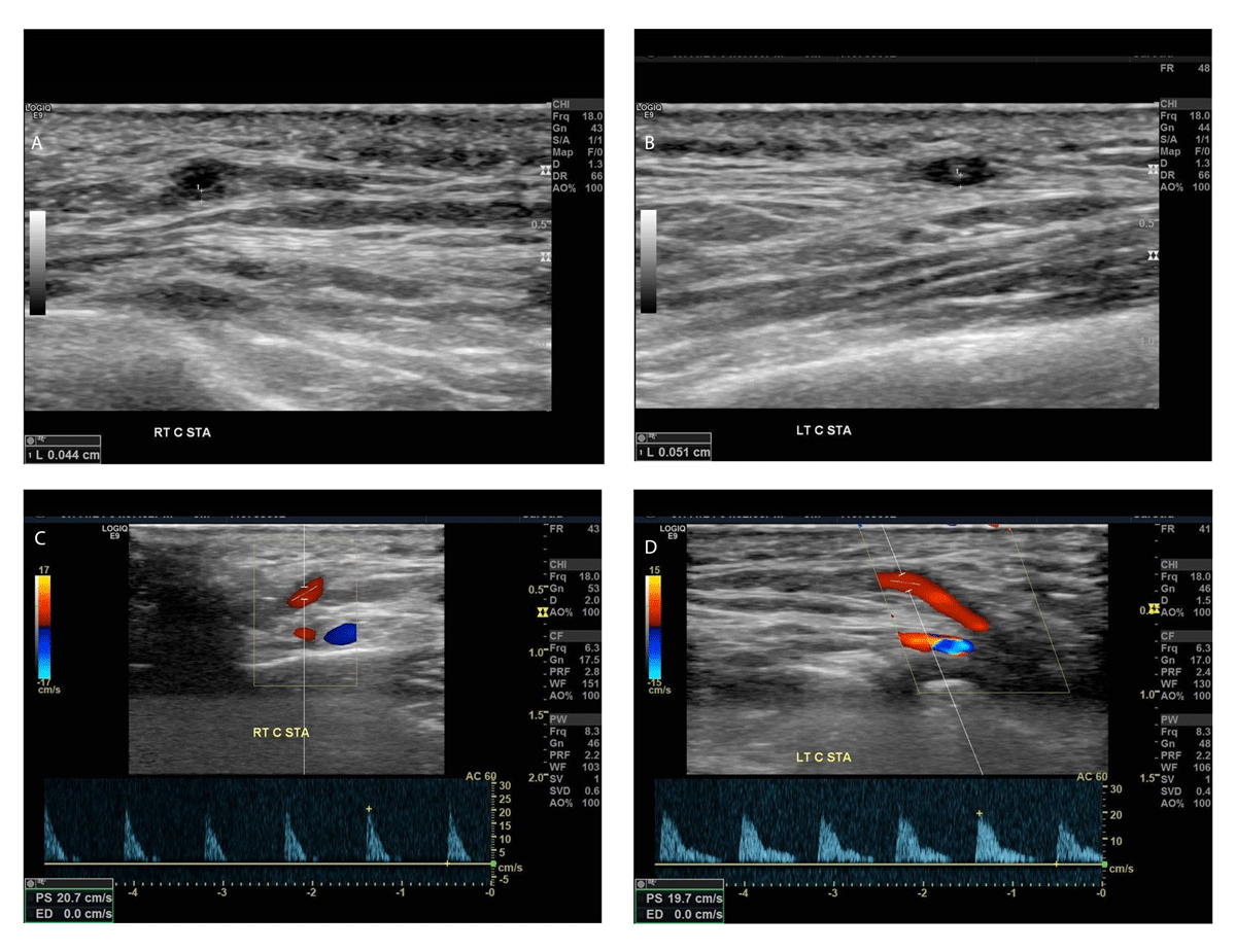
Figure 1
(A) No halo sign is seen in the right superficial temporal artery, (B) whereas a halo sign is present in the left common superficial temporal artery (wall thickness > 0.05 cm). (C, D) Flow velocities are remarkably decreased in both vessels; color Doppler mode increases the visibility of the thickened hypoechoic vessel wall.
The TAB was successfully performed a week later and confirmed the initial TAUS finding, a positive histological result for GCA. The intima of the vessel was infiltrated by lymphocytes, histiocytes, and rare eosinophils. Inflammation was also observed in the muscular layer. Only a few large, multinucleated, giant cells were present, likely due to the initiation of steroid therapy. Features of recanalization were seen in the intima with formation of small-caliber vessels (Figure 2). The patient remained on oral corticosteroid therapy at a lower dose with the addition of anti-interleukin-6 treatment (tocilizumab). Her blood pressure remained under control, and jaw pain and ophthalmic symptoms did not recur by the 6-month follow-up.
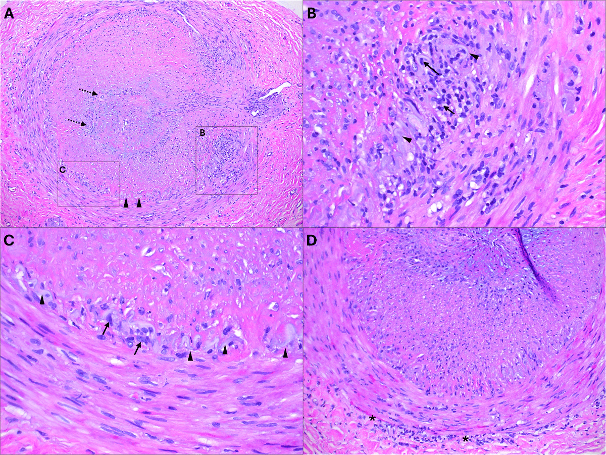
Figure 2
(A) Low-power view of a cross-section of the temporal artery showing complete obstruction by a hyperplastic intima with a mild mixed inflammatory infiltrate and areas of recanalization (dashed arrows). The media also exhibits foci of inflammation (square “B”). The elastic lamina (arrowheads) is focally present and associated with histiocytes (square “C”). (B) Higher magnification image of an area of active inflammation, where the elastic lamina (arrowheads) is obscured by lymphocytes and histiocytes (arrows). (C) Higher magnification image showing an area with less inflammation around the elastic lamina (arrowhead); however, histiocytes remain evident (arrows). (D) Low-power view of a cross-section of the temporal artery displaying a mild lymphocytic infiltrate at the media-adventitia junction (asterisk), indicating healing/treatment effect.
Discussion
Historically, temporal artery TAB has been considered the gold standard procedure for diagnosing GCA and to this day is the most frequently performed test.3,7 The side to be biopsied is selected based on clinical symptoms, and the specimen must be at least 20 mm. GCA is confirmed by inflammation in the media and intima and is characterized by the presence of giant cells.3 The sensitivity of TAB ranges from 39% to 80%, and its specificity is 100%. Due to the lack of sensitivity, a negative TAB does not exclude the presence of the disease.5,6 This means that in cases with mild clinical symptoms, proper diagnosis may be missed or delayed, and if the disease worsens, serious consequences, including vision loss, may occur. Thus, in clinical practice, steroid treatment is typically initiated for patients if symptoms are intensely present, even if the results from superficial TAB are negative.8 This empirical approach creates the risk of overtreatment with potential side effects, including hypertensive episodes and hyperglycemia. The elderly are at particularly high risk for these side effects.9 Therefore, it is important to routinely use other diagnostic tools to facilitate the recognition of GCA in all cases and to avoid the unnecessary use of steroids.
The frequent false-negative results in TAB procedures often stem from three main factors: sampling error (biopsy from an unaffected area), altered histopathology due to pre-biopsy steroid treatment, and inadequate biopsy size. In suspected cases of GCA, the TAB is typically performed on the symptomatic side because the likelihood of finding histological evidence of arteritis is higher on the side where symptoms are present. If the symptoms are bilateral or if the first biopsy is negative, a contralateral biopsy may be considered. This approach, however, may subject the patient to another invasive procedure and may increase the potential for biopsy-related complications, such as unintended injury to veins and nerves, postoperative hematoma, scalp necrosis, wound infection, facial nerve damage, and eyebrow drooping. Additionally, cosmetic issues such as scar widening and foreign body reactions to trapped hair may occur.
The common superficial temporal artery, located superficially just in front of the ear at a depth of approximately 4 mm to 5 mm, is readily accessible for TAUS evaluation. In healthy individuals, the temporal artery appears as a thin-walled structure on gray-scale imaging. In contrast, the halo sign—a hypoechoic, homogeneous, noncompressible thickening of the intima-media complex (IMC) surrounding the arterial lumen—reflects the edematous wall swelling caused by the granulomatous inflammation characteristic of GCA. A halo thickness exceeding 0.5 mm in the common superficial temporal artery (with variations depending on the arterial branch) is considered diagnostic.10,12 During this patient’s TAUS examination, the presence of the halo sign was clearly established (Figure 1).
TAUS is noninvasive, inexpensive, and can be performed quickly, allowing for efficient and prompt diagnosis. These features make it potentially more desirable than TAB for routine screening of GCA. TAUS has high sensitivity (77%) and specificity (96%) for GCA.1 Moreover, the TAUS examination can be easily performed bilaterally, which increases sensitivity, and if the halo sign is present on both sides, the specificity is close to 100%.11 However, both sensitivity and specificity are highly operator dependent. This is because a halo sign also may be present for other diseases, including arteriosclerosis, amyloidosis, eosinophilic granulomatosis with polyangiitis, and juvenile temporal arteritis. However, the halo sign is a rare finding in these conditions, and the clinical picture for each of these diseases differs significantly from that seen in GCA. Additionally, in certain conditions, TAUS may reveal images similar to the halo sign. For example, arteriosclerosis generally presents asymmetric IMC thickening and a hyperechoic signal or mixed echogenicity rather than the hypoechoic, concentric halo sign often seen in GCA.3,7
In 2022, the American College of Rheumatology classification for GCA was updated to include a halo sign seen via TAUS as a criterion (Table 1). In the revised classification, TAUS and TAB have the same diagnostic weight. A cumulative score of 6 or greater meets the classification criteria for GCA (Table 1).10
Table 1
American College of Rheumatology classification for giant cell arteritis updated in 2022. ESR: erythrocyte sedimentation rate; CRP: C-reactive protein; TAUS: temporal artery ultrasound; FDG-PET: fluorodeoxyglucose-positron emission tomography
| ABSOLUTE REQUIREMENT | |
| Age ≥ 50 years at time of diagnosis | |
| ADDITIONAL CLINICAL CRITERIA | |
| Morning stiffness in shoulders/neck | +2 |
| Sudden visual loss | +3 |
| Jaw or tongue claudication | +2 |
| New temporal headache | +2 |
| Scalp tenderness | +2 |
| Abnormal examination of the temporal artery | +2 |
| LABORATORY, IMAGING, AND BIOPSY CRITERIA | |
| Maximum ESR ≥ 50 mm/hour or maximum CRP ≥ 10 mg/liter | +3 |
| Positive temporal artery biopsy or halo sign on TAUS | +5 |
| Bilateral axillary involvement | +2 |
| FDG-PET activity throughout aorta | +2 |
The two diagnostic methods do not exclude each other; in fact, performing both helps ensure an accurate diagnosis and the selection of the appropriate therapy for the patient. However, the correct sequence is critical. TAUS should always be performed before TAB. First, pre-biopsy TAUS can help to identify sites of active vessel wall inflammation, which can reduce the chance of sample collection from an unaffected area, ultimately increasing the sensitivity of the biopsy. Second, after a biopsy, the vessels may become occluded, which prevents proper visualization of the halo sign in a subsequent TAUS examination.
For decades, TAB was the gold standard for diagnosing GCA, which is why it remains the first test ordered by physicians in clinical practice. Today, TAUS is increasingly requested alongside TAB, but the correct sequence of the two examinations is often overlooked. TAUS is suitable not only for diagnosis but also for monitoring the effectiveness of medical therapy. This noninvasive procedure allows continuous assessment of the degree of inflammation, providing insight into the progression of disease and whether the therapy is benefiting the patient.
Although TAUS now has the same diagnostic power as TAB according to the scoring system, its clinical application has not yet become widespread. Creating a separate criterion for TAUS in the American College of Rheumatology’s scoring table and assigning distinct scores for TAB and TAUS would help promote its use.
Conclusions
Temporal artery ultrasound (TAUS) effectively diagnoses giant cell arteritis (GCA) with the presence of a “halo sign,” serving as a noninvasive, quick, and cost-effective complement or possible alternative to temporal artery biopsy (TAB).
TAUS should always be performed prior to TAB. Conducting a TAB first leads to vessel occlusion, which can obscure significant findings on subsequent TAUS.
Early diagnosis and treatment of GCA are crucial to prevent severe complications such as permanent blindness. Prompt treatment based on clinical suspicion and TAUS improves patient outcomes.
Competing Interests
The authors have no competing interests to declare.