
Table 1
Characteristics of intravascular ultrasound catheters.
| CHARACTERISTICS | BOSTON SCIENTIFIC OPTICROSS | PHILLIPS VOLCANO EAGLE EYE | PHILLIPS VOLCANO REVOLUTION | PHILLIPS VOLCANO REFINITY | ACIST KODAMA | TERUMO VIEWIT |
|---|---|---|---|---|---|---|
| Transducer frequency | 40 MHz (50 MHz HD also available) | 20 MHz | 45 MHz | 45 MHz | 60 MHz | 40 MHz |
| Distal shaft profile | 3.1F | 3.3F | 3.2F | 3F | 3.2F | 2.6F |
| Proximal shaft profile | 3.1F | 2.9F | 3.5F | 3F | 3.6F | 3.2F |
| Transducer to tip length | 20 mm | 10 mm (2.5 mm for short-tip version) | 30 mm | 20.5 mm | 20 mm | 29 mm |
| Guiding catheter | ≥ 5F | ≥ 5F | ≥ 6F | ≥ 5F | ≥ 6F | ≥ 5F |
Table 2
Characteristics of recent meta-analyses of randomized controlled trials. IVUS: intravascular ultrasound; OCT: optical coherence tomography; MI: myocardial infarction; TLR: target-lesion revascularization
| AUTHOR/ YEAR | NO. OF TRIALS AND PATIENTS | OUTCOMES |
|---|---|---|
| Khan at al./ 20234 | 20 trials and 11,698 patients comparing intravascular imaging guided PCI vs angiography guided PCI | Cardiac death, MI, stent thrombosis, target vessel revascularization, and TLR |
| Kuno et al./ 202344 | 32 trials and 22,684 patients comparing intravascular imaging guided, functionally guided vs angiography guided PCI | Trial defined MACE (composite of cardiovascular death, MI, and TLR) |
| Giacoppo et al./ 202413 | 24 trials (15,489 patients: IVUS vs angiography, 46.4%; OCT vs angiography: 32.1%; IVUS vs OCT: 21.4%) | The two co-primary outcomes were TLR and MI |
| Stone et al./202431 | 22 trials and 15,694 patients comparing IVUS vs OCT vs angiography-guided PCI | TLR, defined as the composite of cardiac death, target vessel MI, or TLR |
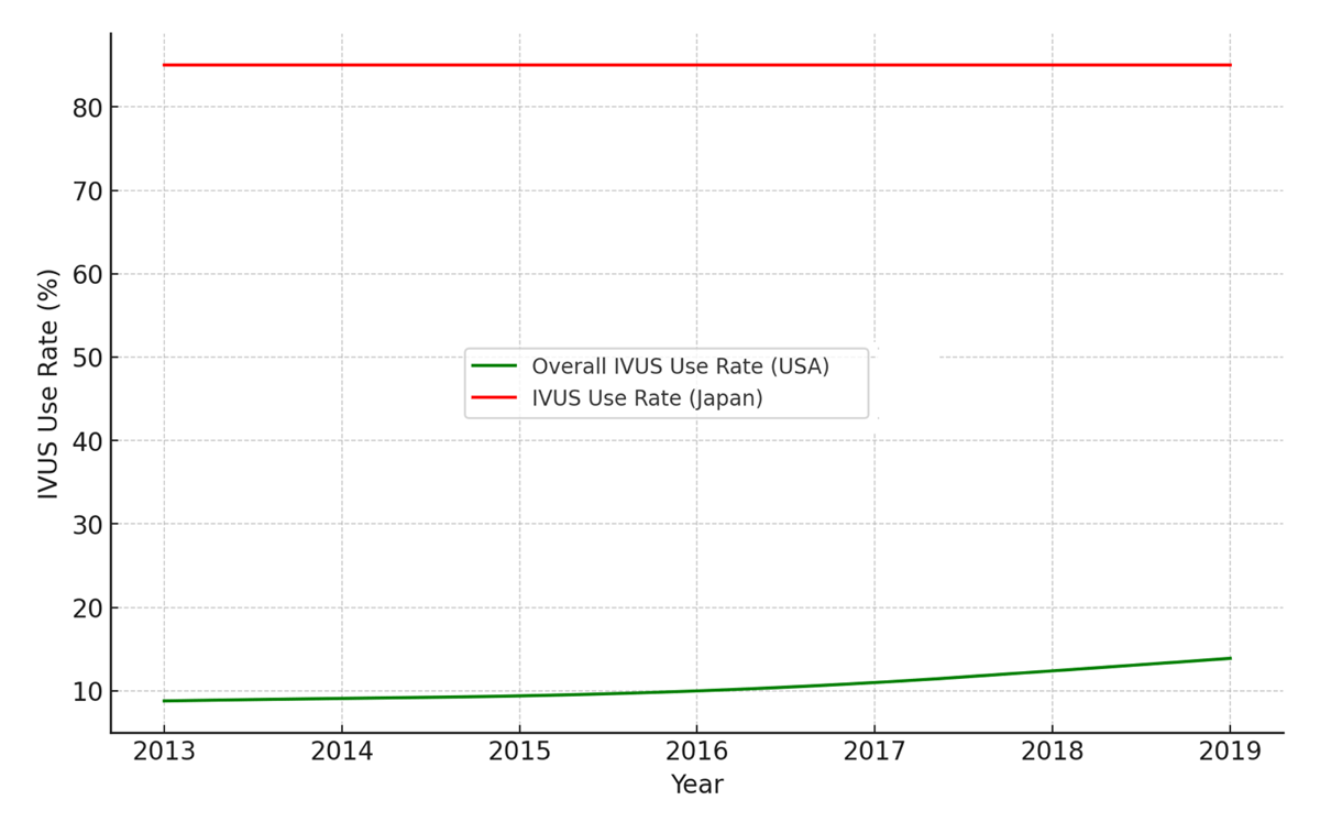
Figure 1
Trends of intravascular ultrasound (IVUS) use in the United States and Japan. To compare the IVUS use rates in percutaneous coronary intervention between the USA and Japan from 2013 to 2019, we extracted data points from provided graphical charts in prior publications.14 For Japan, the data covered up to 2017, which we extrapolated to 2019 using linear extrapolation based on the last observed rate.15 Both datasets were interpolated for smooth transitions between years.
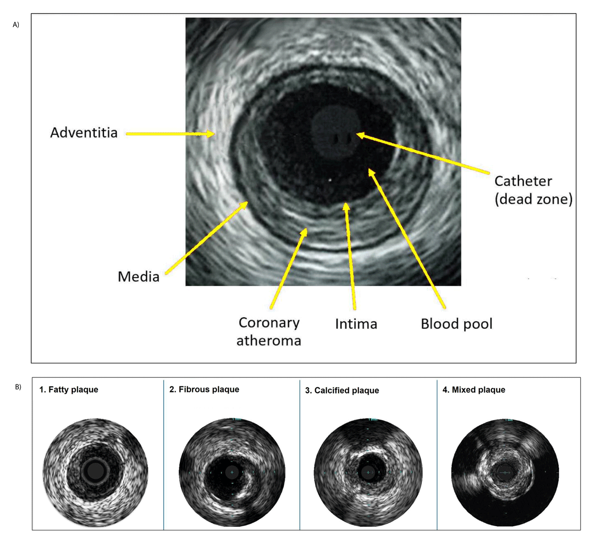
Figure 2
Intravascular ultrasound images of (A) vessel structure and (B) different plaque types.
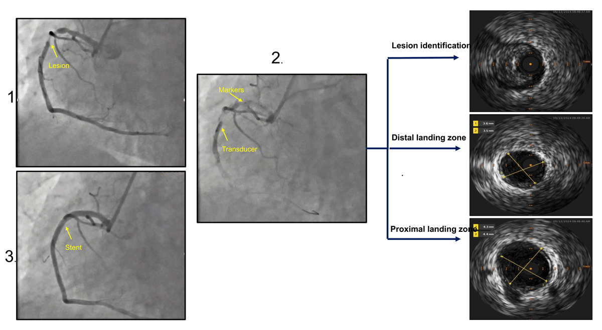
Figure 3
Stepwise approach of SEE IVUS technique. (1) Angiographic evaluation of lesion in the right coronary artery; (2) identification of stent length using IVUS after evaluating lesion, distal and proximal healthy landing zones; and (3) angiogram after stent placement. IVUS: intravascular ultrasound
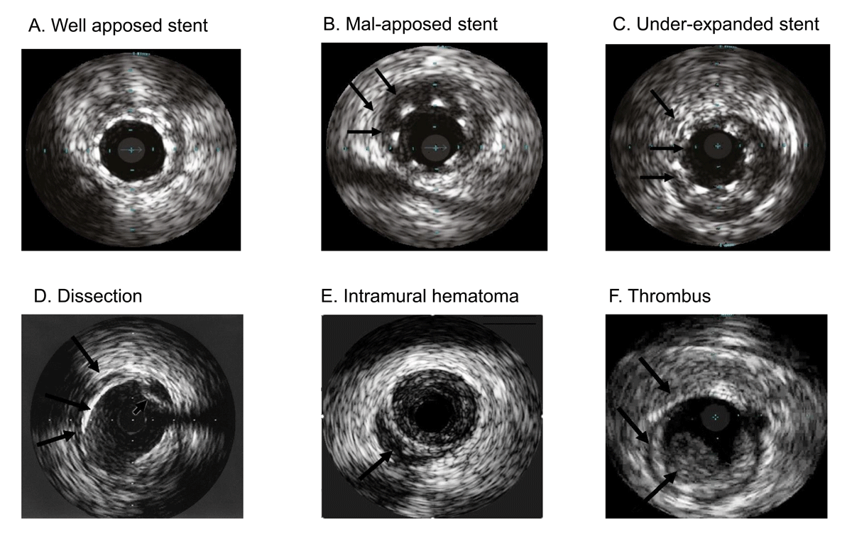
Figure 4
Intravascular ultrasound images of coronary stents and complications.
Table 3
Post stenting IVUS criteria used in landmark studies. MSA: minimal stent area; MLA: minimal lumen area
| STUDY | CRITERIA |
|---|---|
| MUSIC30 | 1. Complete strut apposition 2. Symmetrical stent expansion: minimum/maximum lumen diameter ≥ 0.7 3. Adequate stent expansion
|
| IVUS-XPL42 | 1. Minimal luminal cross-sectional area greater than the luminal cross-sectional area at the distal reference segment 2. Adequate stent expansion
|
| ULTIMATE12 | 1. An MLA in the stented segment > 5.0 mm2 or 90% of the MLA at the distal reference segments 2. Plaque burden 5 mm proximal or distal to the stent edge < 50%; and 3. No edge dissection involving the media with length more than 3 mm 4. Adequate stent expansion
|
| RENOVATE-COMPLEX PCI45 | Sufficient stent expansion without major stent malapposition to the vessel wall or edge dissection
|